Abstract
Drosophila Armadillo and its mammalian homologue β-catenin are scaffolding proteins involved in the assembly of multiprotein complexes with diverse biological roles. They mediate adherens junction assembly, thus determining tissue architecture, and also transduce Wnt/Wingless intercellular signals, which regulate embryonic cell fates and, if inappropriately activated, contribute to tumorigenesis. To learn more about Armadillo/β-catenin's scaffolding function, we examined in detail its interaction with one of its protein targets, cadherin. We utilized two assay systems: the yeast two-hybrid system to study cadherin binding in the absence of Armadillo/β-catenin's other protein partners, and mammalian cells where interactions were assessed in their presence. We found that segments of the cadherin cytoplasmic tail as small as 23 amino acids bind Armadillo or β-catenin in yeast, whereas a slightly longer region is required for binding in mammalian cells. We used mutagenesis to identify critical amino acids required for cadherin interaction with Armadillo/β-catenin. Expression of such short cadherin sequences in mammalian cells did not affect adherens junctions but effectively inhibited β-catenin–mediated signaling. This suggests that the interaction between β-catenin and T cell factor family transcription factors is a sensitive target for disruption, making the use of analogues of these cadherin derivatives a potentially useful means to suppress tumor progression.
INTRODUCTION
Vertebrate β-catenin and its Drosophila homologue Armadillo (Arm) play critical roles in both cell adhesion and signal transduction (reviewed by Gumbiner, 1996; Willert and Nusse, 1998). These proteins are key effectors of Wingless (Wg)/Wnt signal transduction, interacting with DNA-binding proteins of the TCF/LEF family to form bipartite transcription factors that activate Wnt responsive genes (reviewed by Wodarz and Nusse, 1998). β-Catenin and Arm are also core components of the cadherin-catenin complex, which mediates cell-cell adhesion at adherens junctions and connects these junctions to the actin cytoskeleton (reviewed by Ben-Ze'ev and Geiger, 1998; Provost and Rimm, 1999). These quite distinct biological functions of β-catenin/Arm most probably rest on a similar biochemical role: β-catenin/Arm mediates assembly of multiprotein complexes. Thus, in adherens junctions, it simultaneously binds cadherins and α-catenin, whereas in the nucleus it links TCF/LEF proteins to the basal transcriptional machinery (reviewed by Zhurinsky et al., 2000a).
In addition to these roles in normal development and physiology, β-catenin is also a critical target in the development of a variety of human tumors (reviewed by Peifer and Polakis, 2000). In normal cells, β-catenin/Arm's role in signal transduction is kept off by targeting the protein for rapid proteolytic destruction. β-Catenin/Arm is targeted for destruction by a multiprotein complex, which includes two scaffolding proteins, APC and axin/conductin, and a kinase, GSK3β. Assembly of this complex leads to phosphorylation of β-catenin/Arm, and its subsequent ubiquitination and destruction. If this complex is disrupted by mutations in either APC (reviewed by Peifer and Polakis, 2000) or axin/conductin (Liu et al., 2000; Satoh et al., 2000) the Wnt pathway is activated. This can lead to cell proliferation and tumor initiation. Finally, β-catenin binds to a diverse set of other proteins, including the presenilins, the epidermal growth factor (EGF) receptor, the actin-binding protein fascin, and the transcription factor Teashirt (reviewed by Zhurinsky et al., 2000a). In most of these cases, the function of the interaction remains a mystery.
To understand the roles β-catenin/Arm plays in embryonic development and oncogenesis, we must understand in detail how it functions as a scaffold. Furthermore, if we understood in molecular detail how β-catenin/Arm binds to individual partners, we might be able to use this information to design inhibitors that could interfere with β-catenin's interaction with individual partners. For example, a specific inhibitor of the β-catenin/TCF interaction might hold promise as a therapeutic agent in colorectal and other types of cancer. β-Catenin/Arm protein is composed of a series of protein-protein interaction motifs that allow it to function as a scaffold. The N-terminal domain contains the binding site for α-catenin, as well as phosphorylation sites recognized by GSK3β, whereas the C terminus contains the transcriptional activation domain and the binding site for Teashirt (reviewed by Zhurinsky et al., 2000a). The central two thirds of β-catenin/Arm is composed of twelve 42-amino acid Arm repeats. Many partners bind to this region, including TCF/LEF, cadherins, APC, and axin. Because these latter partners play key roles in cell adhesion, Wnt signaling, or the destruction complex, their interactions with β-catenin/Arm have been studied in some detail. These studies examined which regions of β-catenin/Arm are sufficient for binding to the partner and also which region of the partners are sufficient for binding to β-catenin/Arm. In each case, the minimum region of the partner that is sufficient for interaction with β-catenin/Arm is relatively small. The minimal fragments thus far tested range from 70 amino acids for mammalian E- or N-cadherin (Sadot et al., 1998), 41 amino acids for Drosophila E-cadherin (DE-cadherin; Pai et al., 1996), 31 amino acids for Drosophila APC2 (McCartney et al., 1999), 17 amino acids for mammalian LEF-1 (von Kries et al., 2000), and 25 amino acids for human axin (Nakamura et al., 1998).
When the interacting regions of cadherins, TCF/LEF, and APC are aligned, there is only modest sequence similarity, although all are rich in acidic amino acids and serines. Phosphorylation of these serines, as is thought to happen in APC (Rubinfeld et al., 1996), axin (Jho et al., 1999) and cadherin (Stappert and Kemler, 1994; Lickert et al., 2000), would increase the net negative charge further. This charge distribution is intriguing in light of the structure of β-catenin (Huber et al., 1997). The Arm repeats form a superhelix, with a large groove on the surface lined by basic amino acids. In another Arm repeat protein, the nuclear localization signal receptor, the nuclear localization signal peptide binds in an extended conformation in the groove (Conti et al., 1998).
Previous mutational studies of β-catenin/Arm partners have begun to define the sequence requirements for binding. Mutation of three conserved serines in one of the 20-amino acid β-catenin-binding sites of human APC reduced the ability of the mutated fragment to down-regulate β-catenin levels, suggesting reduced binding to β-catenin (Rubinfeld et al., 1997). Clustered point mutations in LEF-1 (Hsu et al., 1998; von Kries et al., 2000) and TCF4 (Omer et al., 1999) identified critical amino acids that are either required for binding or contribute to it. Mutational analysis of the β-catenin-binding site in E-cadherin focused on a series of serine residues that are phosphorylated in vivo (Stappert and Kemler, 1994; Lickert et al., 2000). Mutation of individual serines had a modest effect on binding, whereas mutation of all eight conserved serines abolished binding in vivo.
These data suggest a testable model for the interaction between β-catenin/Arm and its partners, in which charge-based and other interactions mediate the binding between the Arm repeats of β-catenin/Arm and short regions of its partner proteins, potentially binding as extended peptides in the basic Arm repeat groove. Here, we test this model for the interaction between β-catenin/Arm and its partners, by carrying out a detailed analysis of the sequence requirements for interaction between cadherins and β-catenin/Arm.
MATERIALS AND METHODS
Cadherin Constructs and Yeast Two-Hybrid Experiments
The Arm R1–12 construct in pCK2 was described by Pai et al. (1996; it was previously called Arm R1–13, but the subsequent crystal structure of β-catenin led to reassessment of repeat number and boundaries). Similar constructs containing Arm repeats 2–10 (ArmR2–10: amino acids 177–596) and the corresponding fragments of mouse β-catenin (R1–12: amino acids 119–708; R2–10: amino acids 169–583) were generated for this work. DE-cadherin fragments were generated by polymerase chain reaction (PCR) with flanking BamHI and EcoRI restriction sites and cloned into pCK4 (Pai et al., 1996). The amino acids included in each fragment are diagrammed in Figure 1, A and B. All constructs included a stop codon after the final amino acid of DE-cadherin. All clones were sequenced in their entirety to confirm their sequence. The DE-cadherin mutants (DEC) were generated by a two-step PCR procedure. Primers for each strand containing the desired mutant sequence were used in two separate PCR reactions with flanking primers to amplify the N- and C-terminal portions of the DE-cadherin cytoplasmic domain. Products from these two reactions were mixed and used as a template for another PCR reaction containing only the flanking primers. This reaction generated a full-length DE-cadherin cytoplasmic domain with flanking BamHI and EcoRI sites containing the desired mutations, which was cloned into pCK4. Mutation DECM2 was introduced into the smaller DEC30 fragment by amplifying the relevant portion of the longer mutant clone with DEC30 primers. All mutations were confirmed by sequencing and are diagrammed in Figure 6A. Two-hybrid assays were performed as described by Pai et al. (1996). Arm or β-catenin fragments were fused to the LexA DNA-binding domain in pCK2, and DE-cadherin fragments were fused to the Gal4 activation domain in pCK4. The two plasmids were transformed simultaneously into the yeast strain L40. β-Galactosidase values are the averages from duplicate assays performed on at least three independent transformants.
Figure 1.
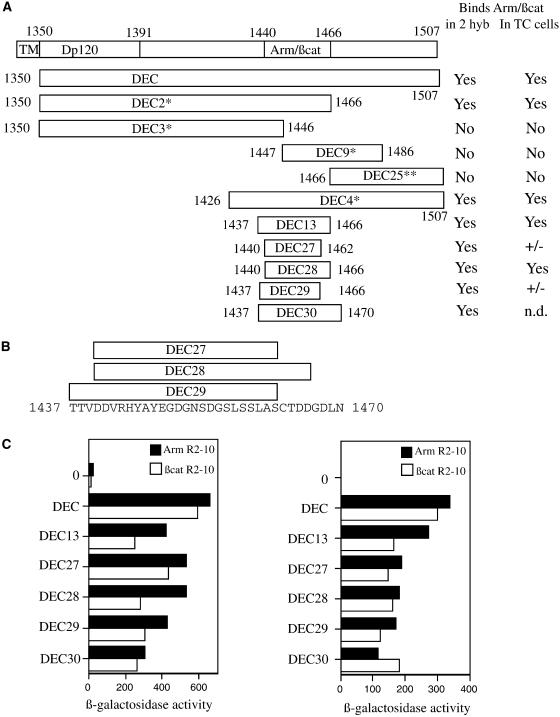
Mapping the minimal binding site on DEC cytoplasmic tail for β-catenin (βcat) and Arm using the yeast two-hybrid (2 hyb) system. (A) Schematic representation of the DEC derivatives used in our analyses, with ability to bind Arm/β-catenin in either yeast or mammalian cells summarized in the right-hand columns. TM, transmembrane; TC, tissue culture. *Data from Pai et al. (1996). (B) Sequence of the minimal binding region of DE-cadherin, with the boundaries of the smallest DEC derivatives indicated. (C) All of the DEC derivatives bind to both fragments of Arm and β-catenin in yeast. The full-length DE-cadherin cytoplasmic domain (DEC), or smaller derivatives of DEC (diagrammed in A and B), fused to the Gal4 transcriptional activation domain, were transformed into yeast cells along with portions of Arm or β-catenin fused to the LexA DNA-binding domain. Average β-galactosidase values are shown for each DEC derivative together with the full Arm repeat region of Arm or β-catenin (Arm R1–12 or βcat R1–12, left), or a smaller fragment of the Arm repeat region (Arm R2–10 or βcat R2–10, right). 0, background level of β-galactosidase activity with no DEC fragment fused to Gal4. **DEC 25 was tested against only Arm R1–12. Its β-galactosidase value was 14.4 U, compared with 18.3 U for the negative control.
Figure 6.
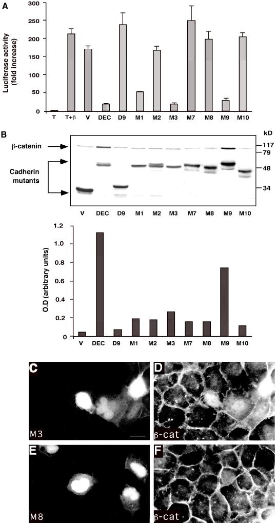
Analysis of the effect of clustered point mutations in the minimal Arm-binding domain of DEC on its capacity to interact with β-catenin, protect it from degradation, inhibit β-catenin/LEF-mediated transactivation, and affect β-catenin organization. (A) The ability of clustered point mutations (diagrammed in Figure 5A) to affect β-catenin/LEF-1–mediated transactivation in 293T cells was examined as described in Figure 2B. (B) The ability to protect β-catenin from degradation was examined in CHO cells, and the levels of β-catenin were quantified as described in Figure 2C. Because the samples were originally analyzed on the same gel with the samples shown in Figure 2C, the control samples (V, DEC, and D9) are shown again. (C–F) MDCK cells were transfected with GFP-tagged DECM3 (C, M3) and DECM8 (E, M8), and the organization of the endogenous β-catenin (β-cat) in the respective samples (D and F) was determined by double fluorescence microscopy. Bar, 10 μm.
Cell Lines and Transfections
Chinese hamster ovary (CHO), 293T and MDCK cells were maintained in DMEM with 10% calf serum. Transient transfections with Drosophila E-cadherin (DEC) constructs were carried out using the calcium phosphate precipitation method with 293T cells and by Lipofectamine (GIBCO, Grand Island, NY) with CHO cells. For recloning the various mutant DEC sequences from the pCK4 plasmid into the pEGFP-C1 plasmid (Clontech, Palo Alto, CA), the DEC inserts were amplified by PCR using primers designed to contain pCK4 plasmid sequences (in the HA-tag domain) that were linked to the multicloning site, ACCTAGATCTTACCCATACGATGTTCCAG, and the terminator sequence, CGATGCAC AGTTGAAGTGAACTTGC, downstream of the multicloning site of pCK4. The amplified sequences were excised by BglII and EcoRI digestion and inserted into pEGFP-C1 at the same BglII/EcoRI sites. The green fluorescence protein (GFP) tag was localized at the N terminus of these DEC constructs. For LEF/TCF-dependent transactivation analysis, cells were transfected with the pCGN-HA expression vector containing the S33Y β-catenin mutant (Shtutman et al., 1999) and the TOPFLASH and FOPFLASH luciferase reporter vectors (van de Wetering et al., 1997), as previously described (Zhurinsky et al., 2000b). A β-galactosidase–expressing vector was cotransfected as an internal control for transfection efficiency. After 24 h, the cells were lysed, and both luciferase and β-galactosidase activities were determined by enzyme assay kits (Promega, Madison, WI). For Western blots and immunoprecipitations, cells were harvested 24 h after transfection and lysed in either Laemmli's sample buffer or immunoprecipitation buffer (see below), respectively.
Immunoblotting and Immunoprecipitation
Equal amounts of total protein from the different transfected cells were separated by SDS-PAGE and subjected to Western blotting using the following antibodies: monoclonal anti-HA (clone 12CA5; Boehringer Mannheim, Indianapolis, IN), polyclonal anti-HA (a gift from M. Oren, Weizmann Institute of Science, Rehovot, Israel), polyclonal anti-β-catenin (Sigma, St. Louis, MO), and monoclonal anti-GFP antibody (Roche Molecular Biochemicals, Burlington, NC). For coimmunoprecipitation, cells transfected with the GFP-DEC constructs and the S33Y β-catenin were lysed in immunoprecipitation buffer containing 20 mM Tris-HCl, pH 8.0, 1% Triton X-100, 140 mM NaCl, 10% glycerol, 1 mM EGTA, 1.5 mM MgCl2, 1 mM dithiothreitol, 1 mM sodium orthovanadate, and 50 μg/ml phenylmethylsulfonyl fluoride. Equal amounts of protein were incubated with 2 μl of polyclonal anti-β-catenin antibody and 20 μl of protein A/G-agarose beads (Santa Cruz Biotechnology, Santa Cruz, CA) for 4 h at 4°C. The beads were washed five times with 20 mM Tris-HCl buffer, pH 8.0, containing 150 mM NaCl and 0.5% Nonidet P-40, and the immune complexes were recovered by boiling in Laemmli's sample buffer and resolved by SDS-PAGE. To detect the coprecipitated GFP-DEC constructs, the blots were incubated with anti-GFP antibody. Blots were developed using the ECL method (Amersham, Arlington Heights, IL). Autoradiograms were scanned with a GS-700 imaging densitometer (Bio-Rad, Hercules, CA) and quantitated using the FotoLook PS 2.07.2 software. The intensity of the bands was quantitated using the National Institutes of Health image 1.61 software.
Immunofluorescence Microscopy
Cells were cultured on glass coverslips, fixed with 3% paraformaldehyde in phosphate-buffered saline, and permeabilized with 0.5% Triton X-100. Monoclonal or polyclonal antibodies to β-catenin were used to label the endogenous β-catenin. The secondary antibodies were Cy3 goat anti-mouse or anti-rabbit immunoglobulin G (Jackson ImmunoResearch Laboratories, West Grove, PA). The transfected GFP-DEC constructs were detected in the fluorescein isothiocyanate channel. The samples were visualized using an Axiovert S100 microscope (Zeiss, Germany).
RESULTS
Mapping the Minimal Arm/β-Catenin–interacting Region of DE-Cadherin
The binding site for Arm on the DE-cadherin cytoplasmic tail was previously mapped to the C-terminal portion of the cadherin tail (Pai et al., 1996). To determine the minimum region essential for binding, we first used the yeast two-hybrid system to assess binding between the Arm repeat region of both Arm and β-catenin and smaller fragments of the DE-cadherin tail (Figure 1). A series of fragments, ranging in size from 23–34 amino acids, were tested, and all bound both Arm and β-catenin as assessed by the two-hybrid system (Figure 1C).
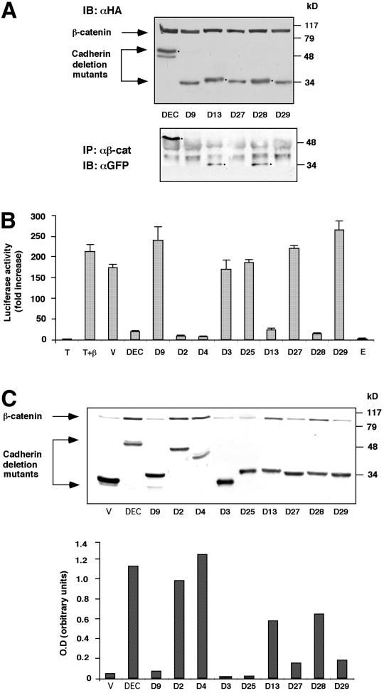
We then examined whether these minimal binding fragments, when fused to GFP at their N termini, retained the ability to interact with β-catenin in mammalian cells. To do so, we made use of several assays. First, we assessed the ability of the GFP–DE-cadherin tail and its fragments to coimmunoprecipitation with β-catenin (Figure 2A). Second, we tested the ability of these fragments to block the interaction of β-catenin with endogenous LEF/TCF, as measured by their ability to block LEF/TCF-mediated transactivation (Figure 2B). Finally, we assessed the capacity of these fragments to block the interaction of β-catenin with endogenous APC or axin, thus stabilizing β-catenin by blocking its targeting to the proteasome (Figure 2C). These assays generally paralleled the results in the yeast two-hybrid system (Figure 1C). One difference was noted however: whereas DEC28, which is 27 amino acids in length, binds by all three assays to β-catenin (Figures 1), DEC27 and DEC29, the smallest constructs that bound Arm and β-catenin in yeast (Figure 1, A and C), failed to detectably coimmunoprecipitate with β-catenin (Figure 2A) and also failed to block LEF/TCF-mediated gene expression (Figure 2B). DEC27 and DEC29, which are 4 amino acids shorter than DEC28 at their C termini (DEC29 also has three extra N-terminal amino acids), exhibited a reduced ability to stabilize β-catenin, although they retained some activity in this assay (Figure 2C).
Figure 2.
Analysis of the ability of different fragments of the DEC cytoplasmic tail to interact with β-catenin, affect its stability, and inhibit β-catenin–mediated transactivation. (A) The ability of selected GFP-DEC derivatives to coimmunoprecipitate with cotransfected HA-tagged β-catenin was determined by immunoprecipitation (IP) from 293T cells transfected with HA-tagged β-catenin and GFP-tagged DEC constructs with anti-GFP antibody, followed by Western blotting with anti-HA antibody. The total level of transfected β-catenin and DEC constructs was determined by immunoblotting (IB) with anti-HA-antibody. (B) 293T cells were transfected with GFP-tagged derivatives of the DEC cytoplasmic tail (DEC) or the full-length mammalian E-cadherin tail (E), along with β-catenin (β), a LEF/TCF reporter plasmid (T), and Lac Z. Luciferase activity was determined from duplicate plates as fold activation after normalizing for transfection efficiency by measuring β-galactosidase activity. T, cells were transfected with the reporter plasmid alone; V, cells transfected with the reporter plasmid, HA-tagged β-catenin and the GFP-vector used for the construction of the cadherin derivatives. (C) The cadherin derivatives used in B were transfected into CHO cells, and their ability to protect the endogenous β-catenin from degradation was determined by analyzing the level of β-catenin expressed in the DEC mutant-transfected cells by Western blotting with anti-β-catenin antibody. The level of expression of DEC constructs was determined by immunoblotting with an antibody against the GFP tag. Quantitation of the β-catenin level expressed in CHO cells was carried out by normalizing the intensity of the β-catenin bands shown to those of the DEC band for each derivative.
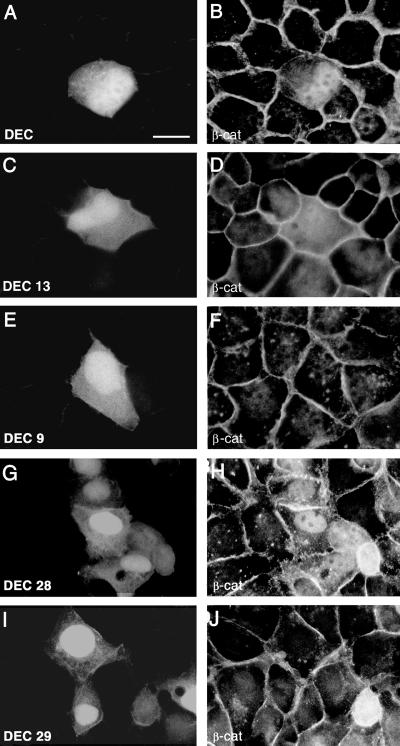
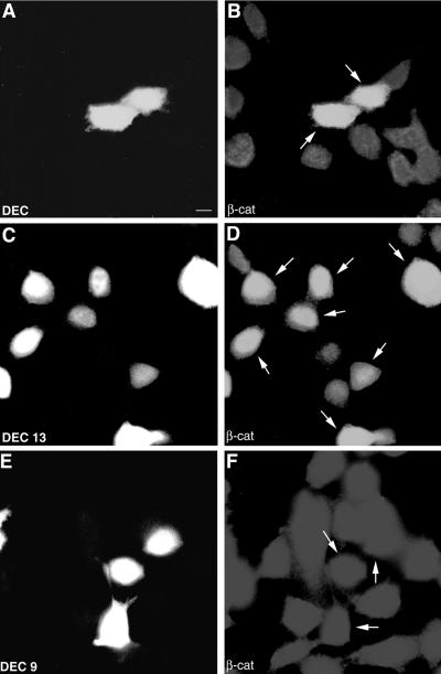
A subset of these DEC fragments was also tested for the effect on endogenous β-catenin localization and levels by immunofluorescence (Figure 3). Transfection of the control GFP expression vector into MDCK cells gave a diffuse distribution of GFP in the cytoplasm and the nucleus, without affecting the organization of the endogenous β-catenin in the transfected cells. In contrast, expression of the GFP-tagged DEC tail in these cells (DEC, Figure 3A) resulted in partial disruption of adherens junctions and the accumulation of β-catenin in the cytoplasm and the nuclei (Figure 3B). Expression of the shorter (30 amino acid) cadherin tail fragment DEC13 in MDCK cells (Figure 3C) resulted in the accumulation and diffuse distribution of β-catenin (Figure 3D) but without a detectable effect on its organization in adherens junctions. In contrast, DEC9, which was unable to bind β-catenin in the assays described above (Figures 1 and 2), had no effect on the accumulation or organization of endogenous β-catenin in MDCK cells (Figure 3, E and F). It is noteworthy that DEC13 was positive in Arm/β-catenin binding in the two-hybrid screen (Figure 1, A and B) and by coimmunoprecipitation in mammalian cells (Figure 2A) and effectively protected β-catenin from degradation in 293 cells (Figure 2C). In CHO cells that express only very low levels of N-cadherin (and thus do not form adherens junctions), transfection of DEC (Figure 4A) or DEC13 (Figure 4C) brought about the accumulation of β-catenin in the nuclei of these cells (Figure 4, B and D), whereas DEC9 expression (Figure 4E), as expected, had no effect on the endogenous β-catenin (Figure 4F). The transfection into MDCK cells of DEC27 and DEC29 (Figure 3I), which did not bind to β-catenin in the assays described above (Figures 1 and 2), also had no effect on the subcellular distribution of β-catenin or the organization of adherens junctions (Figure 3J). In contrast, DEC28 (which bound to β-catenin in the two-hybrid assay and coimmunoprecipitation, Figures 1 and 2), when transfected into MDCK cells (Figure 3G), induced the accumulation of the endogenous β-catenin in the cytoplasm and nuclei of these cells (Figure 3H).
Figure 3.
The effect of DEC cytoplasmic domain derivatives on the organization of adherens junctions and subcellular distribution of β-catenin (β-cat). MDCK cells were transfected with various GFP-tagged DEC derivatives (diagrammed in Figure 1, A and B), and the distribution of the GFP-tagged DEC derivatives (A, C, E, G, and I) and of the endogenous β-catenin (B, D, F, H, and J) was determined by double fluorescence microscopy using rhodamine-labeled anti-β-catenin antibody. Bar (in A), 10 μm. Note the reduction in junctional β-catenin in DEC-expressing cells but not in cells transfected with other DEC constructs. Also note that DEC9 and DEC29 do not increase the endogenous β-catenin level, whereas DEC13 does.
Figure 4.
Analysis of the ability of DEC derivatives to increase the level and the accumulation of endogenous β-catenin (β-cat) in the nucleus. Some of the GFP-DEC constructs described in Figure 3 were transfected into CHO cells (A, C, and E), and their ability to elevate the endogenous β-catenin and induce its translocation into the nucleus (B, D, and F) was determined by double fluorescence as described in Figure 3. Bar in (A), 10 μm. The arrows mark the transfected cells. Note that, whereas DEC13 and DEC induced the accumulation of endogenous β-catenin in the nucleus, DEC9 was unable to do so.
Defining Amino Acids Critical for the Arm/β-Catenin–DE-Cadherin Interaction
We next set out to determine which amino acids within the minimal DE-cadherin–binding region were essential for the interaction with Arm and β-catenin. Mammalian β-catenin can bind DE-cadherin both in Drosophila (White et al., 1998; Cox et al., 1999) and in cultured mammalian cells (see below), and Arm can also bind mammalian E-cadherin (A. Wodarz and R. Nusse, personal communication). We therefore used the comparison of vertebrate and Drosophila cadherins to determine candidate residues that might contribute to binding. Based on comparisons of the Arm-binding regions of cadherin, APC, and TCF family members, we focused on acidic and serine/threonine residues, although we also mutated other conserved amino acids. Although we focused on residues within the minimal binding region, we introduced our mutations in the context of the full-length DE-cadherin tail, thus mimicking the situation in vivo.
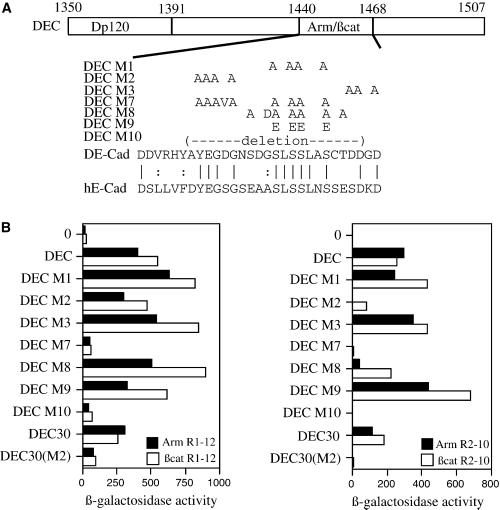
We began with three clustered point mutations that each change three or four nearby residues in different regions of the minimal binding site to alanine (Figure 5A). DECM1 altered four conserved serine residues in the center of the minimal binding region, DECM2 altered four conserved amino acids including one acidic residue in the N-terminal part of the minimal binding region, and DECM3 altered three conserved acidic residues (aspartates) in the C-terminal part of the minimal binding region (Figure 5A). Surprisingly, none of these mutations significantly affected binding to the full-length Arm repeat region of either Arm or β-catenin in the two-hybrid system (Figure 5B, left). Because Arm repeats 3–8 retain the ability to bind several of Arm's partners (Pai et al., 1996), we reasoned that such shorter Arm fragments might be compromised in binding to DE-cadherin derivatives and thus might be more sensitive to mutational changes. We therefore tested the DECM1-DECM3 mutants for their capacity to bind to Arm repeats 2–10 of both Arm and β-catenin (Figure 5B, right). In this assay, there was a substantial reduction in the binding of DECM2 to both Arm and β-catenin, whereas the other two mutations (DECM1 and DECM3) did not substantially affect binding (Figure 5B, right). These data suggested that DECM2 might weaken binding but not enough to be detectable in the context of the full-length DE-cadherin tail binding to the full-length Arm repeat region. Interactions outside the minimal Arm-binding region may normally help stabilize this association and thus could partially compensate for mutations such as DECM2. We therefore introduced the DECM2 mutation into a 34-amino acid peptide centered on the minimal binding region (DEC30; Figure 1A). In this context (rather than in the full-length DEC tail), the DECM2 mutation essentially abolished binding to even the full-length Arm repeat region (DEC30(M2); Figure 5B).
Figure 5.
Analysis of the effect of clustered point mutations in the minimal Arm-binding domain of DEC on its ability to bind Arm and β-catenin (βcat) in the yeast two-hybrid system. (A) Diagram of the DEC tail and sequences of the clustered point mutations used in this study, with the sequences of DE-cadherin (DE-Cad) and human E-cadherin (hE-Cad) in the region of the mutations shown below. All mutations were introduced into and analyzed in the context of the full-length cytoplasmic tail. The mutation DECM2 was also tested in the context of a smaller fragment of the cadherin tail (DEC30; Figure 1A)—this derivative is DEC30(M2). (B) The DE-cadherin mutants diagrammed in A were fused to the Gal4 transcription activation domain and transformed into yeast cells together with the full Arm repeat region of Arm or β-catenin (Arm R1–12 or βcat R1–12, left) or a smaller fragment of the Arm repeat region (Arm R2–10 or βcat R2–10, right), fused to the LexA DNA-binding domain. Average β-galactosidase activities are shown.
Next, we tested this same set of mutants for binding to β-catenin in cultured mammalian cells, using the assays described above. Both DECM1 and DECM3 retained substantial ability to block TCF-directed gene expression (Figure 6A), suggesting that they could block binding of β-catenin to TCF family members. All three mutants were reduced in their ability to stabilize β-catenin (Figure 6B), although all appear to retain a small amount of activity in this assay. Finally, the overexpression of DECM3 in MDCK cells (Figure 6C) resulted in the accumulation of β-catenin in the cytoplasm and nuclei of these cells (Figure 6D).
In addition to these mutations, we also analyzed three mutants with more substantial changes in the minimal binding region. Mutant DECM7 combined the changes found in DECM1 and DECM2 and also mutated an additional amino acid, aspartic acid 1450, to valine (Figure 5A). Mutant DECM8 altered all of the serine and threonine residues in the core of the binding site to alanine (Figure 5A) and also altered the semiconserved residue glycine 1455 to aspartic acid. A subset of these residues is a likely target of phosphorylation in vivo (Stappert and Kemler, 1994). Finally, in DECM10, 20 amino acids were deleted in the core of the minimal binding region (Figure 5A). When tested against the full Arm repeat region of Arm or β-catenin in the two-hybrid system, DECM7 and DECM10 were essentially inactive (Figure 5B). In contrast, DECM8 had little effect on binding to the entire Arm repeat region (Figure 5B, left), although it did reduce binding to Arm repeats 2–10 of both Arm and β-catenin (Figure 5B, right). We also analyzed an additional mutant, DECM9, in which four of the conserved serine residues were changed to glutamic acid (Figure 5A). These serine residues are phosphorylated in vivo, and in some cases, this change mimics phosphorylation. DECM9 retained full ability to bind both Arm and β-catenin in the two-hybrid assays (Figure 5B).
We then studied the interaction of this set of mutants with β-catenin in mammalian cells. In this setting, DECM7, DECM8, and DECM10 all abolished interaction completely, losing both the ability to block TCF/LEF-dependent gene expression (Figure 6A) and to stabilize β-catenin (Figure 6B). The expression of M8 in MDCK cells (Figure 6E) had no effect on adherens junctions or on β-catenin organization (Figure 6F). In contrast, DECM9 preserved the capacity to interact with β-catenin, because it very efficiently protected it from degradation (Figure 6C) and inhibited LEF/TCF-directed transactivation (Figure 6A), in line with the two-hybrid assays. This is in striking contrast to DECM1, in which the same serine residues were changed to alanine rather than glutamic acid.
DISCUSSION
β-Catenin/Arm plays key roles in cell-cell adhesion and Wnt signal transduction. Deregulation of these activities can lead to disease. Activation of β-catenin–mediated signaling contributes to a wide variety of human tumors (reviewed by Zhurinsky et al., 2000a), and dysfunction of cadherin-catenin adhesion is involved in cancer metastasis (reviewed by Christofori and Semb, 1999). β-Catenin/Arm mediates these distinct processes by forming a scaffold upon which different multiprotein complexes are assembled. To unravel β-catenin's normal functions and the alterations in its function in disease, a detailed understanding of its interactions with protein partners is required. This might facilitate a rational approach to design inhibitors of these interactions. For example, an effective, specific inhibitor of the β-catenin–TCF interaction might have therapeutic potential in cancers in which Wnt signaling is activated.
We used the cadherin/β-catenin interaction as a model for investigating this question. We previously found that 71 amino acid derivatives of the cytoplasmic tail of vertebrate N- or E-cadherin inhibit β-catenin/TCF-mediated transactivation when introduced into human colon cancer cells (Sadot et al., 1998; Simcha et al., 1998). Moreover, expression of the N-cadherin tail in human colon cancer cells inhibited the elevated transcription of cyclin D1 (Shtutman et al., 1999), thus potentially suppressing its oncogenic function. In the present study, we analyzed the interaction between DE-cadherin and β-catenin/Arm in detail, using several assays, each of which provided different measures of binding. Using the yeast two-hybrid system, we assessed interaction in the absence of most, if not all, of β-catenin/Arm's normal partners, because yeast lack β-catenin, cadherins, TCFs, APC, and axin. Furthermore, kinases and other proteins that regulate interactions between β-catenin/Arm and its partners, are also likely absent. We also used several assays in mammalian cells, which, in contrast to yeast, possess both a full (or nearly full) complement of β-catenin partners and the normal set of regulatory machinery that modulates the interaction between β-catenin and its partners. This diversity of assays allowed us to discriminate among the binding abilities of cadherin mutants in a more detailed way than was possible in most previous studies of β-catenin/Arm interaction with other partners, which, for the most part, relied on single assays.
Using these assays, we found that quite small fragments of DE-cadherin, including the 23-amino acid DEC27, bind both β-catenin and Arm in yeast. In cultured mammalian cells the criteria for interaction were more stringent. The smallest DE-cadherin peptide that interacted in mammalian cells was DEC28, which is 27 amino acids in length. This difference may reflect the fact that in mammalian cells DEC fragments must compete with endogenous partners for binding—weakened interactions might prevent effective competition. Alternately, it may simply reflect differences in the fusion proteins used in each assay. It is noteworthy, however, that the binding of short DEC fragments, such as DEC28 in mammalian cells, is weaker than binding of the full cytoplasmic tail of DE-cadherin or mammalian E-cadherin, as assessed by their ability to inhibit transcriptional activation by β-catenin (Figure 2B).
Our mutational analysis also revealed critical amino acids in cadherin required for interaction with β-catenin. The β-catenin/Arm-binding site is highly conserved among all classical cadherins. Most of our mutations in conserved residues had parallel effects in yeast and mammalian cells. For example, mutation of three acidic amino acids near the C terminus of the minimal binding region (DECM3) had little effect on either binding in yeast or the ability to block TCF-mediated transactivation, whereas mutation of four more N-terminal conserved residues (DECM2) resulted in a detectable reduction in binding in yeast and a substantial reduction in the ability to block TCF-mediated transactivation. The most extensive mutations, DECM7 and DECM10, completely blocked the binding in all assays.
Surprisingly, the serine residues in the binding site, mutated in DECM1 and DECM8, were largely dispensable for binding in yeast. In contrast, these mutations impaired or eliminated the ability to block TCF-mediated transactivation and to stabilize β-catenin in mammalian cells. One possible explanation for these differential effects is that these serines are phosphorylated in mammalian cells (Stappert and Kemler, 1994; Lickert et al., 2000); this may strengthen binding. Consistent with this possibility, mutation of the four conserved serines to glutamic acid (mutant DECM9), which may mimic phosphorylation, did not block binding to β-catenin in mammalian cells. In fact, DECM9 very effectively protected β-catenin against degradation (Figure 2C), in agreement with recent studies by Lickert et al. (2000). If, in yeast, the relevant kinase(s) are absent, mutation of these serines would not affect binding.
While this paper was under review, two studies appeared that complement our data. Graham et al. (2000) solved the structure of β-catenin bound to XTcf3, thus revealing in full detail how β-catenin binds to one of its partners. XTcf3 binds in the groove on the surface of β-catenin, with the XTcf3 peptide forming a β-hairpin at its N terminus and an α-helix at its C terminus, with an extended peptide in between. From this structure and parallel mutagenesis of β-catenin, they identified two key charge-charge interactions between β-catenin and the extended XTcf3 peptide and a key hydrophobic interaction of β-catenin with the α-helix of XTcf3. They also assessed the ability of cadherin to bind to their β-catenin mutants and, from this, proposed a model for how cadherins bind β-catenin.
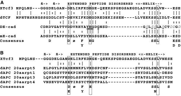
Based on our data, we extended this model, as shown in Figure 7A. In addition to the sequence similarity noted by Graham et al. (2000) in the extended peptide region, we suggest a further sequence alignment in the α-helical region. Notably, the three XTcf3 residues, which they identified as critical for interaction with β-catenin, are conserved in diverse cadherins (boxed in Figure 7A). Although the spacing between the extended peptide and the α-helix differs between TCF and cadherins, this region of XTcf3 is disordered in the structure and may form a flexible loop, and if fully extended, the cadherin peptide could span the gap. We also noted a similar, although less striking, alignment of the 20 amino acid repeats of APC and XTcf3, with all three key residues also conserved (Figure 7B).
Figure 7.
A model for the structure of the β-catenin-binding region of cadherin. (A) The sequence of Xenopus Tcf3 (XTcf3) and Drosophila TCF (dTCF) are aligned below a diagrammatic representation of the structure of XTcf3 as determined by Graham et al. (2000). A β-hairpin motif is indicated by “β ->.” Identical residues are indicated by vertical lines and similar residues are indicated by colons. Below is a proposed alignment of Drosophila E-cadherin (DE-cad) and mouse E-cadherin (mE-cad) with the extended peptide and α-helical regions of XTcf3. A consensus is displayed at the bottom positions (where at least three fourths of the sequences match). The three residues that are key for XTcf3 binding to β-catenin are boxed, and all are conserved in all sequences. The amino acids altered in mutation DECM2 (YEG G), which have a strong effect on DEC binding to β-catenin/Arm in our assays, are bold and underlined. The amino acids altered in mutation DECM3 (DD D), which had the weakest effect on binding, are shown in italics. The serines altered in mutations DECM1 and DECM9 are in italics and underlined. (B) Alignment of the XTcf3 sequence and structure with four 20-amino acid repeats of Drosophila APC (dAPC).
Our mutational analysis can also be examined in light of this structure (Figure 7A). Mutation DECM2, which has a severe effect on binding, alters four amino acids including a glutamic acid predicted by analogy to XTcf3 to mediate one of the key charge-charge interactions with β-catenin (Figure 7A, bold underline). In contrast, mutation DECM3, which had no effect in yeast and the least severe effect in mammalian cell assays, maps to a region predicted by analogy to be outside the structured portion of the binding site (Figure 7A, italics). The analysis of mutations DECM1 and DECM9 is more complex. Mutation of the four serines targeted in DECM1 to alanine has no effect in yeast but substantially reduces binding in mammalian cells. In contrast, mutation DECM9, which altered these serines to glutamic acid, did not affect binding. Of these four serines, the second and third align with serines in XTcf3. The second serine is predicted to be on the face of the α-helix away from β-catenin, whereas the third serine does not contribute to binding. The first serine is a valine in XTcf3, which participates in hydrophobic contacts, whereas the fourth serine is predicted by analogy to XTcf3 to be beyond the end of the α-helix and to have its side chain pointed away from β-catenin. If, as discussed above, these serines are phosphorylated, then the first and third phosphoserines might make charge-charge interactions with lysine 292 of β-catenin; this would also be the case if they were mutated to glutamic acid.
von Kries et al. (2000) also revealed new insights into β-catenin's interaction with its partners. They mutagenized β-catenin to identify amino acids in the Arm repeat region, which are essential for binding to APC, axin/conductin, and TCF/LEF. They found that mutations mapping to distinct Arm repeats blocked binding to individual partners. Thus, LEF-1 binding was inhibited by mutations in Arm repeat 8, whereas conductin binding was inhibited by mutations in Arm repeats 3 and 4. These data suggest that either different partners bind to distinct sites on β-catenin or, if the binding sites coincide, different subsets of the contacts between β-catenin and each its partners provide most of the free energy of binding. In parallel, they also examined whether these β-catenin mutations affected binding to E-cadherin (J.P. von Kries and W. Birchmeier, personal communication). In contrast to their results with the other partners, none of the mutations specifically blocked β-catenin binding to cadherin. Graham et al. (2000) also tested mutant forms of β-catenin for binding to XTcf3, C-cadherin, APC, and axin. XTcf3 binding required two key charge-charge interactions with the extended peptide region and a key hydrophobic interaction with the α-helix. For cadherin, mutations predicted from the structure to block the key charge-charge interactions reduced binding, but mutations in the α-helix–binding region had little effect. These data are of interest in relation to the present study in which, contrary to expectations, none of the first series of clustered point mutations (DECM1, DECM2, and DECM3) abolished DEC binding to β-catenin in yeast. One possible explanation for all these results is that the binding of cadherins to β-catenin differs from that of the other partners, with strong contacts made throughout the binding region. Thus, point mutations in either cadherin or β-catenin would have a lesser effect on binding. This might also explain the apparently higher affinity of cadherin for Arm in vivo, as assessed by competition for the limiting pool of Arm present in arm mutant embryos (Cox et al., 1996).
We assessed our mutations in the full DE-cadherin cytoplasmic tail. We also assessed DECM2 in a second context, introduced into a 34-amino acid fragment centered on the minimal binding region. In this context, DECM2 had a much more severe effect on β-catenin binding in yeast than it did when present in the full DE-cadherin tail. This result is consistent with the possibility that β-catenin binding is stabilized by interactions with regions of the cadherin tail outside the minimal binding domain or that the entire tail folds into a conformation that facilitates β-catenin binding.
To affect one function of β-catenin without affecting the others, one must design inhibitors that specifically interfere with a particular protein-protein interaction. Our results provide some insight into this issue. Both wild-type and mutant cadherin peptides were more effective in blocking interaction of endogenous β-catenin with TCF/LEF than in blocking interactions between endogenous β-catenin and the axin/APC complex or assembly of adherens junctions. This would be the desired outcome for a specific inhibitor that blocked the oncogenic action of β-catenin. Our data also suggest possible peptide candidates for cocrystallization of cadherin and β-catenin. When combined with the β-catenin–TCF structure, this would set the stage for initiating the design of synthetic inhibitors of different protein-protein interactions, which can be tested in cell culture and animal models for efficacy in blocking Wnt signaling or modulating cell adhesion and cancer progression.
ACKNOWLEDGMENTS
We are grateful to Mary Teachey who made some of the DEC constructs and Daniela Salomon for subcloning them into mammalian expression vectors, to Mary Teachey and Amanda Neisch for assistance with β-galactosidase assays, and to J.P. von Kries, W. Birchmeier, and W. Xu for communicating unpublished data and for valuable discussions. This work was supported by an IDEA Award from the Army Breast Cancer Research Program to M.P (DAMD17-98-1-8223) and by start-up funds from the University of Minnesota to C.K. C.K. was supported by the National Cancer Institute of Canada with funds from the Terry Fox Run. G.P. was supported by NIH 5T32 GM07092 and by a predoctoral fellowship from the Army Breast Cancer Research Program (DAMD17-98-1-8220), and M.P. was supported by a Career Development Award from the U.S. Army Breast Cancer Research Program. A.B.-Z. was supported by grants from the German-Israeli Foundation for Scientific Research and Development, the Cooperation Program in Cancer Research between the German Cancer Research Center and the Israeli Ministry of Science and Arts, CaP CURE, the Crown Endowment Fund for Immunological Research, and Yad Abraham Center for Cancer Diagnosis and Therapy.
REFERENCES
- Ben-Ze'ev A, Geiger B. Differential molecular interactions of β-catenin and plakoglobin in adhesion, signaling and cancer. Curr Opin Cell Biol. 1998;10:629–639. doi: 10.1016/s0955-0674(98)80039-2. [DOI] [PubMed] [Google Scholar]
- Christofori G, Semb H. The role of the cell-adhesion molecule E-cadherin as a tumor-suppressor gene. Trends Biochem Sci. 1999;24:73–76. doi: 10.1016/s0968-0004(98)01343-7. [DOI] [PubMed] [Google Scholar]
- Conti E, Uy M, Leighton L, Blobel G, Kuriyan J. Crystallographic analysis of the recognition of a nuclear localization signal by the nuclear import factor karyopherin-α. Cell. 1998;94:193–204. doi: 10.1016/s0092-8674(00)81419-1. [DOI] [PubMed] [Google Scholar]
- Cox RT, Kirkpatrick C, Peifer M. Armadillo is required for adherens junction assembly, cell polarity, and morphogenesis during Drosophila embryogenesis. J Cell Biol. 1996;134:133–148. doi: 10.1083/jcb.134.1.133. [DOI] [PMC free article] [PubMed] [Google Scholar]
- Cox RT, Pai L-M, Kirkpatrick C, Stein J, Peifer M. Roles of the C-terminus of Armadillo in Wingless signaling in Drosophila. Genetics. 1999;153:319–332. doi: 10.1093/genetics/153.1.319. [DOI] [PMC free article] [PubMed] [Google Scholar]
- Graham TA, Weaver C, Mao F, Kimelman D, Xu W. Crystal structure of a β-catenin/Tcf complex. Cell. 2000;103:885–896. doi: 10.1016/s0092-8674(00)00192-6. [DOI] [PubMed] [Google Scholar]
- Gumbiner BM. Cell adhesion: the molecular basis of tissue architecture and structure. Cell. 1996;84:345–357. doi: 10.1016/s0092-8674(00)81279-9. [DOI] [PubMed] [Google Scholar]
- Hsu SC, Galceran J, Grosschedl R. Modulation of transcriptional regulation by LEF-1 in response to Wnt-1 signaling and association with β-catenin. Mol Cell Biol. 1998;18:4807–4818. doi: 10.1128/mcb.18.8.4807. [DOI] [PMC free article] [PubMed] [Google Scholar]
- Huber AH, Nelson WJ, Weis WI. Three-dimensional structure of the armadillo repeat region of β-catenin. Cell. 1997;90:871–882. doi: 10.1016/s0092-8674(00)80352-9. [DOI] [PubMed] [Google Scholar]
- Jho E-H, Lomvardas S, Costantini F. A GSK3β phosphorylation site in axin modulates interaction with β-catenin and TCF-mediated gene expression. Biochem Biophys Res Commun. 1999;266:28–35. doi: 10.1006/bbrc.1999.1760. [DOI] [PubMed] [Google Scholar]
- Lickert H, Bauer A, Kemler R, Stappert J. Casein kinase II phosphorylation of E-cadherin increases E-cadherin/β-catenin interaction and strengthens cell adhesion. J Biol Chem. 2000;275:5090–5095. doi: 10.1074/jbc.275.7.5090. [DOI] [PubMed] [Google Scholar]
- Liu W, Dong X, Mai M, Seelan RS, Taniguchi K, Krishnadath KK, Halling KC, Cunningham JM, Qian C, Christensen E, Roche PC, Smith DI, Thibodeau SN. Mutations in AXIN2 cause colorectal cancer with defective mismatch repair by activating β-catenin/TCF signaling. Nat Genet. 2000;26:146–147. doi: 10.1038/79859. [DOI] [PubMed] [Google Scholar]
- McCartney BM, Dierick HA, Kirkpatrick C, Moline MM, Baas A, Peifer M, Bejsovec A. Drosophila APC2 is a cytoskeletally-associated protein that regulates Wingless signaling in the embryonic epidermis. J Cell Biol. 1999;146:1303–1318. doi: 10.1083/jcb.146.6.1303. [DOI] [PMC free article] [PubMed] [Google Scholar]
- Nakamura T, Hamada F, Ishidate T, Anai K, Kawahara K, Toyoshima K, Akiyama T. Axin, an inhibitor of the Wnt signaling pathway, interacts with β-catenin, GSK-3β and APC and reduces the β-catenin level. Genes Cells. 1998;3:395–403. doi: 10.1046/j.1365-2443.1998.00198.x. [DOI] [PubMed] [Google Scholar]
- Omer CA, Miller PJ, Diehl RE, Kral AM. Identification of Tcf4 residues involved in high-affinity β-catenin binding. Biochem Biophys Res Commun. 1999;256:584–590. doi: 10.1006/bbrc.1999.0379. [DOI] [PubMed] [Google Scholar]
- Pai L-M, Kirkpatrick C, Blanton J, Oda H, Takeichi M, Peifer M. Drosophila α-catenin and E-cadherin bind to distinct regions of Drosophila Armadillo. J Biol Chem. 1996;271:32411–32420. doi: 10.1074/jbc.271.50.32411. [DOI] [PubMed] [Google Scholar]
- Peifer M, Polakis P. Wnt signaling in oncogenesis and embryogenesis: a look outside the nucleus. Science. 2000;287:1606–1609. doi: 10.1126/science.287.5458.1606. [DOI] [PubMed] [Google Scholar]
- Provost E, Rimm DL. Controversies at the cytoplasmic face of the cadherin-based adhesion complex. Curr Opin Cell Biol. 1999;11:567–572. doi: 10.1016/s0955-0674(99)00015-0. [DOI] [PubMed] [Google Scholar]
- Rubinfeld B, Albert I, Porfiri E, Fiol C, Munemitsu S, Polakis P. Binding of GSK-3β to the APC/β-catenin complex and regulation of complex assembly. Science. 1996;272:1023–1026. doi: 10.1126/science.272.5264.1023. [DOI] [PubMed] [Google Scholar]
- Rubinfeld B, Albert I, Porfiri E, Munemitsu S, Polakis P. Loss of β-catenin regulation by the APC tumor suppressor protein correlates with loss of structure due to common somatic mutations of the gene. Cancer Res. 1997;57:4624–4630. [PubMed] [Google Scholar]
- Sadot E, Simcha I, Shtutman M, Ben-Ze'ev A, Geiger B. Inhibition of β-catenin-mediated transactivation by cadherin derivatives. Proc Natl Acad Sci USA. 1998;95:15339–15344. doi: 10.1073/pnas.95.26.15339. [DOI] [PMC free article] [PubMed] [Google Scholar]
- Satoh S, Daigo Y, Furukawa Y, Kato T, Miwa N, Nishiwaki T, Kawasoe T, Ishiguro H, Fujita M, Tokino T, Sasaki Y, Imaoka S, Murata M, Shimano T, Yamaoka Y, Nakamura Y. AXIN1 mutations in hepatocellular carcinomas, and growth suppression in cancer cells by virus-mediated transfer of AXIN1. Nat Genet. 2000;24:245–250. doi: 10.1038/73448. [DOI] [PubMed] [Google Scholar]
- Shtutman M, Zhurinsky J, Simcha I, Albanese C, D'Amico M, Pestell R, Ben-Ze'ev A. The cyclin D1 gene is a target of the β-catenin/LEF-1 pathway. Proc Natl Acad Sci USA. 1999;96:5522–5527. doi: 10.1073/pnas.96.10.5522. [DOI] [PMC free article] [PubMed] [Google Scholar]
- Simcha I, Shtutman M, Salomon D, Zhurinsky J, Sadot E, Geiger B, Ben-Ze'ev A. Differential nuclear translocation and transactivation potential of β-catenin and plakoglobin. J Cell Biol. 1998;141:1433–1448. doi: 10.1083/jcb.141.6.1433. [DOI] [PMC free article] [PubMed] [Google Scholar]
- Stappert J, Kemler R. A short core region of E-cadherin is essential for catenin binding and is highly phosphorylated. Cell Adhes Commun. 1994;2:319–327. doi: 10.3109/15419069409014207. [DOI] [PubMed] [Google Scholar]
- van de Wetering M, Cavallo R, Dooijes D, van Beest M, van Es J, Loureiro J, Ypma A, Hursh D, Jones T, Bejsovec A, Peifer M, Mortin M, Clevers H. Armadillo co-activates transcription driven by the product of the Drosophila segment polarity gene dTCF. Cell. 1997;88:789–799. doi: 10.1016/s0092-8674(00)81925-x. [DOI] [PubMed] [Google Scholar]
- von Kries JP, Winbeck G, Asbrand C, Schwarz-Romond T, Sochnikova N, Dell'Oro A, Behrens J, Birchmeier W. Hot spots in β-catenin for interactions with LEF-1, conductin and APC. Nat Struct Biol. 2000;7:800–807. doi: 10.1038/79039. [DOI] [PubMed] [Google Scholar]
- White P, Aberle H, Vincent J-P. Signaling and adhesion activities of mammalian β-catenin and plakoglobin in Drosophila. J Cell Biol. 1998;140:183–195. doi: 10.1083/jcb.140.1.183. [DOI] [PMC free article] [PubMed] [Google Scholar]
- Willert K, Nusse R. β-catenin: a key mediator of Wnt signaling. Curr Opin Gene Dev. 1998;8:95–102. doi: 10.1016/s0959-437x(98)80068-3. [DOI] [PubMed] [Google Scholar]
- Wodarz A, Nusse R. Mechanisms of Wnt signaling in development. Annu Rev Cell Dev Biol. 1998;14:59–88. doi: 10.1146/annurev.cellbio.14.1.59. [DOI] [PubMed] [Google Scholar]
- Zhurinsky J, Shtutman M, Ben-Ze'ev A. Plakoglobin and β-catenin: protein interactions, regulation and biological roles. J Cell Sci. 2000a;113:3127–3139. doi: 10.1242/jcs.113.18.3127. [DOI] [PubMed] [Google Scholar]
- Zhurinsky J, Shtutman M, Ben-Ze'ev A. Differential mechanisms of LEF/TCF-family dependent transcriptional activation by β-catenin and plakoglobin. Mol Cell Biol. 2000b;20:4238–4252. doi: 10.1128/mcb.20.12.4238-4252.2000. [DOI] [PMC free article] [PubMed] [Google Scholar]


