Abstract
Purpose/Background:
Abnormalities in glenohumeral rhythm and neuromuscular control of the upper trapezius (UT), middle trapezius (MT), lower trapezius (LT) and serratus anterior (SA) muscles have been identified in individuals with shoulder pain. Upper extremity diagonal or proprioceptive neuromuscular facilitation (PNF) patterns have been suggested as effective means of activating scapular muscles, yet few studies have compared muscular activation during diagonal patterns with varying modes of resistance. The purpose of this study is to determine which type of resistance and PNF pattern combination best elicits electromyographic (EMG) activity of the scapular muscles.
Methods:
Twenty one healthy subjects with no history of scapulohumeral dysfunction were recruited from a population of convenience. Surface electrodes were applied to the SA, UT, MT and LT and EMG data collected for each muscle as the subject performed resisted UE D1 flexion, UE D1 extension, UE D2 flexion and UE D2 extension with elastic resistance and a three pound weight.
Results:
No significant differences were found between scapular muscle activity during D1 flexion when using elastic resistance and when using a weight. UT, MT and LT values were also not significantly different during D2 flexion when using elastic resistance vs. using a weight. The activity of the SA remained relatively the same during all patterns. The LT activity was significantly greater during D2 flexion with elastic resistance than during the D1 flexion and D1 extension with elastic resistance. MT activity was significantly greater during D2 flexion with elastic resistance as compared to all other patterns except D2 flexion with a weight. UT activity was significantly greater during flexion patterns than extension patterns.
Conclusions:
The upper extremity PNF pattern did significantly affect the mean UT, MT and LT activity but was not found to significantly affect SA activity. The type of resistance did not significantly change muscle activity when used in the same diagonal patterns.
Keywords: elastic resistance, electromyography, proprioceptive neuromuscular facilitation, scapular musculature
INTRODUCTION
In the normal shoulder complex with full function; the humerus and the scapula must move in a predictable pattern or demonstrate “a normal scapulohumeral rhythm.” When viewed in the scapular plane; a position midway between a sagittal and frontal plane; scapulohumeral rhythm is a progressive upward rotation of the scapula accompanied by a decrease in internal rotation and movement from an anteriorly tipped position to a posteriorly tipped position of the scapula as the humerus elevates during overhead movement. In a patient with dysfunction of the shoulder complex; scapulohumeral rhythm may be altered and has been shown to be present in patients with shoulder pathologies such as impingement; shoulder instability; and frozen shoulder syndromes.1–4 Given that dysfunction of the shoulder complex is estimated to effect approximately 7-36% of the population; a clear understanding of shoulder movement is important to health care professionals and their patients.5
The presence or absence of normal scapulohumeral rhythm is largely dependent on the appropriate firing pattern of the muscles providing dynamic support about the shoulder complex. Muscles which are crucial to scapular movement include the upper trapezius; middle trapezius; lower trapezius; rhomboids; and serratus anterior muscles while the rotator cuff muscles (supraspinatus; infraspinatus; teres minor; and subscapularis) are important to humeral stability. Some evidence documents altered recruitment in the scapular stabilizing musculature in patients with shoulder pathologies2–4;6 but it is unclear as to whether the alterations in patterns precede the development of the pathology or occur as a result of the altered mechanics of the shoulder complex.7
Physical therapy interventions designed to restore normal scapulohumeral rhythm frequently include exercises that focus on the scapular muscles. Movements recommended for strengthening the upper trapezius include rowing; the military press and shoulder shrugging while exercises that maximize EMG activity of the middle trapezius include horizontal abduction; shoulder extension; overhead arm raise in a prone position; and rowing. Shoulder abduction; rowing; horizontal abduction and overhead arm raises in prone or sidelying maximize lower trapezius activity while shoulder flexion; shoulder abduction; scapular plane abduction above shoulder height maximize serratus anterior activity.8;9
Diagonal patterns of the upper extremity are often included in exercises thought to affect recruitment of the scapular muscles. Traditional proprioceptive neuromuscular facilitation (PNF) interventions focus on functional diagonal patterns of movement to improve both muscular strength and flexibility as well as utilize sensory cues such as cutaneous; visual; and auditory stimuli to improve neuromuscular control and function. Incorporation of the elementary movements of these patterns into shoulder rehabilitation programs may also be effective in increasing scapular muscle activation.9–11
While the selection of the type of exercise movement or pattern to incorporate into a rehabilitation program to activate the desired muscle(s) is an important consideration; the selection of resistance to achieve the appropriate intensity and type of muscle activation is also vital. Training intensities of greater than 60% are often recommended and multiple methods of resistance exist for achieving that intensity. Common in both clinical and home settings are the use of free weights and/or the use of elastic tubing to provide the resistance needed to approach the prescribed level of muscle activation. Traditionally; weights have provided concentric and eccentric resistance with torque on a muscle varying with positional changes. Use of elastic tubing has been suggested as an alternative to weights due to low cost; portability; and increasing resistance that occurs throughout the exercise movement. Andersen et al12 used surface electromyography (EMG) to compare the effects of free weights and elastic tubing on the upper trapezius; middle deltoid; and infraspinatus EMG output. The authors reported that during a standing lateral raise of the arm and during sidelying external rotation of the glenohumeral joint; normalized EMG output was not significantly different between conditions using free weight or elastic tubing. However; Page and associates13 suggest that elastic resistance may have some advantages over free weights. They measured isokinetic torque output at 60°/sec during a D2 flexion and extension pattern and found greater output following an exercise program where subjects trained using elastic resistance as compared to those who trained using free weights.
Because diagonal patterns are commonly used clinically to recruit scapular muscles and resistance during these patterns can be applied using weights or elastic resistance; studying both methods of resistance during diagonal patterns would be of assistance when choosing an exercise to strengthen selected scapular muscles. The aim of this study was to investigate the level of muscle activation that occurred in the scapular muscles during diagonal patterns using weights and elastic resistance; as measured by surface EMG. The null hypothesis was that the levels of EMG activity of the scapular muscles would be similar in all patterns with both types of resistance.
Methods
Participants
The study was performed on a population of convenience. Twenty-one volunteers (6 male and 15 female) whose age ranged from 21-37 years (mean = 25.3 yrs) participated in the study. All subjects signed an informed consent form that had been approved by the Institutional Review Board at the authors' research facility. Subjects were not compensated for participation. Information regarding subject medical history was obtained through a self-report survey and questioning by the investigators. Exclusion criteria included a history of cardiovascular problems requiring medical treatment; and any individuals with medical conditions or problems which would prevent them from putting forth maximal effort. Subjects who reported pain in the shoulder or scapular region within the last 6 weeks; those who had a history of biomechanical problems of the shoulder including dislocation; subluxation of the glenohumeral joint or dyskinesis of the scapula; had a history of rotator cuff tear or repair; unhealed fracture; current nerve damage to the shoulder; or were currently undergoing diagnostic testing or treatment for shoulder problems were excluded.
Procedures
As a warm up; subjects rode an upper body ergometer for 4 minutes. A reviewer then led the subject in the performance of stretches of the neck and shoulder. Stretches were held for 15 seconds at the end ranges of right and left cervical sidebending (to stretch the upper trapezius); scapular protraction (to stretch the rhomboids and middle trapezius); shoulder horizontal adduction (to stretch the posterior capsule); shoulder horizontal abduction (to stretch the pectoralis major); shoulder flexion with the elbow flexed (to stretch the triceps) and shoulder extension with the elbow extended (to stretch the biceps).
The skin was cleaned using an alcohol wipe and then EMG surface electrodes were placed on the upper trapezius; middle trapezius; lower trapezius; and serratus anterior muscles on the self- reported non-dominant shoulder of the subjects as described by Michener et al.14 Manual muscle tests (MMTs) were performed on the upper trapezius; middle trapezius; lower trapezius; and serratus anterior muscles to determine maximal volitional contraction of the non-dominant arm. The upper trapezius MMT was performed as described by Kendall et al.15 In a sitting position; the subject elevated the arm and posterolaterally extended the neck as the examiner applied pressure against the shoulder toward depression and against the head toward anterolateral flexion. The MMT for the middle trapezius was performed with the subject prone; arm abducted to 90°; shoulder laterally rotated and the scapula adducted. The examiner applied pressure to the arm; proximal to the elbow; in a downward direction.15;16 The lower trapezius MMT was performed with the subject prone; arm in 145° of abduction; shoulder laterally rotated and the scapula adducted. The examiner applied pressure on the arm proximal to the elbow in a downward direction.15;16 The serratus anterior MMT was performed with the subject sitting in a chair with the arm flexed to 125°. As the examiner monitored the inferior angle of the scapula for internal rotation or anterior tipping of the scapula; pressure was applied on the arm; proximal to the elbow in a downward motion. Pressure was adjusted throughout the test for any changes in scapular position to assure that the subject held the appropriate position.15;16 Each MMT was performed three times with maximal resistance given for 5 seconds with a 20 second rest period between tests. The order of testing was randomized prior to subject recruitment using a randomization table. The mean EMG activity of the three trials was used to determine the maximal volitional contraction. All EMG data was collected using a Biometrics DataLink (Biometrics Ltd; Gwent; United Kingdom).
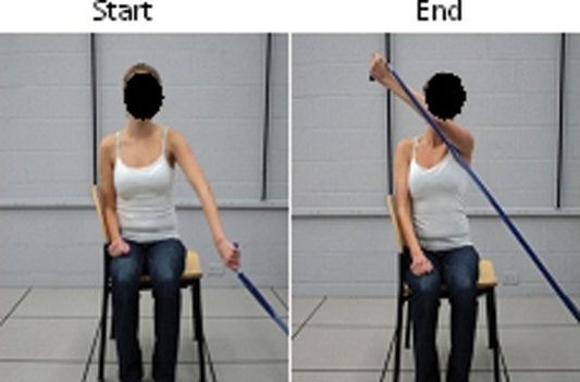
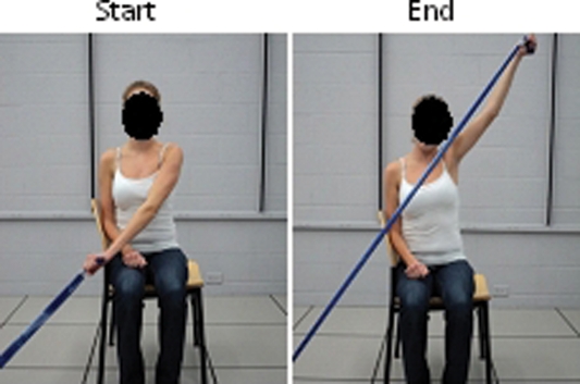
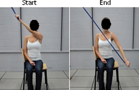
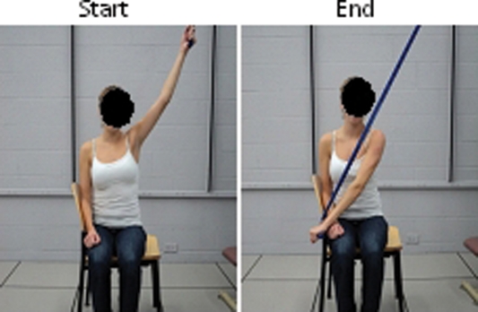
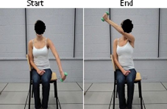
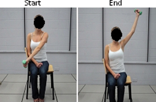
Following the MMTs; subjects were instructed in how to perform D1 and D2 flexion and D1 and D2 extension PNF patterns as per typical PNF theory. To perform the D1 flexion pattern; the subject started with the arm down by the ipsilateral side and brought it up toward the ear of the contralateral side moving through glenohumeral motions of flexion; adduction; and external rotation. The subject performed the D1 extension pattern by starting with their arm by the contralateral ear and bringing it down toward the ipsilateral side of the waist moving through the glenohumeral motions of extension; abduction; and internal rotation. The pattern of D2 flexion was performed with the subject starting with their arm on the contralateral side of their waist and bringing arm up above the ipsilateral side of the head; a movement which consisted of glenohumeral flexion; abduction; and external rotation. D2 extension was performed with the subject starting with their arm above the ipsilateral side of the head and bringing it down toward the contralateral waist; a motion that involved extension; adduction and internal rotation. Throughout all patterns subjects were instructed to keep the elbow in as much extension as possible and all instruction and trials were performed in a sitting position. Subjects continued to receive instruction until they could complete the patterns independently and without verbal cues.
Following mastery of the patterns; the subjects performed the patterns without resistance and then progressed to resistance using blue elastic resistance (The Hygenic Corporation; Akron OH). Blue elastic resistance was utilized by Lister et al17 and represents the midrange of resistances. To standardize the length of the elastic resistance for each individual; the distance between the anchor and the hand was equal to the length of the distance between the floor and the greater trochanter as measured with the individual in a standing position. For flexion patterns; one end of the elastic resistance was anchored to the floor by a research assistant and the other end held by the subject (see Figure 1 for D1 flexion with elastic resistance; see Figure 2 for D2 flexion with elastic resistance). For extension patterns; one end of the elastic resistance was attached above the subject to an adjustable ceiling bracket and the other end held in the hand (see Figure 3 for D1 Extension with elastic resistance; see Figure 4 for D2 Extension with elastic resistance). At the start of the extension pattern with the arm in a position of flexion; abduction and external rotation; the length of the elastic resistance between the bracket and the hand was adjusted to equal the measurement of the torso (floor to greater trochanter) taken previously. Each pattern was performed three times at a self selected speed with a 20 second rest between each repetition.
Figure 1.
D1 Flexion with Elastic Resistance.
Figure 2.
D2 Flexion with Elastic Resistance.
Figure 3.
D1 Extension with Elastic Resistance.
Figure 4.
D2 Extension with Elastic Resistance.
In addition to elastic resistance; subjects performed the D1 flexion and the D2 flexion patterns with a three pound dumbbell. Because the goal of this study was to measure activation of the scapular during common exercises and not to result in muscle fatigue; the weight of the dumbbell was selected to avoid excessive loading of the rotator cuff. Carson18 cautions against utilization of weights over 5 pounds due to stress on the rotator cuff and advocates weights of one through 5 pounds for specific rotator cuff rehabilitation exercises. To duplicate typical clinical applications without rotator cuff fatigue; a midpoint of this suggested range (3 pounds) was used by all subjects in this study. Patterns performed with a weight involved the same motions as described above (see Figure 5 for D1 flexion with a weight and see Figure 6 for D2 flexion with a weight) with the subjects completing three trials of each pattern with a 20 second rest between each trial.
Figure 5.
D1 Flexion with Weight.
Figure 6.
D2 Flexion with Weight.
Similar to the MMT; the order for patterns and resistance was randomized before beginning the study using a randomization table. All data was collected during a single session.
Data Reduction
Following collection; all raw EMG signals were full-wave rectified and processed using a root-mean-square algorithm with a 4-ms moving window. The mid three second time period of each five second MMT was recorded for each trial and the three trials averaged to provide the MVC for each muscle. The start and stop of each resisted activity was marked on the processed EMG signal and the mean EMG activity of each muscle during that time was recorded. To normalize values during the exercises; the EMG value during each exercise trial was expressed as a percent of the MVC value recorded during MMT (%MVC). The %MVC for each of the three trials was averaged and used for analysis.
Statistical Methods
Separate one way ANOVAs were performed for UT; MT; LT and SA muscle activity to examine the differences in the percent maximal contractions between exercises. When a significant effect (p <.05) was found; comparisons of least square means with Bonferroni adjustment were completed to compare the various patterns and identify significant differences due to the type of pattern and the type of resistance used. Further analysis on each of the percent mean values was completed to examine additional factors that could have influenced the muscle activity including pattern; body mass index (BMI); age and gender. Backward regression analysis was used to further explain differences in the percent maximal activity of each of the scapular muscles. All analyses were completed using SAS Version 9.1 (SAS Institute Inc.; Cary; NC).
Results
Subject demographics are found in Table 1. Mean age was 25.4 years and the majority of the subjects were right hand dominant. The average BMI of 25.3 for the males is slightly higher than the 50th percentile of American males while the average BMI for females (22.6) falls within the normal range as proposed by the Centers for Disease Control and Prevention.19
Table 1.
Subject Demographics
| All (n=21) | Males (n=6) | Females (n=15) | Range | |
|---|---|---|---|---|
| Mean Age (years) | 25.4 | 26.8 | 24.7 | 21-37 |
| Body Mass Index | 23.3 | 25.3 | 22.6 | 19.6-28.2 |
Mean ± standard error of the mean (SEM) values of the percent maximal volitional contraction (PMVC) for each muscle during each pattern are presented in Table 2.
Table 2.
Percent of Maximal Volitional Contraction (% of MVC) of Scapular Muscles During Exercise Patterns with Associated Standard Error of the Mean (SEM).
| Pattern | Upper Trapezius % of MVC (SEM) | Middle Trapezius % of MVC (SEM) | Lower Trapezius % of MVC (SEM) | Serratus Anterior % MVC (SEM) |
|---|---|---|---|---|
| D1ET | 27.6 (15.5) | 15.9 (15.3) | 25.2 (26.1) | 42.5 (20.9) |
| D1FT | 53.0 (29.7) | 16.7 (15.0) | 23.9 (16.3) | 50.0 (28.1) |
| D1FW | 39.6 (26.4) | 18.4 (15.6) | 23.7 (18.4) | 43.7 (22.7) |
| D2ET | 27.1 (17.1) | 24.2 (23.1) | 29.3 (35.3) | 43.1 (21.5) |
| D2FT | 68.5 (47.9) | 45.3 (20.7) | 44.9 (31.2) | 48.7 (21.6) |
| D2FW | 53.2 (39.7) | 33.2 (14.7) | 32.0 (13.9) | 44.6 (21.6) |
D1ET = D1 Extension with elastic resistance; D1FT = D1 Flexion with elastic resistance; D1FW = D1 Flexion with a 3 pound weight; D2ET = D2 Extension with elastic resistance; D2FT = D2 Flexion with elastic resistance; D2FW = D2 Flexion
Upper Trapezius
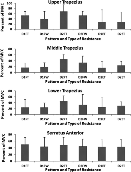
One way ANOVA tests confirmed that upper trapezius %MVC values were significantly affected by pattern (p=0.00). There were; however; no significant differences between the upper trapezius %MVC during D1 flexion with a weight and during D1 flexion with elastic resistance. Similarly there were no significant differences between %MVC of the upper trapezius between D2 flexion with a weight and D2 flexion with elastic resistance. In addition; there were no significant differences for upper trapezius %MVC between D1 and D2 patterns; D1 flexion with a weight was comparable to D2 flexion with a weight; D1 flexion with elastic resistance was comparable to D2 flexion with elastic resistance; and D1 extension with elastic resistance was comparable to D2 extension with elastic resistance. The %MVC of the upper trapezius; however; did show significant variation in activation between extension and flexion patterns. The %MVC of the upper trapezius during D1 extension with elastic resistance and during D2 extension with elastic resistance were both significantly less (p=0.00) than D1 and D2 flexion with elastic resistance while only D2 extension with elastic resistance was significantly different than D2 flexion with a weight (see Figure 7).
Figure 7.
Electromyographic (EMG) Activity of the Scapular Muscles during Exercises. EMG activity of each scapular muscle is expressed as a percent of the mamimal volitional contraction (MCV) as measured during manual muscle testing of that muscle. Values are reported for each of the exercises: D1 Extension with elastic resistance (D1ET); D1 Flexion with elastic resistance (D1FT); D1 Flexion with a 3 pound weight (D1FW); D2 Extension with elastic resistance (D2ET); D2 Flexion with elastic resistance (D2FT) and D2 Flexion with a 3 pound weight.
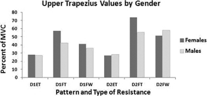
The additional factors of BMI; gender; and age were examined in order to determine their potential effects on UT mean EMG output values. A MANOVA and regression analysis were used to examine all data; demonstrating that BMI (p=0.00) and gender (p=0.04); independent of pattern; were significantly associated with UT mean values. As illustrated in Figure 8; female UT EMG activity was significantly greater than male activity during flexion patterns with elastic resistance.
Figure 8.
Electromyographic (EMG) Activity of the Upper Trapezius (UT) by Gender. EMG activity of the UT is expressed as a percentage of the mamimal volitional contraction (% of MVC) of the UT. The % of MVC is reported for each of the exercises: D1 Extension with elastic resistance (D1ET); D1 Flexion with elastic resistance (D1FT); D1 Flexion with a 3 pound weight (D1FW); D2 Extension with elastic resistance (D2ET); D2 Flexion with elastic resistance (D2FT) and D2 Flexion with a 3 pound weight. Female values were significantly higher than male value during D1FT and D2FT.
Middle Trapezius
Type of exercise significantly affected the %MVC output produced by the middle trapezius (p=0.00) with the differences associated with the pattern; but not the type of resistance. There were no significant differences in EMG output between the flexion patterns when subjects used different types of resistance; D1 flexion with a weight was comparable to D1 flexion with elastic resistance and D2 flexion with a weight was comparable to D2 flexion with elastic resistance. Similarly; output during D1 extension with elastic resistance was not significantly different from D2 extension with elastic resistance. However; there were significant differences between the %MVC output of the middle trapezius when different patterns were performed. D1 flexion with a weight was significantly less than D2 flexion with a weight and D1 flexion with elastic resistance was significantly less than D2 flexion with elastic resistance (p=0.00). In a comparison of flexion and extension patterns; the %MVC output of the middle trapezius was significantly greater during D2 flexion with elastic resistance than during D1 or D2 extension with elastic resistance. In contrast; D1 extension with elastic resistance and D2 extension with elastic resistance were not significantly different than D1 flexion with elastic resistance; D1 flexion with a weight; or D2 flexion with a weight (p=0.00).
Regression analysis revealed that age (p=0.78); gender (p=.0.11) and BMI (p=0.46) were not significantly associated with middle trapezius %MVC output during the exercises.
Lower Trapezius
Similar to the other parts of the trapezius; there were no significant differences between the %MVC output of the lower trapezius when similar patterns were performed with different types of resistance. D1 flexion with a weight was similar to D1 flexion with elastic resistance and D2 flexion with a weight was similar to D2 flexion with elastic resistance. However; the lower trapezius %MVC activity was significantly less during D1 flexion with elastic resistance and D2 flexion with elastic resistance (p=0.05). Likewise; lower trapezius %MVC activity during D1 flexion with a weight was also significantly less than D2 flexion with a weight (p=0.05). The extension patterns did not significantly vary between D1 extension with elastic resistance and D2 extension with elastic resistance. As with the middle trapezius; regression analysis revealed that age (p=0.17); gender (p=0.39) and BMI (0.88) were not significantly associated with lower trapezius %MVC output during the exercises.
Serratus Anterior
Unlike the other scapular muscles; the PNF pattern did not significantly affect the serratus anterior %MVC output. There were no significant differences between %MVC output during any patterns or between the types of resistance (p=0.85). Values for the serratus anterior %MVC output had a relatively narrow range with a low of 42.5% of the MVC during D1 extension with elastic resistance to a high of 50.0% of the MVC during D1 flexion with elastic resistance. Although the type of pattern and resistance did not explain variations in serratus anterior %MVC; regression analysis did reveal that gender was a significant factor (p=0.00) in explaining differences among SA values.
DISCUSSION
The use of elastic resistance and free weights during shoulder exercises is widely recommended as part of shoulder rehabilitation programs20–22 yet little evidence is available to guide the clinician in determining which type of resistance or pattern of exercise may be optimal. The results of this study indicate that the pattern of the exercise chosen to be performed during shoulder complex diagonal exercises may be more crucial to the muscular activity produced by the scapular muscles than the selection of a weight or of elastic tubing as the type of resistance. In the current study; the %MVC output was not significantly different for any of the scapular muscles tested when a weight was used versus elastic resistance. However; the different diagonal patterns did alter the EMG output of the various portions of the trapezius muscle; indicating that attention be paid to the type of diagonal pattern that is selected.
The finding that the %MVC of the scapular muscles was similar when using a weight and when using elastic resistance is consistent with past studies.12;23–25 In the current study; this was demonstrated when performing PNF diagonal patterns of the upper extremity. While little research exists which directly compares two types of resistance during diagonal patterns several studies have compared the output of muscles when exercising with weights and with elastic resistance.12;23 Measuring EMG output; Andersen et al reported no significant differences in the trapezius; medial deltoid; or infraspinatus activity when subjects performed a lateral raise with a dumbbell and with elastic tubing; demonstrating only an increase in activity as the resistance of both were increased.12 Measuring torque output using an isokinetic dynamometer; Page et al also found no difference in the production of eccentric torque at faster isokinetic speeds (180°/sec) in a group of subjects trained with dumbbells when compared with a group of subjects trained with elastic resistance.23 Differences in torque output were reported at the slower speed of 60°/sec23 suggesting a varied effect of exercising with elastic resistance and exercising with weights. The use of isokinetic torque production by Page and associates as an outcome measure is difficult to compare to the isolated EMG %MVC output of specific scapular muscles used in the current study. In addition; it is difficult to determine if isolated scapular activity would show similar or different results after a training period. Further investigation; however; of the changes in the scapular muscle %MVC outputs after training with various resistive methods is warranted.
Differences in scapular muscle EMG activity when using cuff weights versus theraband were reported by Lister et al.17 They reported significantly greater activity of the upper trapezius; lower trapezius; and serratus anterior during shoulder flexion to 90° and shoulder abduction to 90° when elastic resistance (blue) was used as opposed to a cuff weight (1.1 kg). While the amount of resistance may be similar between the current study and the study by Lister et al; the motions which were examined and the methods used to normalize EMG values differed. The previous study modified traditional manual muscle tests for the lower trapezius and the serratus anterior by performing both in standing while the current study utilized the descriptions from Kendall et al.15 Such a change could alter the percent of the MVC output and prevent a direct comparison between the values reported in that study and the current investigation.
Values of the %MVC output for the scapular muscles found in this study ranged from a low of 15.9% of the MVC of the middle trapezius during D1 extension with theraband to a high of 68.5% of the MVC of the upper trapezius during D2 flexion with elastic resistance. In a 1992 study by Mosely et al; elevation of the humerus in the scapular plane resulted in %MVC values of 54%; 60%; 91% and 84% for the upper trapezius; lower trapezius; middle serratus anterior and lower serratus anterior respectively when using a self-selected free weight resistance which varied between 3 and 30 pounds.8 As expected; the output produced by Mosley's subjects are not similar to the 53.2%; 32% and 44.6% for the upper trapezius; lower trapezius and serratus anterior found in this study where the resistance was limited to one level of elastic resistance and the pattern involved different arm movements at the shoulder complex. Although the humeral positions were not monitored in the current study; efforts were made to assure the subject completed the pattern in a true diagonal and not into positions of full flexion or abduction. Movement out of diagonal planes could potentially alter the amount work required by the scapula and influence the stabilization efforts required to maintain its position. Differences in muscle activity during elevation in the scapular plane and D2 flexion would be expected.
Myers et al studied a movement that was multiplanar.10 Referred to as throwing acceleration; this movement started with the glenohumeral joint in full external rotation and abduction with the elbow at 90° of flexion. Elastic tubing was held in the hand and surface EMG recordings were taken as the subject moved the arm across the body as in the acceleration phase of throwing and then returned the arm to the original position. Described by the investigators of that study as being a D2 flexion pattern; measurements were expressed as %MVC which was determined by performing maximal volitional contractions of each muscle prior to the measurement during exercise. The EMG activity for the serratus anterior was reported to be 55.5% of the MVC.10 This is slightly higher than the amount recorded in the current study but as indicated above; the starting and ending positions were different as was the resistance used during each study. Ekstrom et al9 also reported %MVC of the trapezius and serratus anterior muscles in a position similar to D2F. After determining a five repetition maximum (5 RM) for each subject; each subject lifted 85%–90% of their 5-repetition maximum during an overhead arm raise in the prone position. The overhead arm raise used in their study; a traditional MMT position for the lower trapezius; included testing the arm in line with the fibers of the lower trapezius; a position that is similar to D2F in that the end position includes shoulder flexion; abduction; and external rotation. Ekstrom et al found %MVC values during their tested movement at 79%; 101%; 97% and 43% for the upper trapezius; middle trapezius; lower trapezius and serratus anterior respectively. These values are considerably higher than the measurements in the current study but are likely explained by changes in the amount of resistance. The three pound weight used in the current study would not have approached the five repetition maximum in the study by Ekstrom and associates. In addition; the variation in the positioning of the body may account for differences in reported %MVC. The values reported by Ekstrom and associates were obtained in a prone position while the current study was performed with subjects in a sitting position. The effects of gravity on the arm could have altered the activation patterns of the scapular muscles contributing to the lack of consensus between the two studies.
While a true D1F pattern has not been investigated in other studies to the authors' knowledge; Ekstrom et al studied upper and lower serratus anterior EMG activity as maximal manual resistance was applied in a position of shoulder flexion; horizontal adduction and external rotation. A %MVC of 66% for the upper serratus anterior and a %MVC of 73% for the lower serratus anterior were reported for this static position. This position is similar to the end position of the D1F pattern which; in the current study; resulted in a mean %MVC of 50% for the serratus anterior (not assessed as upper or lower portions). Variations in EMG placement could explain the differences in activity as well as differences in the amount of pressure and the performance of a maximal end range contraction versus a dynamic contraction.26
Application to clinical practice
While some discrepancies with previous literature are noted due to varying position and resistance; the results of this study can assist in the selection of diagonal patterns that maximize or minimize EMG activity.
The D2 flexion pattern with elastic resistance consistently activated all of the scapular muscles at levels that were 40% of the MVC. In addition; because D2 flexion with a weight was not significantly different from D2 flexion with elastic resistance; it could be utilized to achieve similar levels of muscular activity regardless of type of resistance. While activation of all scapular muscles may be desired; in some clinical conditions; inhibition of the upper trapezius may be recommended to avoid excessive scapular elevation during overhead movements.27;28 If the goal is to minimize upper trapezius activity; while eliciting activation of the remaining scapular muscles; the use of D2 extension with elastic resistance may be the preferred clinical choice. The D2 extension with elastic resistance yields among the lowest upper trapezius %MVC output yet is accompanied by serratus anterior activity that is comparable to all of the other patterns. In addition; D2 extension using elastic resistance as performed in the current study resulted in the third highest %MVC for the middle trapezius and lower trapezius.
Interestingly; the patterns of activation of all three trapezius muscles between patterns and between types of resistance were remarkably similar (see Figure 7). The upper; middle and lower trapezius exhibited consistently higher %MVC output during the D2 flexion pattern with either elastic resistance or a weight; intermediate output during the D1 flexion pattern with either elastic resistance or a weight and the lowest level output during the D1 and D2 extension patterns. This is in contrast to the serratus anterior; where similar %MVC output values were produced regardless of pattern or resistance. Selection of a pattern to activate the serratus anterior may be guided by the need to maximize or to minimize activity of the remaining scapular muscles. For the clinician seeking to isolate the serratus anterior; the results of this study suggest that a D1 extension pattern with elastic resistance would provide equal %MVC to the D1 flexion pattern but without the higher levels of trapezius activity.
Limitations
The data collected in this study is limited by the population studied; the use of surface EMG as a measurement of muscle activity and the type of resistance used. The population included healthy subjects with no attempt to determine the influence of regular exercise; past sports related activities; or current fitness levels. However; muscle activity was compared as a % of an individuals' MVC which can compensate for differences in baseline strength and capacity.
The selection of the amount of resistance in this study may have also influenced the results. Clearly; a three pound weight would not be a maximal amount of isotonic resistance for all of the subjects nor would blue elastic resistance be maximal elastic resistance. However; the amount of resistance used likely falls within the ranges of resistance utilized during rehabilitation of individuals with scapular and shoulder dysfunction. Future studies could standardize this amount by using a dynamometer to objectively determine the force production of the muscles of each subject; and then incorporate a calculated isotonic resistance during performance of the movements being studied. Some variation; even with this alternative methodology; would be expected as diagonal patterns are three dimensional and consistent resistance in all three planes would be questionable. The submaximal amount of resistance in this study may have been adequate in identifying contraction patterns and to be used in support of exercise selection.
In regards to the use of surface EMG to measure muscle activity; this methodology certainly exposes data collection to the possibility of cross talk between muscles. However; the use of EMG to record superficial muscles is well accepted29 and common in rehabilitative research.9;10;12
CONCLUSIONS
This study supports the use of a D2 flexion pattern with elastic resistance or with a weight to achieve the greatest activation of the upper trapezius; middle trapezius; lower trapezius and serratus anterior muscles. If minimization of upper trapezius activity with maximization of the lower trapezius and serratus anterior is desired; a D2 extension pattern with elastic resistance is recommended. Isolated activation of the serratus anterior with minimization of activation of all parts of the trapezius muscle occurred during D1 extension with elastic resistance.
REFERENCES
- 1.Borstad JD, Ludewig PM. Comparison of scapular kinematics between elevation and lowering of the arm in the scapular plane. Clin Biomech (Bristol; Avon). 2002;17:650–659 [DOI] [PubMed] [Google Scholar]
- 2.Lin JJ, Wu YT, Wang SF, Chen SY. Trapezius muscle imbalance in individuals suffering from frozen shoulder syndrome. Clin Rheumatol. 2005;25(569–575). [DOI] [PubMed] [Google Scholar]
- 3.Ludewig PM, Cook TM, Nawoczenski DA. Three dimensional scapular orientation and muscle activity at selected positions of humeral elevation. JOSPT. 1996;24:57–65 [DOI] [PubMed] [Google Scholar]
- 4.Matias R, Pascoal AG. The unstable shoulder in arm elevation: a three dimensional and electromyographical study in subjects with glenohumeral instability. Clin Biomech (Bristol; Avon). 2006;21S:52–58 [DOI] [PubMed] [Google Scholar]
- 5.Ferreira PH, Ferreira ML, Hodges PW. Changes in the recruitment of the abdominal muscles in people with low back pain: ultrasound measurement of muscle activity. Spine. 2004;29:2560–2566 [DOI] [PubMed] [Google Scholar]
- 6.Herbert LJ, Moffet H, McFadyen BJ, Dionne C. Scapular behavior in shoulder impingement syndrome. Arch Phys Med Rehabil. 2002;83:60–69 [DOI] [PubMed] [Google Scholar]
- 7.Ludewig PM, Cook TM. Alterations in shoulder kinematics and associated muscle activity in people with symptoms of shoulder impingement. Phys Ther. Mar 2000;80(3):276–291 [PubMed] [Google Scholar]
- 8.Moseley JB, Jr., Jobe FW, Pink M, Perry J, Tibone J. EMG analysis of the scapular muscles during a shoulder rehabilitation program. Am J Sports Med. Mar-Apr 1992;20(2):128–134 [DOI] [PubMed] [Google Scholar]
- 9.Ekstrom RA, Donatelli RA, Soderberg GL. Surface electromyographic analysis of exercises for the trapezius and serratus anterior muscles. J Orthop Sports Phys Ther. May 2003;33(5):247–258 [DOI] [PubMed] [Google Scholar]
- 10.Myers J, Pasquale M, Laudner K, Sell T, Bradley J, Lephart S. On-the-field resistance-tubing exercises for throwers: An electromyographic analysis. J Athl Train. 2005;40(1):15–22 [PMC free article] [PubMed] [Google Scholar]
- 11.Townsend H, Jobe FW, Pink M, Perry J. Electromyographic analysis of the glenohumeral muscles during a baseball rehabilitation program. Am J Sports Med. May-Jun 1991;19(3):264–272 [DOI] [PubMed] [Google Scholar]
- 12.Andersen LL, Andersen CH, Mortensen OS, Poulsen OM, Bjornlund IB, Zebis MK. Muscle activation and perceived loading during rehabilitation exercises: comparison of dumbbells and elastic resistance. Phys Ther. Apr 2010;90(4):538–549 [DOI] [PubMed] [Google Scholar]
- 13.Page PA, Lamberth J, Abadie B, Boling R, Collins R, Linton R. Posterior rotator cuff strengthening using theraband(r) in a functional diagonal pattern in collegiate baseball pitchers. J Athl Train. Winter 1993;28(4):346–354 [PMC free article] [PubMed] [Google Scholar]
- 14.Michener LA, Boardman ND, Pidcoe PE, Frith AM. Scapular muscle tests in subjects with shoulder pain and functional loss: reliability and construct validity. Phys Ther. Nov 2005;85(11):1128–1138 [PubMed] [Google Scholar]
- 15.Kendall FP, McCreary EK, Provance PG, Rodgers MM, Romani WA. Muscles Testing and Function with Posture and Pain. 5th ed. Baltimore: Lippincott Williams and Wilkins; 2005 [Google Scholar]
- 16.Hislop HJ, Montgomery J. Muscle Testing: Techniques of Manual Examination. 8th ed. St. Louis: Elsevier; 2007 [Google Scholar]
- 17.Lister JL, Del Rossi G, Ma F, et al. Scapular stabilizer activity during bodyblade; cuff weights and thera-band use. J Sport Rehabil. 2007;16:50–67 [DOI] [PubMed] [Google Scholar]
- 18.Carson WG. Rehabilitation of the throwing shoulder. Clin Sports Med. 1989;8(4):657–689 [PubMed] [Google Scholar]
- 19.Flegal KM, Carroll MD, Ogden CL, Curtin LR. Prevalence and trends in obesity among US adults; 1999–2008. JAMA. 2010;303(3):235–241 [DOI] [PubMed] [Google Scholar]
- 20.Lombardi I, Jr., Magri AG, Fleury AM, Da Silva AC, Natour J. Progressive resistance training in patients with shoulder impingement syndrome: a randomized controlled trial. Arthritis Rheum. May 15 2008;59(5):615–622 [DOI] [PubMed] [Google Scholar]
- 21.O'shea SD, Taylor NF, Paratz JD. A predominantly home-based progressive resistance exercise program increases knee extensor strength in the short-term in people with chronic obstructive pulmonary disease: a randomized controlled trial. Aust J Physiother. 2007;53(4):229–237 [DOI] [PubMed] [Google Scholar]
- 22.McNeely ML, Parliament MB, Seikaly H, et al. Effect of exercise on upper extremity pain and dysfunction in head and neck cancer survivors: a randomized controlled trial. Cancer. Jul 1 2008;113(1):214–222 [DOI] [PubMed] [Google Scholar]
- 23.Page PA, Lamberth J, Abadie B, Boling R, Collins R, Linton R. Posterior rotator cuff strengthening using theraband in a functional diagonal pattern in collegiate baseball pitchers. J Athl Train. 1993;28(4):346–354 [PMC free article] [PubMed] [Google Scholar]
- 24.Melchiorri G, Rainoldi A. Muscle fatigue induced by two difference resistances: Elastic tubing versus weight machines. J Electromyogr Kinesiol. 2011;Sep 13. [DOI] [PubMed] [Google Scholar]
- 25.Colado JC, Garcia-Masso X, Pellicer M, Alakhdar Y, Benavent J, Cabeza-Ruiz R. A comparison of elastic tubing and isotonic resistance exercises. Int J Sports Med. Nov 2010;31(11):810–817 [DOI] [PubMed] [Google Scholar]
- 26.Ekstrom RA, Bifulco KM, Lopau CJ, Andersen CF, Gough JR. Comparing the function of the upper and lower parts of the serratus anterior muscle using surface electromyography. J Orthop Sports Phys Ther. May 2004;34(5):235–243 [DOI] [PubMed] [Google Scholar]
- 27.Lin JJ, Hsieh SC, Cheng WC, Chen WC, Lai Y. Adaptive patterns of movement during arm elevation test in patients with shoulder impingement syndrome. J Orthop Res. 2011;29(5):653–657 [DOI] [PubMed] [Google Scholar]
- 28.Ludewig PM, Cook TM. Alterations in shoulder kinematics and associated muscle activity in people with symptoms of shoulder impingement. Phys Ther. 2000;80(3):276–291 [PubMed] [Google Scholar]
- 29.Ekstrom RA, Soderberg GL, Donatelli RA. Normalization procedures using maximum voluntary isometric contractions for the serratus anterior and trapezius muscles during surface EMG analysis. J Electromyogr Kinesiol. 2005;15:418–428 [DOI] [PubMed] [Google Scholar]


