Abstract
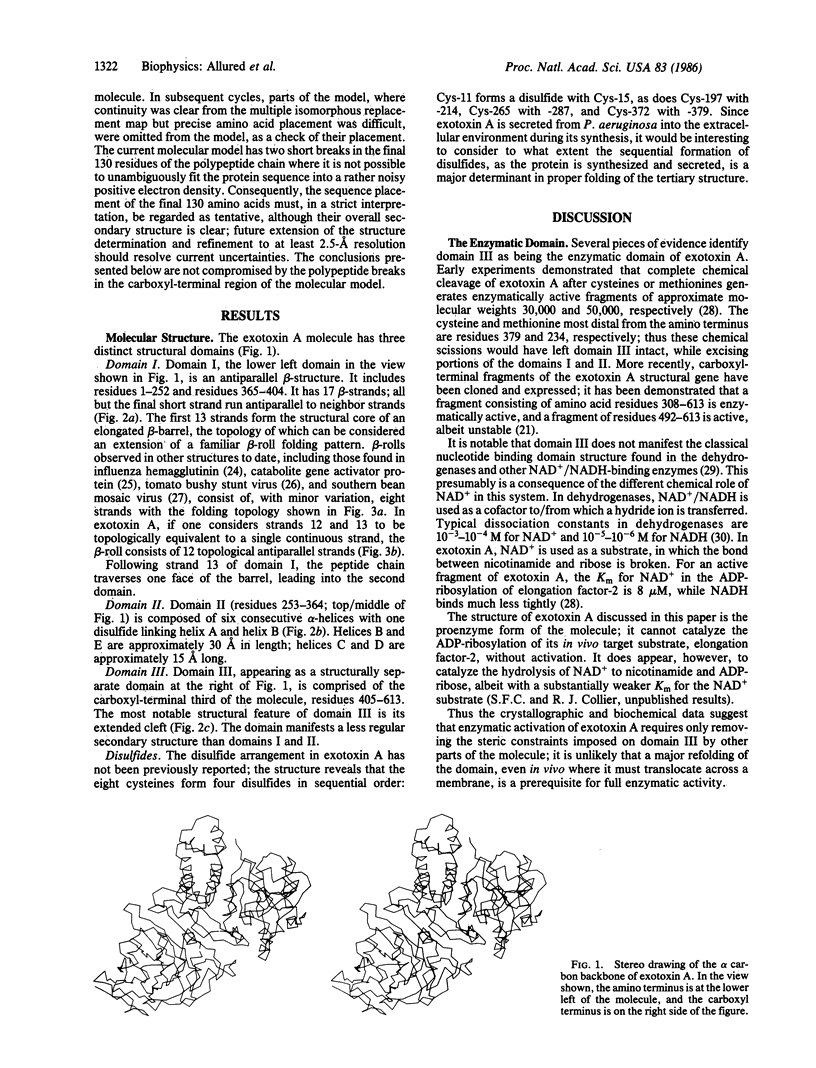
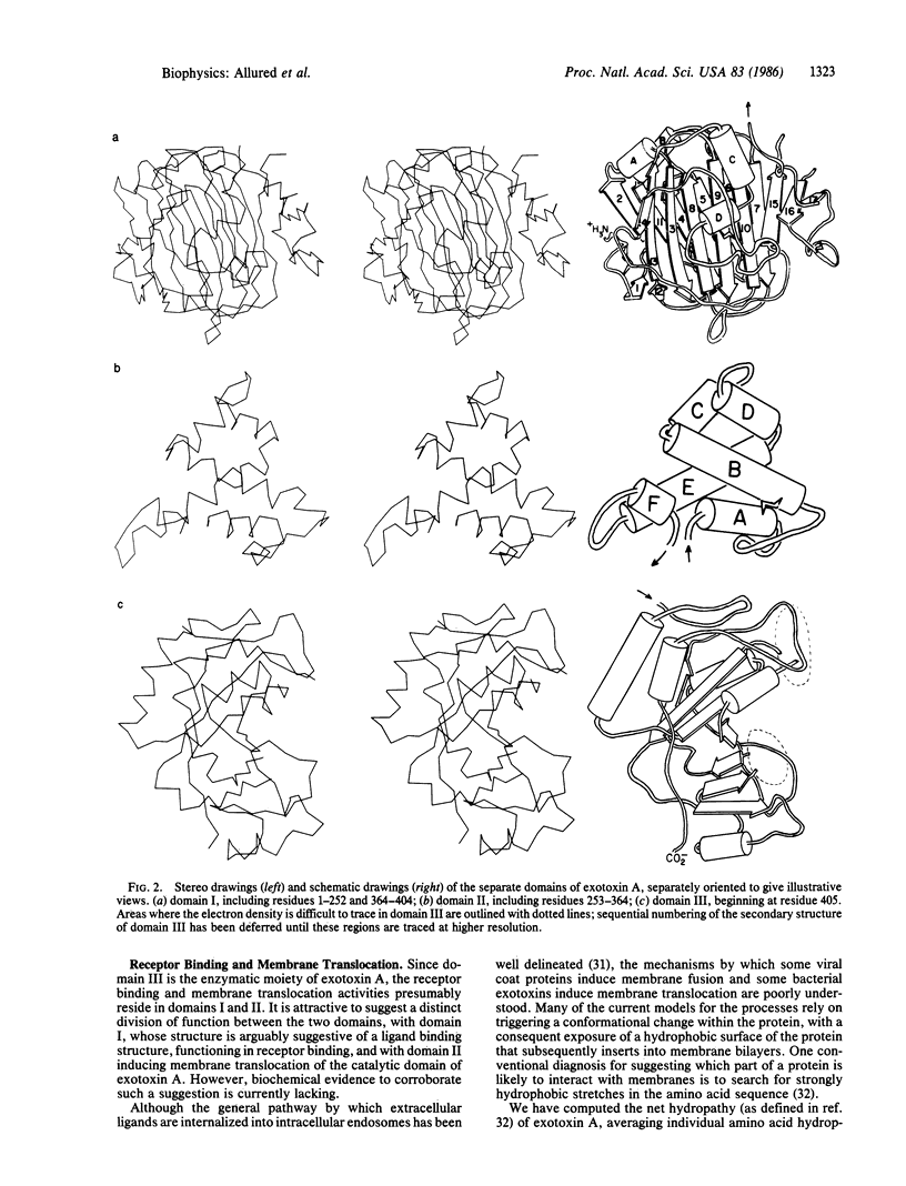
Exotoxin A of Pseudomonas aeruginosa is a secreted bacterial toxin capable of translocating a catalytic domain into mammalian cells and inhibiting protein synthesis by the ADP-ribosylation of cellular elongation factor 2. The protein is a single polypeptide chain of 613 amino acids. The x-ray crystallographic structure of exotoxin A, determined to 3.0-A resolution, shows the following: an amino-terminal domain, composed primarily of antiparallel beta-structure and comprising approximately half of the molecule; a middle domain composed of alpha-helices; and a carboxyl-terminal domain comprising approximately one-third of the molecule. The carboxyl-terminal domain is the ADP-ribosyltransferase of the toxin. The other two domains are presumably involved in cell receptor binding and membrane translocation.
Full text
PDF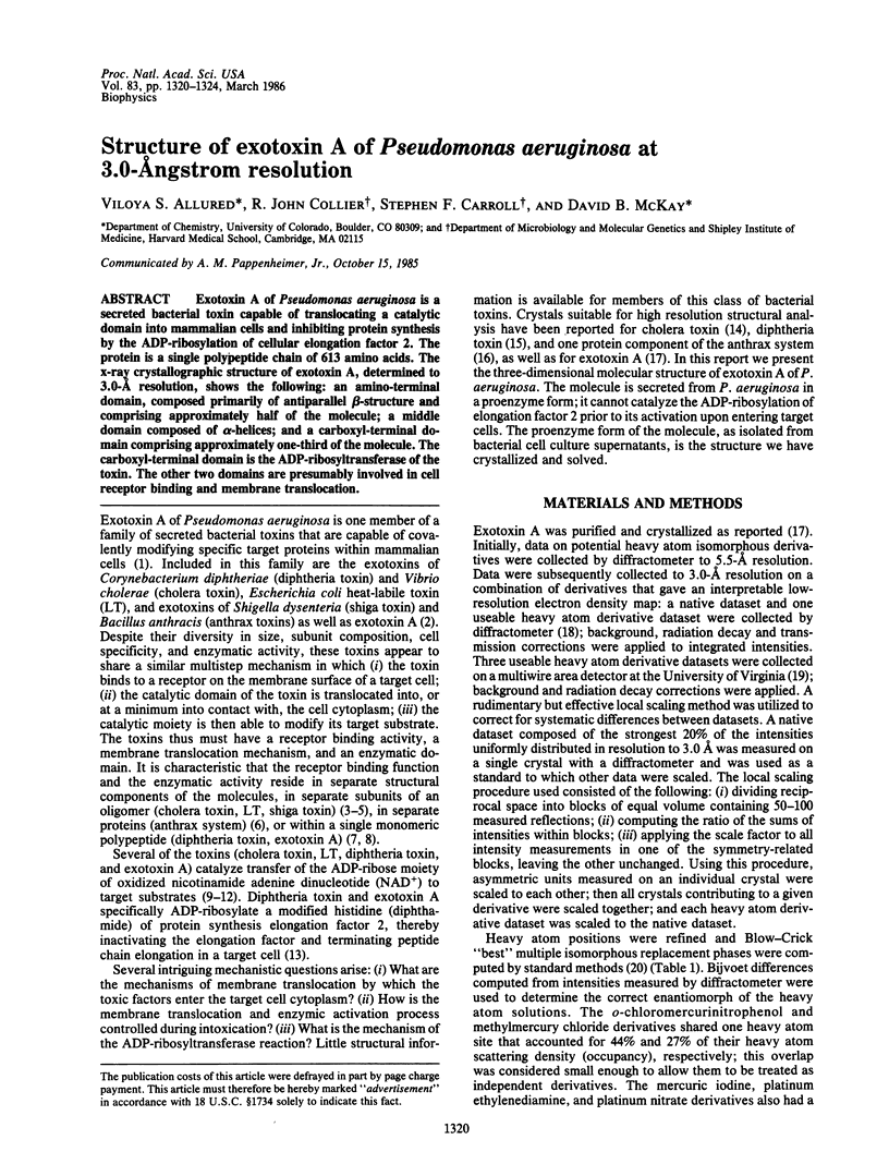
Selected References
These references are in PubMed. This may not be the complete list of references from this article.
- Allured V. S., Case L. M., Leppla S. H., McKay D. B. Crystallization of the protective antigen protein of Bacillus anthracis. J Biol Chem. 1985 Apr 25;260(8):5012–5013. doi: 10.21236/ada148938. [DOI] [PubMed] [Google Scholar]
- BEALL F. A., TAYLOR M. J., THORNE C. B. Rapid lethal effect in rats of a third component found upon fractionating the toxin of Bacillus anthracis. J Bacteriol. 1962 Jun;83:1274–1280. doi: 10.1128/jb.83.6.1274-1280.1962. [DOI] [PMC free article] [PubMed] [Google Scholar]
- Clements J. D., Yancey R. J., Finkelstein R. A. Properties of homogeneous heat-labile enterotoxin from Escherichia coli. Infect Immun. 1980 Jul;29(1):91–97. doi: 10.1128/iai.29.1.91-97.1980. [DOI] [PMC free article] [PubMed] [Google Scholar]
- Collier B., Mitchell J. F. The central release of acetylcholine during consciousness and after brain lesions. J Physiol. 1967 Jan;188(1):83–98. doi: 10.1113/jphysiol.1967.sp008125. [DOI] [PMC free article] [PubMed] [Google Scholar]
- Collier R. J., Kandel J. Structure and activity of diphtheria toxin. I. Thiol-dependent dissociation of a fraction of toxin into enzymically active and inactive fragments. J Biol Chem. 1971 Mar 10;246(5):1496–1503. [PubMed] [Google Scholar]
- Collier R. J., McKay D. B. Crystallization of exotoxin A from pseudomonas aeruginosa. J Mol Biol. 1982 May 15;157(2):413–415. doi: 10.1016/0022-2836(82)90243-1. [DOI] [PubMed] [Google Scholar]
- Collier R. J., Westbrook E. M., McKay D. B., Eisenberg D. X-ray grade crystals of diphtheria toxin. J Biol Chem. 1982 May 10;257(9):5283–5285. [PubMed] [Google Scholar]
- Gill D. M., Richardson S. H. Adenosine diphosphate-ribosylation of adenylate cyclase catalyzed by heat-labile enterotoxin of Escherichia coli: comparison with cholera toxin. J Infect Dis. 1980 Jan;141(1):64–70. doi: 10.1093/infdis/141.1.64. [DOI] [PubMed] [Google Scholar]
- Goldstein J. L., Anderson R. G., Brown M. S. Coated pits, coated vesicles, and receptor-mediated endocytosis. Nature. 1979 Jun 21;279(5715):679–685. doi: 10.1038/279679a0. [DOI] [PubMed] [Google Scholar]
- Gray G. L., Smith D. H., Baldridge J. S., Harkins R. N., Vasil M. L., Chen E. Y., Heyneker H. L. Cloning, nucleotide sequence, and expression in Escherichia coli of the exotoxin A structural gene of Pseudomonas aeruginosa. Proc Natl Acad Sci U S A. 1984 May;81(9):2645–2649. doi: 10.1073/pnas.81.9.2645. [DOI] [PMC free article] [PubMed] [Google Scholar]
- Greenfield L., Bjorn M. J., Horn G., Fong D., Buck G. A., Collier R. J., Kaplan D. A. Nucleotide sequence of the structural gene for diphtheria toxin carried by corynebacteriophage beta. Proc Natl Acad Sci U S A. 1983 Nov;80(22):6853–6857. doi: 10.1073/pnas.80.22.6853. [DOI] [PMC free article] [PubMed] [Google Scholar]
- Iglewski B. H., Kabat D. NAD-dependent inhibition of protein synthesis by Pseudomonas aeruginosa toxin,. Proc Natl Acad Sci U S A. 1975 Jun;72(6):2284–2288. doi: 10.1073/pnas.72.6.2284. [DOI] [PMC free article] [PubMed] [Google Scholar]
- Kyte J., Doolittle R. F. A simple method for displaying the hydropathic character of a protein. J Mol Biol. 1982 May 5;157(1):105–132. doi: 10.1016/0022-2836(82)90515-0. [DOI] [PubMed] [Google Scholar]
- Lai C. Y. The chemistry and biology of cholera toxin. CRC Crit Rev Biochem. 1980;9(3):171–206. doi: 10.3109/10409238009105434. [DOI] [PubMed] [Google Scholar]
- Lambotte P., Falmagne P., Capiau C., Zanen J., Ruysschaert J. M., Dirkx J. Primary structure of diphtheria toxin fragment B: structural similarities with lipid-binding domains. J Cell Biol. 1980 Dec;87(3 Pt 1):837–840. doi: 10.1083/jcb.87.3.837. [DOI] [PMC free article] [PubMed] [Google Scholar]
- Liu P. V. The roles of various fractions of Pseudomonas aeruginosa in its pathogenesis. 3. Identity of the lethal toxins produced in vitro and in vivo. J Infect Dis. 1966 Oct;116(4):481–489. doi: 10.1093/infdis/116.4.481. [DOI] [PubMed] [Google Scholar]
- McKay D. B., Steitz T. A. Structure of catabolite gene activator protein at 2.9 A resolution suggests binding to left-handed B-DNA. Nature. 1981 Apr 30;290(5809):744–749. doi: 10.1038/290744a0. [DOI] [PubMed] [Google Scholar]
- Olsnes S., Eiklid K. Isolation and characterization of Shigella shigae cytotoxin. J Biol Chem. 1980 Jan 10;255(1):284–289. [PubMed] [Google Scholar]
- Ratti G., Rappuoli R., Giannini G. The complete nucleotide sequence of the gene coding for diphtheria toxin in the corynephage omega (tox+) genome. Nucleic Acids Res. 1983 Oct 11;11(19):6589–6595. doi: 10.1093/nar/11.19.6589. [DOI] [PMC free article] [PubMed] [Google Scholar]
- Remington S., Wiegand G., Huber R. Crystallographic refinement and atomic models of two different forms of citrate synthase at 2.7 and 1.7 A resolution. J Mol Biol. 1982 Jun 15;158(1):111–152. doi: 10.1016/0022-2836(82)90452-1. [DOI] [PubMed] [Google Scholar]
- Sigler P. B., Dryan M. E., Kiuefer H. C., Finkelstein R. A. Cholera toxin crystals suitable for x-ray diffraction. Science. 1977 Sep 23;197(4310):1277–1279. doi: 10.1126/science.197.4310.1277-a. [DOI] [PubMed] [Google Scholar]
- Van Ness B. G., Howard J. B., Bodley J. W. ADP-ribosylation of elongation factor 2 by diphtheria toxin. NMR spectra and proposed structures of ribosyl-diphthamide and its hydrolysis products. J Biol Chem. 1980 Nov 25;255(22):10710–10716. [PubMed] [Google Scholar]
- Vasil M. L., Kabat D., Iglewski B. H. Structure-activity relationships of an exotoxin of Pseudomonas aeruginosa. Infect Immun. 1977 Apr;16(1):353–361. doi: 10.1128/iai.16.1.353-361.1977. [DOI] [PMC free article] [PubMed] [Google Scholar]
- Wilson I. A., Skehel J. J., Wiley D. C. Structure of the haemagglutinin membrane glycoprotein of influenza virus at 3 A resolution. Nature. 1981 Jan 29;289(5796):366–373. doi: 10.1038/289366a0. [DOI] [PubMed] [Google Scholar]
- Wyckoff H. W., Doscher M., Tsernoglou D., Inagami T., Johnson L. N., Hardman K. D., Allewell N. M., Kelly D. M., Richards F. M. Design of a diffractometer and flow cell system for X-ray analysis of crystalline proteins with applications to the crystal chemistry of ribonuclease-S. J Mol Biol. 1967 Aug 14;27(3):563–578. doi: 10.1016/0022-2836(67)90059-9. [DOI] [PubMed] [Google Scholar]
- van Heyningen S. Cholera toxin. Biol Rev Camb Philos Soc. 1977 Nov;52(4):509–509. doi: 10.1111/j.1469-185x.1977.tb00858.x. [DOI] [PubMed] [Google Scholar]


