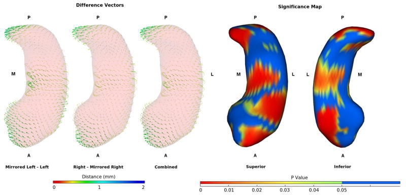
Figure 4.
Difference vectors between hippocampi segmented in mirrored and original images and the significance map for the combined data. Data is averaged across all subjects and raters. Vectors are lengthened by a factor of 2 for display clarity. The right and mirrored left segmentations are shifted medially at the head and tail while the bodies are shifted laterally, giving them a more curved appearance than the corresponding segmentations performed on the left side of the image.

