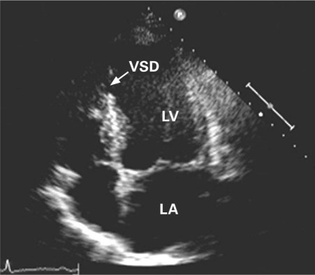
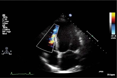
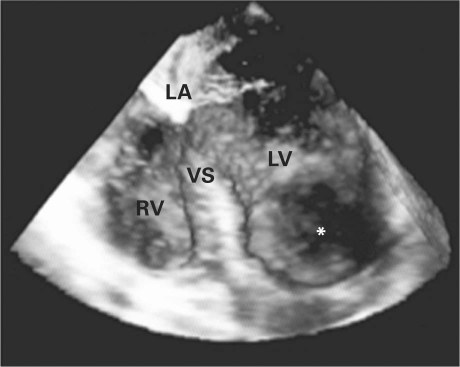
A 55-year-old man with known hypertension presented emergently with severe, acute-onset chest pain of 3 to 4 hours' duration, associated with vomiting and diaphoresis. The diagnosis was acute anterolateral-wall MI, and streptokinase was given as thrombolysis. On the 3rd day of hospitalization, the patient developed orthopnea and acute pulmonary edema. Examination revealed an apical pansystolic murmur. Transthoracic echocardiography (TTE) showed an apical ventricular septal defect (VSD) (size, 5–6 mm) with a peak gradient of 70 mmHg (Figs. 1 and 2). The patient refused surgery in favor of medical management. His condition improved with decongestive measures, and he was hemodynamically stable with intra-aortic balloon pump support. He was discharged from the hospital after 2 weeks in stable condition with strict instructions regarding follow-up after 1 week and monthly thereafter. He was placed on regular therapy with low-dose angiotensin-converting enzyme inhibitors, and he remained in stable condition in New York Heart Association (NYHA) functional class II. At the 6-year follow-up visit, the murmur had disappeared, and TTE revealed that a left ventricular clot had closed the VSD. The patient was monitored for another year. He remained in NYHA class II. Three-dimensional transesophageal echocardiography confirmed that the VSD was covered from the left ventricular side by the clot (Fig. 3).
Fig. 1 Transthoracic echocardiogram (apical 4-chamber view) shows a ventricular septal defect (VSD) (arrow).
LA = left atrium; LV = left ventricle
Fig. 2 Two-dimensional transthoracic color-flow Doppler echocardiogram shows left-to-right flow through the ventricular septal defect.
Fig. 3 Three-dimensional transesophageal echocardiogram (4-chamber view) shows the closure of the ventricular septal defect by a clot (asterisk).
LA = left atrium; LV = left ventricle; RV = right ventricle; VS = ventricular septum
Comment
Ventricular septal rupture is a rare complication after acute myocardial infarction (MI). The spontaneous closure of a ventricular septal rupture after MI has been reported by Williams and Ramsey1 and Huang and colleagues.2 In another patient, a residual VSD closed spontaneously 10 months after the surgical repair of a ventricular septal rupture that had complicated an MI.3 To our knowledge, ours is the 4th report of spontaneous closure of a ventricular septal rupture after an acute MI. Our patient's 7-year survival was probably because the VSD was small, with a gradient of 70 mmHg and pulmonary-to-systemic flow ratio (Qp/Qs) of >2:1.
Footnotes
Address for reprints: Naved Aslam, DM, Department of Cardiology, Dayanand Medical College & Hospital, Tagore Nagar, Civil Lines, Ludhiana 141001, India
E-mail: cm.mittal@yahoo.com
References
- 1.Williams RI, Ramsey MW. Spontaneous closure of an acquired ventricular septal defect. Postgrad Med J 2002;78(921):425–6. [DOI] [PMC free article] [PubMed]
- 2.Huang G, Antonini-Canterin F, Pavan D, Piazza R, Cassin M, Burelli C, Nicolosi GL. Spontaneous closure of postinfarction ventricular septal rupture. A case report. Ital Heart J 2003;4(7):484–7. [PubMed]
- 3.Vogel JH, McFadden RB, Fisher GU, Jahnke EJ. Spontaneous closure of residual ventricular septal defect following surgical repair of ventricular septal defect complicating acute myocardial infarction. Adv Cardiol 1980;27:343–5. [DOI] [PubMed]


