The crystallization of the OmpA periplasmic domain from A. baumannii is described.
Keywords: OmpA, Acinetobacter baumannii, peptidoglycan
Abstract
Outer membrane protein A from Acinetobacter baumannii (AbOmpA) is a major outer membrane protein and a key player in the bacterial pathogenesis that induces host cell death. AbOmpA is presumed to consist of an N-terminal β-barrel transmembrane domain and a C-terminal periplasmic OmpA-like domain. In this study, the recombinant C-terminal periplasmic domain of AbOmpA was overexpressed in Escherichia coli, purified and crystallized using the vapour-diffusion method. A native diffraction data set was collected to a resolution of 2.0 Å using synchrotron radiation. The space group of the crystal was P21, with unit-cell parameters a = 58.24, b = 98.59, c = 97.96 Å, β = 105.92°. The native crystal contained seven or eight molecules per asymmetric unit and had a calculated Matthews coefficient of 2.93 or 2.56 Å3 Da−1.
1. Introduction
The bacteria of the genus Acinetobacter are Gram-negative microorganisms that can be isolated from fresh water, soil and clinical models of animal and human origin (Bergogne-Bérézin & Towner, 1996 ▶). One of the 32 named and unnamed species belonging to Acinetobacter is A. baumannii, which is frequently found in clinical isolates from nosocomial infections. A. baumannii induces the apoptosis of epithelial cells in various types of opportunistic infections, for example, wound infections, pneumonia, bacterial meningitis and urinary-tract infections (Choi et al., 2005 ▶; Jyothisri et al., 1999 ▶). The evolution of multi-drug-resistant strains of A. baumannii has increased the threat posed by this bacterium among clinically important nosocomial pathogens.
In a previous study on the mechanism of host-cell apoptosis by A. baumannii, an outer membrane protein was isolated (Choi et al., 2005 ▶). This major outer membrane protein in A. baumannii (AbOmpA, or Omp38 after its molecular weight of 38 kDa) is composed of an N-terminal β-barrel transmembrane domain and a C-terminal periplasmic domain termed the OmpA-like domain. AbOmpA induces apoptosis in host epithelial cells through mitochondrial and nuclear targeting. There is a mitochondrial localization sequence in the N-terminal β-barrel and a putative nuclear localization signal sequence in the C-terminal periplasmic domain. When AbOmpA directly binds to the surfaces of host cells, the mitochondrial targeting of the porin in AbOmpA induces the activation of caspase cascades, and the nuclear targeting of the periplasmic domain leads to translocation to the host nucleus and the degradation of DNA (Choi et al., 2005 ▶, 2008 ▶).
The OmpA-like domain is a conserved motif that was originally identified in OmpA-related outer membrane proteins and MotB and has the important function of binding to the peptidoglycan layer or to peptidoglycan-associated lipoproteins, thus supporting the integrity of the outer membrane (De Mot & Vanderleyden, 1994 ▶; Koebnik et al., 2000 ▶). The structures of OmpA-like domains such as Neisseria meningitidis RmpM, Haemophilus influenza Pal and Helicobacter pylori MotB (Grizot & Buchanan, 2004 ▶; Parsons et al., 2006 ▶; Roujeinikova, 2008 ▶) have been determined, from which the molecular mechanism of peptidoglycan binding is emerging. Even though the OmpA-like domain of AbOmpA has several important novel functions as described above, its detailed atomic structure is not yet available. Here, we report the crystallization of the OmpA-like domain of AbOmpA for structural analysis in order to understand the unique properties of this interesting domain.
2. Materials and methods
2.1. Cloning and overexpression
The nucleotide sequence corresponding to the C-terminal periplasmic domain comprised of residues 221–339 (AbOmpA-PD) of AbOmpA (GenBank accession No. AY485227) was amplified using genomic DNA from A. baumannii ATCC 19606 with the primers 5′-ACTCATATGGAGTTAACTGAAGACCTTAACATGG-3′ and 5′-ACTCTCGAGTTAACGGCTACCAGTGATTGTCG-3′ by PCR and subsequently cloned into pET-28a(+) (Novagen) using NdeI and XhoI. The resulting plasmid was transformed into the expression host Escherichia coli BL21 (DE3) and the transformant colonies were inoculated into Luria–Bertani medium containing 50 µg ml−1 kanamycin and grown at 310 K until the OD600 reached 0.5–0.6. The expression of recombinant AbOmpA-PD was induced with 0.5 mM isopropyl β-d-1-thiogalactopyranoside at 291 K for 18 h. The cells were then harvested by centrifugation at 5000 rev min−1 for 20 min at 277 K.
2.2. Purification
The harvested cells were resuspended in 50 ml lysis buffer consisting of 50 mM sodium phosphate pH 7.0, 300 mM NaCl, 1 mM PMSF and disrupted by sonication (Vibra-Cell, Sonics & Materials Inc., USA) on ice. The supernatant containing the target protein was separated from the crude lysate by centrifugation (15 000 rev min−1 for 45 min at 277 K) and loaded onto a 5 ml HisTrap column (GE Healthcare, USA) pre-equilibrated with buffer A (50 mM sodium phosphate pH 7.0, 300 mM NaCl). The protein was eluted with a linear gradient from 0 to 500 mM imidazole in buffer B (50 mM sodium phosphate pH 7.0, 300 mM NaCl, 500 mM imidazole). To remove the His6 tag, the eluted fraction was dialyzed overnight with buffer A at 277 K and then reacted with 0.5 units ml−1 thrombin for 2 h at 293 K. The thrombin-treated target protein was loaded onto a 5 ml HisTrap column pre-equilibrated with buffer A. The unbound fractions were concentrated by filtration using an Amicon Ultra 10 kDa cutoff filter (Millipore, USA). In the final step, the concentrated AbOmpA-PD was passed through a Superdex 75 HiLoad 16/60 prep-grade gel-filtration chromatography column (GE Healthcare, USA) equilibrated with buffer C (20 mM Tris–HCl pH 6.8, 100 mM NaCl). The flow rate was 1.0 ml min−1. The eluted fractions containing AbOmpA-PD were detected using Coomassie Brilliant Blue on a 15% SDS–PAGE gel. The purified protein was concentrated to a final concentration of 33 mg ml−1.
2.3. Crystallization
Initial crystallization screening was performed by the sitting-drop vapour-diffusion method using various screening kits from Hampton Research (SaltRX, PEG/Ion, PEG/Ion 2, Crystal Screen HT and Additive Screen). Crystallization conditions that yielded crystals were further optimized. In the final condition, 1 µl drops of protein solution consisting of 16.5 mg ml−1 protein in 20 mM Tris–HCl pH 6.8, 100 mM NaCl were mixed with 1 µl reservoir buffer and equilibrated against the reservoir solution by vapour diffusion. After several days, rod-shaped crystals of AbOmpA-PD were obtained using a reservoir buffer consisting of 0.2 M ammonium sulfate, 0.1 M HEPES pH 7.8, 10 mM glycine and between 16 and 20%(w/v) PEG 3350.
2.4. X-ray diffraction analysis
A crystal of suitable size was transferred to cryoprotectant buffer consisting of crystallization buffer containing 20%(v/v) glycerol and mounted on a CryoLoop (Hampton Research, USA) for diffraction studies under cryogenic conditions. X-ray diffraction tests of native crystals were performed using a MicroMax-007 HF microfocus X-ray generator and an R-AXIS IV++ imaging-plate area detector (Rigaku, Japan). The final diffraction data were collected on the 6C1 beamline of the Pohang Accelerator Laboratory (PAL; Pohang, Republic of Korea). The best crystal diffracted to a resolution of 2.0 Å. The diffraction images were integrated and scaled using MOSFLM (Leslie, 2006 ▶) and SCALA (Evans, 2011 ▶).
3. Results
The OmpA-like domain of AbOmpA (residues 221–339, based on multiple sequence alignment) was cloned with a His6 tag fused to its N-terminus followed by a thrombin cleavage site. The recombinant AbOmpA-PD was expressed in soluble form and could be overproduced in the expression host E. coli BL21 (DE3). The protein was purified using nickel-affinity chromatography and subsequent thrombin digestion to remove the N-terminal His6 tag. The protein was further purified to electrophoretic homogeneity by gel-filtration chromatography. The molecular weight (13.6 kDa) and purity of the AbOmpA-PD were verified by SDS–PAGE and the results of gel-filtration chromatography indicated that the AbOmpA-PD protein exists mainly as a monomer in solution.
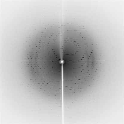
Initial screening of the crystallization conditions was performed by the sitting-drop vapour-diffusion method using various screening kits as described above. Conditions containing 0.2 M ammonium sulfate or ammonium tartrate as salt and PEG 3350 as precipitant yielded crystals and after rounds of optimization the best crystallization conditions were found to be a buffer consisting of 0.2 M ammonium sulfate, 0.1 M HEPES pH 7.8 and between 16 and 20%(w/v) PEG 3350. Additive Screen (Hampton Research) was also used and it was found that adding glycine (to a final concentration of 10 mM) improved the crystal quality. The mixture was equilibrated against 500 µl reservoir buffer with the same composition as the precipitant solution. Single crystals grew within one week and were suitable for X-ray analysis (Fig. 1 ▶). An X-ray diffraction experiment using a native AbOmpA-PD crystal was performed on the macromolecular beamline 6C1 at PAL. A complete data set was obtained to a resolution of 2.0 Å (Fig. 2 ▶). By processing the X-ray diffraction data, the crystal was found to belong to space group P21, with unit-cell parameters a = 58.24, b = 98.59, c = 97.96 Å, β = 105.9° (Table 1 ▶). The Matthews coefficient was 2.93 or 2.56 Å3 Da−1 and the solvent content was 57.99% or 51.98% assuming the presence of seven or eight molecules in the asymmetric unit, respectively. Two major peaks (θ = 177.6°, ϕ = 0° and θ = 0°, ϕ = −148.8°) from noncrystallographic twofolds were found in the self-rotation function (κ = 180°; Fig. 3 ▶), while no significant peaks were found in the native Patterson function (data not shown). These preliminary X-ray data will be useful for further structural characterization of AbOmpA-PD.
Figure 1.
Single crystal of the native OmpA periplasmic domain from A. baumannii.
Figure 2.
X-ray diffraction image obtained from the crystal of the native AbOmpA periplasmic domain. The edge of the detector corresponds to a resolution of 2.0 Å.
Table 1. X-ray data-collection statistics.
Values in parentheses are for the highest resolution bin.
| Beamline | 6C1, PAL |
| Wavelength (Å) | 1.0000 |
| Space group | P21 |
| Unit-cell parameters (Å, °) | a = 58.24, b = 98.59, c = 97.96, β = 105.92 |
| Resolution range (Å) | 50–2.0 |
| No. of observations | 229900 |
| No. of unique reflections | 67969 |
| Rmerge† | 0.065 (0.363) |
| Multiplicity | 3.4 |
| Completeness (%) | 94.8 (80.0) |
| Mean I/σ(I) | 10.7 (2.1) |
R
merge = 
 .
.
Figure 3.
Self-rotation function calculated using MOLREP (Vagin & Teplyakov, 2010 ▶). The κ = 180° section shows peaks corresponding to noncrystallographic twofolds.
Acknowledgments
We thank the staff of beamlines 4A and 6C1 at Pohang Accelerator Laboratory for help with the X-ray data collection. This work was supported by the Korean Membrane Protein Initiative program (to HYK, Korea Basic Science Institute) of the Korean Ministry of Education, Science and Technology.
References
- Bergogne-Bérézin, E. & Towner, K. J. (1996). Clin. Microbiol. Rev. 9, 148–165. [DOI] [PMC free article] [PubMed]
- Choi, C. H., Lee, E. Y., Lee, Y. C., Park, T. I., Kim, H. J., Hyun, S. H., Kim, S. A., Lee, S.-K. & Lee, J. C. (2005). Cell. Microbiol. 7, 1127–1138. [DOI] [PubMed]
- Choi, C. H., Lee, J. S., Lee, Y. C., Park, T. I. & Lee, J. C. (2008). BMC Microbiol. 8, 216. [DOI] [PMC free article] [PubMed]
- De Mot, R. & Vanderleyden, J. (1994). Mol. Microbiol. 12, 333–334. [DOI] [PubMed]
- Evans, P. R. (2011). Acta Cryst. D67, 282–292. [DOI] [PMC free article] [PubMed]
- Grizot, S. & Buchanan, S. K. (2004). Mol. Microbiol. 51, 1027–1037. [DOI] [PubMed]
- Jyothisri, K., Deepak, V. & Rajeswari, M. R. (1999). FEBS Lett. 443, 57–60. [DOI] [PubMed]
- Koebnik, R., Locher, K. P. & Van Gelder, P. (2000). Mol. Microbiol. 37, 239–253. [DOI] [PubMed]
- Leslie, A. G. W. (2006). Acta Cryst. D62, 48–57. [DOI] [PubMed]
- Parsons, L. M., Lin, F. & Orban, J. (2006). Biochemistry, 45, 2122–2128. [DOI] [PubMed]
- Roujeinikova, A. (2008). Proc. Natl Acad. Sci. USA, 105, 10348–10353. [DOI] [PMC free article] [PubMed]
- Vagin, A. & Teplyakov, A. (2010). Acta Cryst. D66, 22–25. [DOI] [PubMed]


