Abstract
Idiopathic arterial calcification of infancy is a rare condition characterized by extensive calcification and stenosis of large and medium sized arteries. A ten day old female baby developed sudden shortness of breath and was treated with oxygen and antibiotics. Antenatal echocardiography showed calcification of the aorta and pulmonary arteries. Autopsy examination revealed extensive calcification in the walls of major arteries and vessels of several organs. The baby was found to have a karyotype of 47 chromosomes.
Introduction
Calcification of the heart and vessel in fetuses is an extremely rare entity. It may be dystrophic or metastatic. Idiopathic arterial calcification of infancy(IAIC) is an unusual genetically inherited autosomal recessive condition characterized by extensive arterial calcification and stenosis of large and medium sized arteries.1,2 Pathologically, these calcific deposits are seen along the internal elastic membrane of arteries accompanied by fibrous thickening of the intima resulting in luminal narrowing.3 It may also be complicated by severe systemic hypertension and cardiomyopathy.4 Accurate diagnosis is important since follow up with genetic counseling is mandatory. We report here an autopsy case of IACI, associated with a cytogenetic abnormality, which has not been reported so far in literature.
Patient
A ten day old female baby developed sudden shortness of breath and was treated with oxygen and antibiotics, for presumed sepsis. She was the third child of a second-degree consanguineous marriage, born at full term by normal delivery. Antenatal echocardiography had revealed calcification of the aorta and pulmonary artery. In spite of active resuscitation, she died of cardio-respiratory arrest. Family history revealed that the mother's first female baby, full term normal delivery died at forty-two days of age. Her second male baby died at eleven days of age in a similar manner. No further investigations were done in both the cases.
Autopsy on this patient showed significant changes, predominantly in the major arteries of the following organs: heart, lungs, kidneys, stomach, intestine, liver and spleen. Macroscopically, the aortic wall appeared thickened, rope-like and the lumen appeared narrow .The pulmonary arteries were dilated and there was a gritty sensation in the vessel walls of both the arteries. Histological examination revealed extensive calcification mainly along the internal elastic lamina of the aorta (figure 1) and the pulmonary arteries (figure 2).
Figure 1.
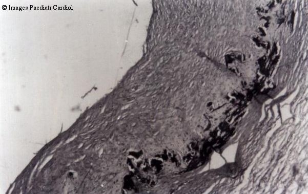
Sections from the aorta showing extensive calcification in the region of the internal elastic lamina. (H&E stain) ×400
Figure 2.
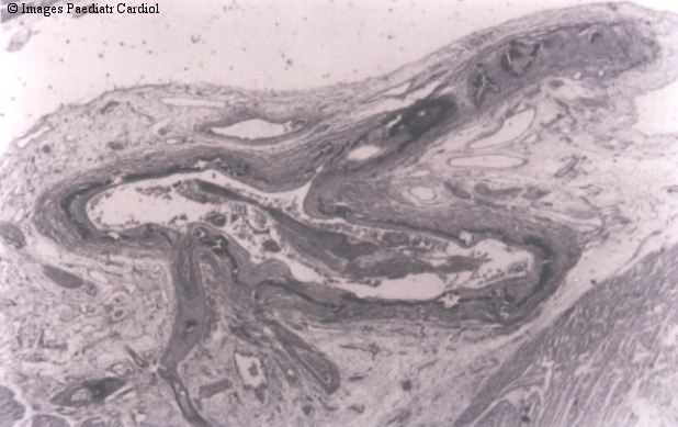
Sections showing the pulmonary vessels with calcification encircling the vessel wall (H&E stain) × 100
Specks of calcification were also seen in the coronaries (figure 3), bronchial arteries (figure 4) and branches of the gastric arteries (figure 5).
Figure 3.
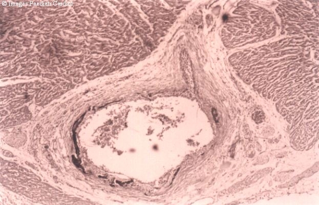
Sections show coronary vessel with partial calcification in the wall with myocardium seen in the vicinity. (H&E stain) ×100
Figure 4.
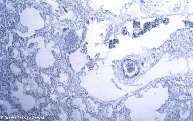
Sections from the lung show alveoli and a bronchiole with cartilage.A bronchial vessel shows a tiny speck of calcification. (H&E stain) ×100
Figure 5.
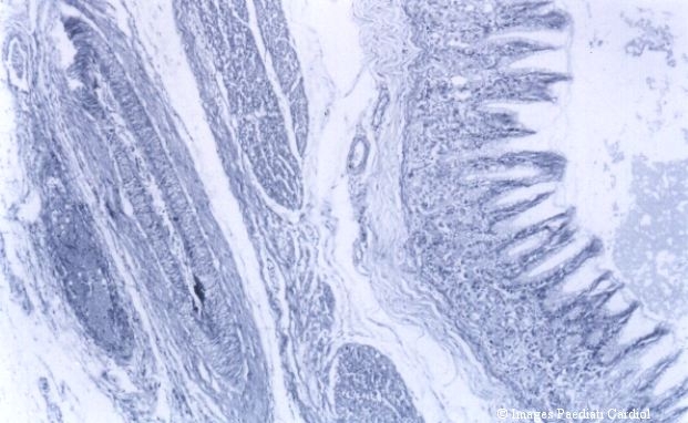
Sections shows gastric mucosa with an elongated vessel in the muscularis propria showing a focus of calcification. (H&E stain) ×100
Both kidneys showed calcification in the tubules, glomeruli (figure 6) and in the vessels. The lungs showed focal intra alveolar hemorrhage, large areas of infarction and evidence of aspiration pneumonia. The liver showed extramedullary haematopoiesis and hyperplasia of blood vessels. Placenta showed extensive calcification, intervillous haemorrhage and focal narrowing of vessels.
Figure 6.
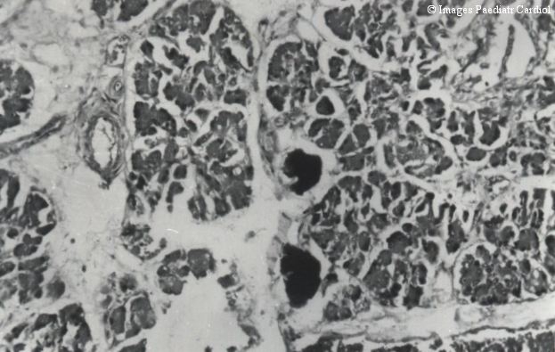
Sections showing renal tissue with calcification obliterating two of the glomeruli. (H&E stain) ×200
Cytogenetic analysis of the cord blood sample of the baby done at Genetech, Hyderabad revealed a karyotype of 47 chromosomes with normal sex chromosomes. (figure 7).
Figure 7.
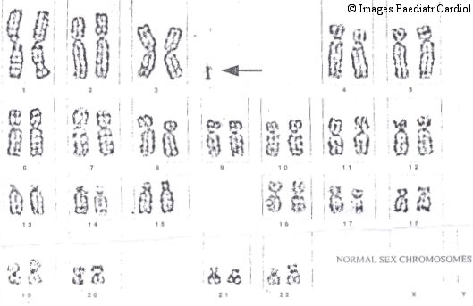
Karyotype of the cord blood sample shows 47 chromosomes.
In view of family history gross and histopathological findings and cytogenetic abnormality a diagnosis of idiopathic arterial calcification of infancy was considered.
Discussion
Idiopathic arterial calcification of infancy represents a clinical spectrum which is associated with calcification of large and medium sized blood vessels and is of unknown etiology.5 One study describes IAIC fundamentally as a defect of the elastic fibres with calcification occurring particularly in the internal elastic lamina. Materials with the staining properties of mucopolysaccarides accumulate around the elastic fibers. This is followed by fine calcium encrustation of the lamina. Later the lamina ruptures and occlusive changes occur in the intima.6 Death from myocardial infarction usually occurs during the first 6 months of life.7 One of the two sibs reported by Anderson et al had an extensive acute panarteritis suggesting that idiopathic arterial calcification of infancy may be the result of an inflammatory or infectious process. Ultrastructural examination confirmed that the deposits were hydroxyapatite and in addition contained iron deposits.8 Recent studies by Rutsch F et al (2001) showed that inorganic pyrophosphate (Ppi) inhibits hydroxyapatite deposition and mice deficient in the Ppi generating nucleoside triphosphate pyrophosphohydrolase plasma cell membrane glycoprotein-1 develop periarticular and arterial calcification in early life. In idiopathic arterial calcification of infancy hydroxyapatite deposition and smooth muscle cell proliferation occur sometimes in association with periarticular calcification.9 Two infants with idiopathic arterial calcification of infancy presented with severe systemic hypertension refractory to therapy. Radiographic examination revealed calcification of major vessels.10 Van Dyck et al described an infant in whom the diagnosis was made at the age of two weeks and therapy with diphosphonate resulted in complete resolution of the vascular calcification. At the age of two years the child was doing well, but required medical treatment for arterial hypertension.11
In our case, the family history, radiographic picture, clinical presentation and post mortem finding of extensive calcification in major arteries and medium sized arteries of various organs, enabled a diagnosis of idiopathic arterial calcification of infancy. Since the inheritance is autosomal recessive, consanguinity as seen in this case increases the risk of developing the disease. The diagnosis is essential for genetic counseling and for screening of siblings at risk for developing the disease.
Although survival to adulthood has been reported by Sholler et al12 in 1984, most patients die withen the first 6 months of life .The present case is being documented for its rarity and its association with a karyotypic abnormality of 47 chromosomes.
Conclusion
Since antenatal diagnosis is possible, idiopathic arterial calcification of infancy should be suspected when there is hyperechogenicity of vessel walls, evidence of polyhydramnios or a past history of early neonatal deaths. This case was rare in its association with a karyotypic abnormality of 47 chromosomes.
References
- 1.Hajdu J, Marton T, Papp C, et al. Calcification of fetal heart-four case reports and a literature review. Prenatal Diagn. 1998;18:1186–90. doi: 10.1002/(sici)1097-0223(199811)18:11<1186::aid-pd423>3.0.co;2-5. [DOI] [PubMed] [Google Scholar]
- 2.Pao DG, De Angelis GA, Lovell MA, et al. Idiopathic arterial calcification of infancy: Sonographic and magnetic resonance finding with pathologic correlation. Pediatr Radiol. 1981;28:561. doi: 10.1007/s002470050344. [DOI] [PubMed] [Google Scholar]
- 3.Moran JJ. Idiopathic arterial calcification of infancy: a clinicopathologic study. Pathol Annu. 1975;10:393–417. [PubMed] [Google Scholar]
- 4.Samon LM, Ash KM, Murdison KA. Aortopulmonary calcification: An unusual manifestation of idiopathic calcification of infancy evident antenatally. Obstet Gynecol. 1995;85:863–865. doi: 10.1016/0029-7844(94)00362-h. [DOI] [PubMed] [Google Scholar]
- 5.Vera J, Lucaya J, Garcia Conesa JA, et al. Idiopathic infantile arterial calcification: Unusual features. Pediatr-Radiol. 1980;20:585–587. doi: 10.1007/BF02129060. [DOI] [PubMed] [Google Scholar]
- 6.Hamazaki M. Idiopathic arterial calcification in a 3-month-old child, associated with myocardial infarction. Acta Pathol Jpn. 1980;30:301–308. doi: 10.1111/j.1440-1827.1980.tb01324.x. [DOI] [PubMed] [Google Scholar]
- 7.Marrott PK, Newcombe KD, Becroft DM, Friedlander DH. Idiopathic infantile arterial calcification with survival to adult life. Pediat Cardiol. 1984;5:119–122. doi: 10.1007/BF02424963. [DOI] [PubMed] [Google Scholar]
- 8.Anderson KA, Busbach JA, Fenton LJ, et al. Idiopathic arterial calcification of infancy in a newborn sibling with unusual light and electron microscopic manifestations. Arch. Path Lab Med J. 1985;109:838–842. [PubMed] [Google Scholar]
- 9.Rutsch F, Vaingankar B, Johnson K, Goldfine I, et al. PC-I nucleoside triphosphate pyrophosphohydrolase deficiency in idiopathic infantile arterial calcification. Am J Pathol. 2001;158:543–554. doi: 10.1016/S0002-9440(10)63996-X. [DOI] [PMC free article] [PubMed] [Google Scholar]
- 10.Milner LS, Heitner R, Thomson PD, et al. Hypertension as the major problem of Idiopathic arterial calcification of infancy. J.Pediatr. 1984;105:934–938. doi: 10.1016/s0022-3476(84)80080-3. [DOI] [PubMed] [Google Scholar]
- 11.Van Dyck N, Proesmans W, Vanhollebeke E. Idiopathic infantile arterial calcification with cardiac renal and central nervous system involvement. Europ J Paediat. 1989;148:374–377. doi: 10.1007/BF00444138. [DOI] [PubMed] [Google Scholar]
- 12.Sholler GF, Yu JS, Bale DM, et al. Generalized arterial calcification of infancy of three case reports, including regression with long-term survival. J. Pediatr. 1984;105:257–260. doi: 10.1016/s0022-3476(84)80123-7. [DOI] [PubMed] [Google Scholar]


