Abstract
Ticks serve as biological vectors for a wide variety of bacterial pathogens which must be able to efficiently colonize specific tick tissues prior to transmission. The bacterial determinants of tick colonization are largely unknown, a knowledge gap attributed in large part to the paucity of tools to genetically manipulate these pathogens. In this study, we demonstrated that Francisella tularensis subsp. novicida, for which a complete two-allele transposon mutant library has been constructed, initially infects the midguts of 100% of acquisition-fed Dermacentor andersoni nymphs, with stable colonization and replication during a subsequent molt. Increased dissemination to and marked replication within the salivary gland was closely linked to a second (transmission) feed and culminated in secretion of bacteria into the saliva and successful transmission. Simultaneous testing of multiple mutants resulted in total bacterial levels similar to those observed for single mutants. However, there was evidence of a bottleneck during colonization, resulting in a founder effect in which the most successful mutant varied when comparing individual ticks. Thus, it is essential to assess mutant success at the level of the tick population rather than in individual ticks. The ability of F. tularensis subsp. novicida to recapitulate the key physiological events by which bacteria colonize and are transmitted by ixodid ticks provides a new genome-wide approach to identify the required pathogen molecules and pathways. The identification of specific genes and, more importantly, conserved pathways that function at the tick-pathogen interface will accelerate the development of new methods to block transmission.
INTRODUCTION
Ticks transmit a wide variety of microbial pathogens, including bacteria in the genera Anaplasma, Borrelia, Ehrlichia, Francisella, and Rickettsia. Despite specific individual differences, bacterial pathogens from these genera share common features regarding the manner in which they colonize and are transmitted from ixodid ticks. These include replication in the tick midgut following initial acquisition feeding on an infected mammalian host, subsequent transit to the salivary gland, and a final burst of replication coordinated with a transmission feed on a second, naïve mammalian host. However, the bacterial determinants for each of these steps in colonization of the tick and subsequent transmission are largely unknown and represent a significant knowledge gap related to improving disease control.
This knowledge gap is, in large part, attributed to the paucity of tools available to genetically manipulate many tick-borne pathogens. As a result, identification of bacterial genes required for invasion, survival, replication, and dissemination within the tick has primarily focused on alternative approaches. Comparative approaches rely on homology to genes in other organisms to predict function, which is particularly limited in tick-borne pathogens where genes of unknown function and without orthologues comprise a significant percentage of their genomes. Unbiased transcriptional and proteomic whole-genome screens have identified genes of unknown function that are specifically upregulated in the tick (7, 11, 13, 18). In both approaches, however, functional significance is only inferred and not confirmed. A genome-wide knockout mutant screen would allow for empirical identification of pathogen molecules and pathways required for colonization and transmission, determinants that may be shared among multiple tick-transmitted bacterial pathogens.
The two-allele transposon mutant library constructed in Francisella tularensis subsp. novicida (5) is a potentially powerful tool, which could be used to identify these determinants. However, to be informative, F. tularensis subsp. novicida needs to recapitulate the key features of colonization and transmission within ixodid ticks. Additionally, reliable detection of individual mutants within a pool is essential for identification of genes required for pathogen infection of the tick host. In the present work, we tested the following four linked hypotheses: (i) upon acquisition by Dermacentor andersoni, F. tularensis subsp. novicida colonizes the tick midgut; (ii) F. tularensis subsp. novicida disseminates in the tick and replicates in the salivary gland in coordination with a second transmission feed; (iii) F. tularensis subsp. novicida is transmitted upon a second feeding to a susceptible mammalian host; and (iv) multiple F. tularensis subsp. novicida mutants can simultaneously colonize ticks.
MATERIALS AND METHODS
Bacteria.
Francisella tularensis subsp. novicida transposon mutant Tnfn1_pw060328p01q108 (containing a kanamycin [Kan] resistance cassette and subsequently referred to as the ΔISftu2 mutant) (generously provided by Colin Manoil, University of Washington) (5) was used in single-mutant infection assays. F. tularensis subsp. novicida ΔchiA (containing a chloramphenicol [Cm] resistance cassette), ΔchiB (containing a Kan resistance cassette), and ΔchiAB (containing both Cm and Kan resistance cassettes) chitinase mutants (8) were used in multiple-mutant infection assays. All mutants were grown at 37°C and maintained on prepared tryptic soy agar (pTSA) (supplemented with 0.1% l-cysteine and the following antibiotic[s] when required: 15 μg/ml kanamycin 3 μg/ml chloramphenicol, or 15 μg/ml kanamycin plus 3 μg/ml chloramphenicol). Growth curve experiments were performed using Bioscreen C (Oy Growth Curves AB Ltd., Helsinki, Finland). Briefly, overnight cultures of wild-type F. tularensis subsp. novicida U112 (generously provided by Colin Manoil, University of Washington) and F. tularensis subsp. novicida mutants were diluted 1:1,000 into fresh tryptic soy broth (TSB) supplemented with 0.1% cysteine and grown at 25°C or 37°C with shaking at 200 rpm. Optical density was measured at 595 nm every hour for 48 h. Growth curves for each mutant were determined from cultures of six independent clones per mutant (each culture run in quadruplicate).
Ticks and animals.
Dermacentor andersoni (Reynolds Creek) nymphs were obtained from a colony maintained by USDA-ARS-ADRU (Pullman, WA). All nymphs were fed on female C57BL/6 mice that were >8 weeks old and obtained from Simonsen Laboratory (Gilroy, CA). To prepare for tick feeding, mice were first sedated by intraperitoneal administration of 60 mg/kg ketamine and 8 mg/kg xylazine. The feeding sites were prepared by shaving the back from the shoulder blades to the midthorax and attaching two capsules (fabricated from the upper screw-top portion of a 5-ml tube) caudal to the shoulder blades with a bead of superglue and a 3:1 (wt/wt) mixture of rosin and beeswax. Ten D. andersoni nymphs were deposited in each capsule, with an additional 10 ticks allowed to freely feed around the head and neck of the mouse. This purposeful distribution reduced tick crowding in a single area and yielded a greater number of fully replete ticks. After the nymph-to-adult molt, adult ticks were fed on either mice or rabbits. For adult tick feedings on mice, two ticks were deposited into each capsule. Because of the variable success of adult tick feeding on a mouse host, male New Zealand White rabbits (Western Oregon Rabbit Company, Philomath, OR) were also used as hosts for adult tick feedings. To prepare for tick infestation, rabbits were manually restrained; the fur on their ears, head, and neck was shaved down to the skin, and a piece of 2-in. stockinette was placed over each ear. The stockinette was attached to the area around each ear base with nontoxic water-soluble glue and a piece of Vetrap bandaging tape to form ear bags. To prevent ticks from entering the ear canal, small pieces of cotton were inserted in each ear. Up to 15 adult ticks were deposited into each ear bag, and the top of each bag was sealed. Elizabethan collars were used on the rabbits to prevent removal of ear bags by grooming. All animals infested with ticks were individually housed. All animal studies were conducted using an approved Institutional Animal Care and Use Committee protocol on file at Washington State University, Pullman, WA.
Experimental design.
To determine if F. tularensis subsp. novicida could recapitulate the key features of pathogen colonization within the tick host, the F. tularensis subsp. novicida ΔISftu2 mutant (representative of the wild type) was used in tick feeding assays. Nymphs were exposed to F. tularensis subsp. novicida by feeding on bacteremic mice. Bacteremia was determined in each mouse after completion of tick feeding. Fully fed nymphs were collected, and F. tularensis subsp. novicida infection level was assessed in a portion of nymphs immediately (<24 h postrepletion) and 4 days after feeding. The remaining nymphs were allowed to molt to adults. One portion of these adult ticks was assessed for F. tularensis subsp. novicida infection prior to ingestion of adult blood meal through quantification of F. tularensis subsp. novicida organisms in individual tick midguts and salivary glands. A second portion of these adult ticks were allowed to feed on a naïve rabbit for 7 days, after which the F. tularensis subsp. novicida infection level was determined in individual tick midguts, salivary glands, and saliva. To serve as a control for F. tularensis subsp. novicida replication in the absence of tick feeding, a replicate experiment was performed, and the F. tularensis subsp. novicida infection level in unfed adult ticks was determined alongside that of the transmission-fed ticks.
To determine if multiple mutants can concurrently infect ticks, three F. tularensis subsp. novicida chitinase mutants (ΔchiA, ΔchiB, and ΔchiAB) were simultaneously inoculated in mice infested with nymphs. With the exception of examining nymphs immediately after acquisition feeding, F. tularensis subsp. novicida infection was quantified in ticks in the same tissues and at the same time points as those used in the single-mutant assays. Differentiation and quantification of individual chitinase mutants were possible by replica plating of samples on different antibiotic-selective plates. For comparison with single-mutant assays, the total bacterial load in the multiple-mutant assay was determined by replica plating of samples on antibiotic-free pTSA.
Infection of nymphal ticks with F. tularensis subsp. novicida.
F. tularensis subsp. novicida mutants were subcultured from glycerol stocks onto pTSA plates and incubated overnight. A total of 5 to 10 resulting colonies were further subinoculated into prepared tryptic soy broth (pTSB) (supplemented with 0.1% l-cysteine and the appropriate selective antibiotic at the same concentrations for plate cultures) and incubated overnight at 37°C and 225 rpm. To prepare the inoculum, a mid-log-phase culture was generated by subinoculating 500 μl of overnight broth culture into 15 ml of pTSB and incubated for 3 h at 37°C and 225 rpm. Based on the optical density at 600 nm (OD600) of the culture, mutants were diluted in 1× phosphate-buffered saline (PBS) to a final concentration of 1.0 × 10⁴ CFU/ml. Three days after tick infestation, mice were intraperitoneally inoculated with 100 μl of inoculum (approximately 1,000 CFU) for F. tularensis subsp. novicida single-mutant infections (ΔISftu2 mutant). For F. tularensis subsp. novicida multiple-mutant infections (ΔchiA, ΔchiB, and ΔchiAB), mid-log-phase cultures of each mutant were diluted to 1.0 × 10⁴ CFU/ml, equal volumes of each mutant were combined into a single tube, and mice were inoculated with 100 μl of the mutant cocktail (approximately 333 CFU of each mutant). Inoculum concentration was verified by plating in duplicate on pTSA plates and enumerating the resulting CFU. Mice were monitored twice daily and euthanized when signs of advanced clinical disease were first evident. Advanced clinical signs included a hunched posture, ocular or nasal discharge, lethargy, and/or piloerection. After euthanasia, blood was collected by cardiac puncture, and bacteremia was determined by plating serial 10-fold dilutions on pTSA plates and enumerating the resulting CFU. Timing of peak bacteremia corresponded to nymph repletion (D. andersoni nymphs take approximately 5 days to feed to repletion), and fed nymphs were collected and either processed immediately or incubated at 25°C.
Quantification of F. tularensis subsp. novicida in ticks.
Midguts from fed nymphs were examined for F. tularensis subsp. novicida colonization <24 h after feeding (Δisftu2 mutant infection only) or 4 days after feeding. Adult ticks, exposed to F. tularensis subsp. novicida as nymphs, were examined for infection after molting but before feeding and immediately after 7 days of feeding. Only fully fed nymphs and 7-day-fed adults (defined by having well-developed reproductive tissues) were included for analysis. F. tularensis subsp. novicida infection load was determined in nymphs by triturating individual midguts in 0.2% saponin with sterile pestles, plating serial 10-fold dilutions of lysate (dilutions prepared in 1× PBS) on pTSA, and enumerating the resulting CFU. For adult ticks, the F. tularensis subsp. novicida infection load was assessed in the midguts, salivary glands, and saliva (7-day-fed adults only). Adult tick midguts and salivary glands were individually triturated in 0.2% saponin with sterile pestles, and the F. tularensis subsp. novicida infection load was determined by plating serial 10-fold dilutions (prepared using 1× PBS) of lysates. Previous to trituration, all tick tissue samples were rinsed three times in 1× PBS to reduce the possibility of contamination from other tissues. Saliva was collected from individual ticks subsequent to dopamine injection, as previously described (21), and deposited directly in 100 μl 1× PBS, and the entire volume was plated on pTSA. All samples were plated in duplicate (except for saliva), the resulting colonies were enumerated after 24 to 48 h of incubation at 37°C, and the total F. tularensis subsp. novicida load per tick/tissue was calculated.
Transmission of F. tularensis subsp. novicida by ticks.
Adult ticks, which acquisition fed on F. tularensis subsp. novicida-infected mice as nymphs, were placed on Francisella-naïve mice to transmission feed. Adult ticks were allowed to feed to repletion, feed until the mouse host developed advanced clinical disease, or feed for a maximum of 14 days, at which time the ticks were collected and the host was euthanized. Mice were monitored daily for signs of illness, and blood was cultured from all mice at the end of the experiment to confirm F. tularensis subsp. novicida infection.
Statistical analysis.
F. tularensis subsp. novicida infection levels in ticks or tick tissues were determined by enumeration of CFU and statistically assessed after logarithmic transformation. Analysis of variance (SAS/STAT 9.22 user's guide; SAS Institute Inc., Cary, NC) was used to compare the mean levels of infection in fed nymphs, unfed adults (midguts and salivary glands), and fed adults (midguts and salivary glands). For the multiple-mutant assays, to determine if an individual mutant was predominant at a sampling point, the proportion of each mutant within an individual sample was determined. Analysis of variance was used to compare the mean proportions of each chitinase mutant in fed nymphs, unfed adults (midguts and salivary glands), and fed adults (midguts and salivary glands). Analysis of variance, followed by a pairwise t test of the least-squares means, was used to determine statistical significance. For all comparisons, a P value of <0.05 was considered significantly different.
RESULTS
Acquisition, colonization, and initial F. tularensis subsp. novicida replication.
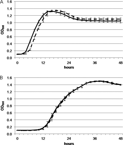
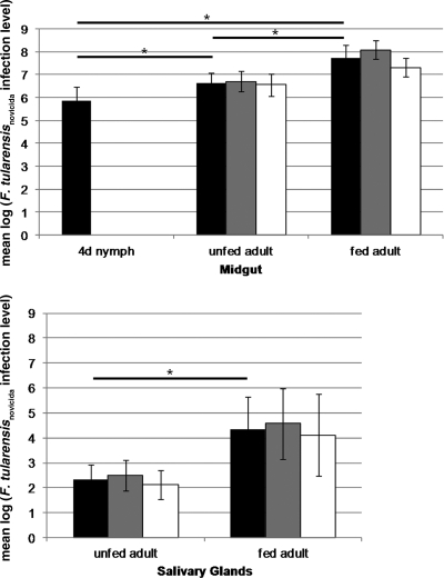
The F. tularensis subsp. novicida ΔISftu2 mutant was utilized in tick infection experiments because the location of the transposon in an insertion element was predicted to have a wild-type phenotype and the Kan-selectable marker allowed for specific differentiation from Francisella-like endosymbionts found in ticks (17). Growth curve analysis confirmed that the replication rates of the ΔISftu2 mutant and wild type were similar at both 37°C and 25°C (Fig. 1). To determine if ticks can naturally acquire F. tularensis subsp. novicida, nymphs were allowed to acquisition feed on bacteremic mice. All mice had bacteremia of >106 CFU/ml blood at the termination of tick feeding. The incidence of F. tularensis subsp. novicida in completely fed nymphs was determined either immediately (<24 h after feeding) (n = 14) or 4 days after feeding (n = 23). In both groups of nymphs, the F. tularensis subsp. novicida infection rate was 100%. The F. tularensis subsp. novicida infection level was quantified in the midguts of nymphs from both groups. In the nymphs assessed immediately after feeding, the mean infection level was 9.4 × 10⁵ CFU/midgut and increased slightly to 1.8 × 10⁶ CFU/midgut after 4 days of incubation (Fig. 2), suggesting that these values reflect colonization rather than the transient presence of bacteria within the blood meal. Similarly, after molting, 100% (20/20) of the unfed adult ticks examined were positive for F. tularensis subsp. novicida infection, confirming successful F. tularensis subsp. novicida colonization and transstadial transmission. Additionally, the mean number of F. tularensis subsp. novicida CFU in the midguts of unfed adult ticks (7.7 × 10⁶ CFU/midgut) was significantly greater than that in the fed nymphs after a 4-day hold (1.8 × 10⁶ CFU/midgut) (Fig. 2), indicating continued replication after feeding.
Fig. 1.
Growth characterization of the F. tularensis subsp. novicida ΔISftu2 mutant. F. tularensis subsp. novicida strain U112 (wild type; solid line) and the ΔISftu2 mutant (dashed line) were cultured in TSB supplemented with 0.1% cysteine and incubated at 37°C (A) and 25°C (B), the temperatures of the mouse host and tick host, respectively. Error bars represent the mean ± one standard deviation.
Fig. 2.
F. tularensis subsp. novicida replication in ticks. Ticks were exposed to F. tularensis subsp. novicida (ΔISftu2 mutant) as nymphs feeding on bacteremic mice. F. tularensis subsp. novicida infection was quantified in nymph (n = 23) midguts 4 days after feeding (4d nymph), unfed adult (n = 20) midguts and salivary glands, and fed adult (n = 20) midguts and salivary glands. For adult-stage ticks, 10 females and 10 males were examined. The mean log F. tularensis subsp. novicida infection level ± standard deviation (SD) in the midgut and salivary gland at the different time points examined are presented for total ticks (black bar), female ticks (gray bar), and male ticks (white bar). The F. tularensis subsp. novicida infection level increased significantly in the midguts of ticks both after the molt and after the adult blood meal. Likewise, the F. tularensis subsp. novicida infection level in the salivary glands increased significantly after the adult blood meal.
Transmission feeding-induced F. tularensis subsp. novicida replication.
Tick-borne bacterial pathogens frequently colonize the salivary gland and undergo replication linked to transmission feeding following the initial acquisition and colonization of the midgut. Following a second feeding, the incidence of F. tularensis subsp. novicida infection remained 100% (20/20) in the midguts and increased from 65% (13/20) to 100% (20/20) in the salivary glands, indicating that signals (e.g., changes in temperature, pH, etc.) associated with tick blood feeding induce further F. tularensis subsp. novicida dissemination to the salivary glands. Additionally, 25% (5/20) of the saliva samples collected from fed adult ticks were positive by culture for F. tularensis subsp. novicida. In both the midguts and salivary glands, the F. tularensis subsp. novicida infection level increased significantly from 7.7 × 10⁶ CFU/midgut to 1.1 × 10⁸ CFU/midgut and from 5.5 × 10² CFU/salivary gland pair to 2.1 × 10⁶ CFU/salivary gland pair (Fig. 2). As the rabbits used for the second feeding were naïve and did not develop bacteremia, the increased levels of F. tularensis subsp. novicida in fed adult ticks were not due to acquisition of bacteria during ingestion of the blood meal.
As a control for F. tularensis subsp. novicida replication through time in the absence of the second (transmission) feeding, the experiment was repeated, with the unfed adult ticks being processed alongside the transmission-fed ticks, both from the same cohort of acquisition-fed ticks. As previously observed, the midguts and salivary glands of transmission-fed adult ticks had significantly greater F. tularensis subsp. novicida infection levels than those observed in unfed adult ticks, indicating that the expansion of F. tularensis subsp. novicida numbers is not merely a function of time but is linked with transmission feeding.
Transmission of F. tularensis subsp. novicida to naïve animals.
To establish whether F. tularensis subsp. novicida colonization and replication in the salivary gland is linked to actual transmission, infected adult ticks were allowed to transmission feed on naïve mice. While mice are not a preferred host for adult D. andersoni, mice were utilized for this assay because of their susceptibility to F. tularensis subsp. novicida infection (6). Naïve mice were infested with four adult ticks per mouse. Although there was reduced adult tick feeding success (not all of the ticks attached or fully fed), 71% (5/7) of naïve mice were successfully infected with F. tularensis subsp. novicida, as determined by culture. Infection following tick transmission resulted in clinical disease, with an endpoint bacteremia similar to that observed in needle-inoculated mice.
Simultaneous colonization of ticks with multiple F. tularensis subsp. novicida mutants.
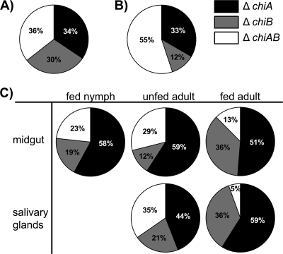
To be useful for future studies (e.g., high-throughput screening of the F. tularensis subsp. novicida two-allele transposon mutant library and competition assays), the tick must be able to simultaneously host multiple F. tularensis subsp. novicida mutants. To address this question, three chitinase mutants (ΔchiA, ΔchiB, and ΔchiAB) (8) were concurrently screened during tick colonization. These mutants were chosen because they could be easily differentiated from each other by culture based on antibiotic susceptibility. In a manner similar to that described above for the single-mutant infection assay, nymphs were allowed to feed on mice inoculated with a mixture containing equal amounts of each chitinase mutant. The infection level and proportion of each mutant in the population were determined at the same time points as those used in the single-mutant assay. The combined infection level of the ΔchiA, ΔchiB, and ΔchiAB mutants in the multiple-mutant assays was comparable at all time points and in all tick tissues to that in the single-mutant assays. The presence of each chitinase mutant in individual tick tissues was determined by plating tick tissue lysates on different selective agars. All of the chitinase mutants were identified in nymphs, unfed adult salivary glands and midguts, and fed adult salivary glands and midguts (Fig. 3). When examining individual ticks, all three mutants were identified in the majority of samples; however, individual mutants were not observed in some samples either due to their true absence or because they were below the assay sensitivity (<2 logs below the total infection level). Despite inoculation of mice with equal proportions of each mutant, mutant ratios were variable at downstream analysis points for individual samples. At the population level, however, all mutants were accounted for at every analysis point. No single mutant was consistently dominant or absent within the population.
Fig. 3.
Detection of multiple F. tularensis subsp. novicida mutants in tick tissues. The mean percentage of each F. tularensis subsp. novicida chitinase mutant was determined in the inoculum (A), mouse blood (B), and tick tissues (C). The tick tissues assessed included the midguts from nymphs held for 4 days after acquisition feeding (n = 9), midguts and salivary glands from unfed adults (n = 12), and midguts and salivary glands from fed adults (n = 9). Mutant composition in individual samples was determined by calculating the proportion of each mutant in the population based on the resulting number of CFU from replica lysate cultures on selective plates. There were no statistically significant differences between the proportions of mutants in each tissue at each time point.
DISCUSSION
The initial F. tularensis subsp. novicida colonization and replication in the tick midgut, followed by dissemination and marked replication in the salivary gland linked to successful transmission feeding, recapitulate key physiological events common among bacterial pathogens transmitted by ixodid ticks. Colonization of the nymphal midgut following acquisition feeding was highly efficient, with a 100% infection rate. The midgut serves as the primary site of colonization for most, if not all, tick-borne bacterial pathogens (2, 20, 21). The midgut presents not only a physical barrier but also an immunological barrier, as midgut epithelial cells are phagocytic and rich in innate and inducible antibacterial molecules (10, 19). Thus, tick-borne pathogens must avoid immediate clearance in order to establish an initial site of infection for subsequent replication and dissemination to other organs (4, 21). For example, the Mississippi strain of Anaplasma marginale can be acquired by D. andersoni, but it fails to replicate in the midgut and thus is unable to be transmitted (21). Importantly, F. tularensis subsp. novicida is able to both successfully colonize this tissue, as evidenced by persistence of infection through the nymph-to-adult molt, and undergo replication over time both before and after the second transmission feeding. Similar efficient acquisition and transmission of F. tularensis subsp. tularensis (type A1b and A2) and F. tularensis subsp. holarctica (type B) in Dermacentor variabilis nymphs and adults have also recently been observed (15).
Dissemination to the salivary glands was not strictly dependent on the second (transmission) feeding. Among unfed adult ticks (following the molt from nymphs), the infection rate in the salivary glands was 65%, with overall bacterial levels being quite low, with 104 fewer bacteria than in the midgut at the same time. In individual ticks, salivary gland colonization was always concurrent with midgut colonization, whereas midgut colonization without dissemination to the salivary glands was frequently observed, consistent with salivary gland infection being contingent on initial midgut colonization. However, the coordination between the second transmission feeding with dissemination to and replication within the salivary glands was remarkable. The salivary gland infection rate increased to 100%, and the infection level increased by >103 bacteria per salivary gland pair. Specifically relevant for transmission, the increased salivary gland levels correlated with detection of F. tularensis subsp. novicida within the saliva of the transmission-fed ticks. The synchronization of bacterial dissemination, replication, and salivary gland secretion indicates that F. tularensis subsp. novicida colonization of D. andersoni is a coordinated event influenced by physiological signals associated with transmission feeding. Colonization and replication to high levels in both the midguts and salivary glands with minimal, if any, detrimental effect on the tick are consistent with a true vector-bacterium relationship rather than nonspecific contamination of a nonessential host. Finally, the successful transmission to a naïve animal completes the cycle. Thus, we accept the three stated hypotheses regarding the physiologically relevant requirements for the D. andersoni/F. tularensis subsp. novicida tick transmission model.
For maximal utility of this model, including high-throughput screening of mutant libraries and competition assays, multiple mutants must be able to be simultaneously tested and recovered following meeting the physiological requirements of initial colonization, dissemination, and replication. We tested this hypothesis using three chitinase mutants, which could be differentiated by growth on selective agar. Infection of ticks with multiple mutants resulted in aggregate infection levels similar to those observed for the single-mutant infections. Importantly, within the tick population, all mutants were detected at each of the sampling points. However, there was variation in the relative frequency of each mutant when comparing individual ticks. This variation is most consistent with a population bottleneck within the tick in which dramatic reductions in population size result in founder effects and predomination of individual genotypes in the subsequent population (12). Thus, it will be critically important to assess mutant success at the level of the tick population rather than in individual ticks. Further testing of individual and multiple chitinase gene deletions will be required to definitively exclude a role for chitinases in tick colonization, especially since additional putative chitinase genes have recently been identified in F. tularensis subsp. novicida (3).
Most bacterial pathogens transmitted by ixodid ticks are obligate intracellular organisms which, using current technologies, are poorly genetically tractable, and knockout approaches to identify required genes are often lethal. Even perhaps the best-studied tick-borne bacterial pathogen, Borrelia burgdorferi, which has the advantage of being cultivable extracellularly in axenic media, remains difficult to manipulate genetically, and a genome-wide mutant library is unavailable. Our results demonstrate an ability of F. tularensis subsp. novicida to infect the tick host that is similar to that of F. tularensis subsp. tularensis (types A1b, A2, and B) (15). This study is the first to describe F. tularensis subsp. novicida infection in a tick host, a recognized biological vector of Francisella spp., via a natural route of acquisition, whereas previous F. tularensis subsp. novicida-arthropod studies have utilized direct infection of fruit flies or insect (e.g., mosquito and fruit fly) cell lines (1, 9, 14, 16). The ability of F. tularensis subsp. novicida to recapitulate the key physiological events by which bacteria colonize and are transmitted by ixodid ticks, coupled with the availability of its complete two-allele transposon mutant library, provides a new approach to identify the pathogen molecules and pathways required to mediate these events. This identification will allow determination of whether the need to mediate common functions within the tick drives convergent evolution among otherwise genetically distinct bacteria. If so, identification of these pathways may provide widely applicable tools to prevent tick-borne disease.
ACKNOWLEDGMENTS
This work was supported by National Institutes of Health grants AI093524 and AI044005 and USDA ARS grant 5348-32000-027-00D/-01S. K. E. Reif was supported by a NIH Postdoctoral Fellowship (T32AI007025). J. J. Marogolis and D. M. Monack were supported by National Institutes of Health grant AI065359.
The technical assistance of Beverly Hunter, Kathy Mason, and Ralph Horn is appreciated. We thank Marc Evans for providing the statistical analyses for this study.
Footnotes
Published ahead of print on 19 September 2011.
REFERENCES
- 1. Asare R., Akimana C., Jones S., Kwaik Y. A. 2010. Molecular bases of proliferation of Francisella tularensis in arthropod vectors. Environ. Microbiol. 12:2587–2612 [DOI] [PMC free article] [PubMed] [Google Scholar]
- 2. Ceraul S. M., et al. 2011. A Kunitz protease inhibitor from Dermacentor variabilis, a vector for spotted fever group rickettsiae, limits Rickettsia montanensis invasion. Infect. Immun. 79:321–329 [DOI] [PMC free article] [PubMed] [Google Scholar]
- 3. Chandler J. C., Molins C. R., Petersen J. M., Belisle J. T. 2011. Differential chitinase activity and production within Francisella species, subspecies and subpopulations. J. Bacteriol. 193:3265–3275 [DOI] [PMC free article] [PubMed] [Google Scholar]
- 4. Dunham-Ems S. M., et al. 2009. Live imaging reveals a biphasic mode of dissemination of Borrelia burgdorferi within ticks. J. Clin. Invest. 119:3652–3665 [DOI] [PMC free article] [PubMed] [Google Scholar]
- 5. Gallagher L. A., et al. 2007. A comprehensive transposon mutant library of Francisella novicida, a bioweapon surrogate. Proc. Natl. Acad. Sci. U. S. A. 104:1009–1014 [DOI] [PMC free article] [PubMed] [Google Scholar]
- 6. Kieffer T. L., Cowley S., Nano F. E., Elkins K. L. 2003. Francisella novicida LPS has greater immunobiological activity in mice than F. tularensis LPS, and contributes to F. novicida murine pathogenesis. Microbes Infect. 5:397–403 [DOI] [PubMed] [Google Scholar]
- 7. Löhr C. V., et al. 2002. Expression of Anaplasma marginale major surface protein 2 operon-associated proteins during mammalian and arthropod infection. Infect. Immun. 70:6005–6012 [DOI] [PMC free article] [PubMed] [Google Scholar]
- 8. Margolis J. J., et al. 2010. Contributions of Francisella tularensis subsp. novicida chitinases and Sec secretion system to biofilm formation on chitin. Appl. Environ. Microbiol. 76:596–608 [DOI] [PMC free article] [PubMed] [Google Scholar]
- 9. Moule M. G., Monack D. M., Schneider D. S. 2010. Reciprocal analysis of Francisella novicida infections of a Drosophila melanogaster model reveal host-pathogen conflicts mediated by reactive oxygen and imd-regulated innate immune response. PLoS Pathog. 6:e1001065. [DOI] [PMC free article] [PubMed] [Google Scholar]
- 10. Nakajima Y., van der Goes van Naters-Yasui A., Taylor D., Yamakawa M. 2002. Antibacterial peptide defensin is involved in midgut immunity of the soft tick, Ornithodoros moubata. Insect Mol. Biol. 11:611–618 [DOI] [PubMed] [Google Scholar]
- 11. Nelson C. M., et al. 2008. Whole genome transcription profiling of Anaplasma phagocytophilum in human and tick host cells by tiling array analysis. BMC Genomics 9:364. [DOI] [PMC free article] [PubMed] [Google Scholar]
- 12. Oberle M., Balmer O., Brun R., Roditi I. 2010. Bottlenecks and the maintenance of minor genotypes during the life cycle of Trypanosoma brucei. PLoS Pathog. 29:e1001023. [DOI] [PMC free article] [PubMed] [Google Scholar]
- 13. Ramabu S. S., Ueti M. W., Brayton K. A., Baszler T. V., Palmer G. H. 2010. Identification of Anaplasma marginale proteins specifically upregulated during colonization of the tick vector. Infect. Immun. 78:3047–3052 [DOI] [PMC free article] [PubMed] [Google Scholar]
- 14. Read A., Vogl S. J., Hueffer K., Gallagher L. A., Happ G. M. 2008. Francisella genes required for replication in mosquito cells. J. Med. Entomol. 45:1108–1116 [DOI] [PubMed] [Google Scholar]
- 15. Reese S. M., et al. 2011. Transmission efficiency of Francisella tularensis by adult American dog ticks (Acari: Ixodidae). J. Med. Entomol. 48:884–890 [DOI] [PubMed] [Google Scholar]
- 16. Santic M., Akimana C., Kouokam J. C., Atay S., Kwaik Y. A. 2009. Intracellular fate of Francisella tularensis within arthropod-derived cells. Environ. Microbiol. 11:1473–1481 [DOI] [PubMed] [Google Scholar]
- 17. Scoles G. A. 2004. Phylogenetic analysis of the Francisella-like endosymbionts of Dermacentor ticks. J. Med. Entomol. 41:277–286 [DOI] [PubMed] [Google Scholar]
- 18. Seo G. M., Cheng C., Tomich J., Ganta R. R. 2008. Total, membrane, and immunogenic proteomes of macrophage- and tick cell-derived Ehrlicihia chaffeensis evaluated by liquid chromatography-tandem mass spectrometry and MALDI-TOF methods. Infect. Immun. 76:4823–4832 [DOI] [PMC free article] [PubMed] [Google Scholar]
- 19. Sonenshine D. E., Hynes W. L., Ceraul S. M., Mitchell R., Benzine T. 2005. Host blood proteins and peptides in the midgut of the tick Dermacentor variabilis contribute to bacterial control. Exp. Appl. Acarol. 36:207–223 [DOI] [PubMed] [Google Scholar]
- 20. Tilly K., Rosa P. A., Stewart P. E. 2008. Biology of infection with Borrelia burgdorferi. Infect. Dis. Clin. North Am. 22:217–234 [DOI] [PMC free article] [PubMed] [Google Scholar]
- 21. Ueti M. W., et al. 2007. Identification of midgut and salivary glands as specific and distinct barriers to efficient tick-borne transmission of Anaplasma marginale. Infect. Immun. 75:2959–2964 [DOI] [PMC free article] [PubMed] [Google Scholar]


