Cell Cycle logoLink to Cell Cycle
. 2010 Jul 15;9(14):2704–2712. doi: 10.4161/cc.9.14.12322

Polycomb group proteins: New targets of anti-cancer therapy

Comment on: Bommi P, et al. Cell Cycle 2010; 9:2663–73

Mo K Kang 1,
PMCID: PMC3233522  PMID: 20676033

Several studies recently unveiled the involvement of the Polycomb group (PcG) proteins, most notably Bmi-1 and EZH2, in the occurrence of various human cancers and maintenance of the invasive phenotype. The PcG proteins are typically involved in the transcriptional silencing of their target genes through epigenetic mechanisms resulting in chromatin compaction. As part of the polycomb repressive complex (PRC) 2, EZH2 trimethylates histone 3 at lysine 27 (H3K27), allowing for the recruitment and assembly of the PRC1 proteins at the regulatory chromatin of the target genes.1 Bmi-1 is a component of the PRC1 required for monoubiquitination of histone 2A (uH2A), which would further promote chromatin compaction and gene silencing. Genome-wide screening revealed nearly 700 genes enriched with uH2A near the transcription start sites, and this correlated well with Bmi-1 enrichment,2 suggesting wide array of molecular pathways under transcriptional repression by Bmi-1. Oncogenic effects seen with the elevated levels of Bmi-1 and EZH2 stem from the transcriptional silencing of tumor suppressors, such as p16INK4A and p57KIP2, among other unknown PcG target genes. Bmi-1 also plays a critical role in maintaining the self-renewal capacity of normal and malignant cancer stem cells (CSCs).3 Recent advances in cancer research revealed importance of CSCs in the perpetuity of the disease due to their unique characteristics, such as enhanced malignant phenotype and resistance to the death signaling by irradiation or chemotherapeutic agents. To that end, effective anti-cancer therapy necessitates selective targeting of the CSCs, which may be accomplished by inhibiting Bmi-1 and EZH2. Success of this approach has been reported in recent articles in which Bmi-1 or EZH2 was targeted by RNAi to result in cell proliferation arrest and loss of self-renewal of cancer cell lines.4,5 However, there has been a notable gap between the principle of this novel anti-cancer approach and its clinical application due to lack of appropriate means to target the PcG proteins.

The article by Bommi et al.6 demonstrated that Bmi-1 expression can be targeted by exposure of cultured cells to sodium butyrate (NaB), a histone deacetylase inhibitor (HDACI).6 This finding is significant because it provides a novel mechanism underlying the anti-cancer effects of HDACIs. To date, the therapeutic benefits HDACIs have been demonstrated in many hematologic malignancies and few solid tumors when they are used as a single agent or in combination with demethylating agent. Several mechanisms have been proposed to involve (1) activating the DNA damage and growth arrest responses through ATM; (2) dowregulating thymidylate synthase, a target of a chemotherapeutic agent 5-fluorouracil; and (3) disrupting the chaperon function of hsp90 by protein acetylation, resulting in the reduced levels of its oncogenic client proteins.7 As shown in the current study, inhibition of the PcG proteins by NaB led to the induction of the growth inhibitory genes, such as p21WAF1 and p57KIP2, and cell death response via premature senescence or apoptosis in the breast cancer cell lines. The authors elegantly demonstrated the transcriptional repression of Bmi-1 by NaB through a mechanism independent of c-Myc, which was previously shown to be necessary for Bmi-1 expression. The time-course experiment also showed that Bmi-1 down-regulation by NaB is indirect, and suggests an unknown factor that mediates the inhibitory effects of the HDACI. Due to the involvement of Bmi-1 in self-renewal and maintenance of CSCs, this study revealed a possibility that HDACIs may target CSCs to elicit their anticancer effect.

Although the relationship between histone acetylation and stem cell phenotype is not well established, a recent study by Lee et al. showed that HDACIs interfere with stem cell function.8 HDACI exposure to human mesenchymal stem cells (hMSCs) isolated from the adipose tissues led to premature senescence by induction of p21WAF1 and loss of pluripotency. Another study by Jung et al. demonstrated that accelerated aging of hMSCs by HDACI treatment was due to downregulation of PcG proteins, namely Bmi-1, EZH2, and SUZ12, while treatment of cells to a histone acetyltransferase (HAT) inhibitor delayed the senescent phenotype.9 These findings plus the current study of Bommie et al. raise the possibility that the therapeutic efficacy of HDACI against cancer lies on its inhibitory effects on CSCs through targeting the PcG expression. Further research is needed to unveil this connection.

References

Cell Cycle. 2010 Jul 15;9(14):2704–2712. doi: 10.4161/cc.9.14.12323

HDAC inhibitors conquer polycomb proteins

Comment on: Bommi P, et al. Cell Cycle 2010; 9:2663–73

Paul Yaswen 1,

Although originally characterized as important developmental regulators in Drosophila embryos, Polycomb group (PcG) proteins have received a lot of bad press in humans lately. In particular, high expression of PcG components BMI1 and EZH2 has been correlated with poor prognosis and treatment failure in a variety of malignancies. In addition, experimental studies have directly implicated these proteins in tumorigenesis. Notably, BMI1 was originally cloned in two different laboratories as an oncogene that can cooperate with c-MYC in B cell transformation.1,2 In rodent fibroblasts, BMI1 inhibits c-MYC-induced apoptosis via downregulation of the tumor suppressor ARF.3 Like c-MYC, ectopically expressed EZH2 genes confer a proliferative advantage in primary murine embryo fibroblasts.4

BMI1 and EZH2 proteins assemble into distinct but interacting Polycomb repressive complexes (PRCs) possessing histone post-translational modification activities. Normally, BMI1, a component of PRC1, is thought to maintain the self-renewal capacity of adult stem cells.57 In addition, BMI1 has recently been shown to regulate self-renewal of cancer stem cell.8 In such cells, high levels of BMI1 may prevent activation of developmental regulatory genes, as well as tumor suppressor genes such as p16INK4a and ARF.9,10 Given the evidence of the contribution of their aberrant expression to malignancy, pharmacological manipulation of BMI1 and EZH2 expression and/or function may have therapeutic value. However, their nuclear localization, and in the case of BMI1, lack of a known enzymatic activity, complicate the development of such strategies.

Alternative therapeutic strategies might indirectly impact the expression and/or function of PcG proteins. In the forthcoming issue of Cell Cycle, Dimri and co-workers describe the ability of broad-spectrum histone deacetylase (HDAC) inhibitors, sodium butyrate and valproic acid, to cause decreased expression of BMI1 and EZH2 mRNA and protein in cultured human breast cancer cells.11 A less comprehensive earlier study has also reported that HDAC inhibitors can cause decreased expression of EZH2 protein and associated PRC2 components in human AML cells.12 In neither case is the mechanism responsible for decreased PcG gene expression entirely clear. However, the Dimri group does show convincingly that HDAC inhibitors exert effects on transcription of the BMI1 and EZH2 genes. These effects, which appear to be relatively late and indirect, can be observed using transiently transfected BMI1 promoter fragments, even those lacking intact c-MYC binding sites. Importantly, exposure to the HDAC inhibitors is sufficient to cause derepression of several growth inhibitory genes ordinarily subject to PRC-mediated silencing, as well as apoptosis or senescence, in different breast cell lines.

Given recent advances in the understanding of the role of PRCs in regulation of normal and cancer stem cells, the work by the Dimri group is timely. Although PcG proteins EZH2 and BMI1 in particular have been previously singled out as prime candidates for potential therapeutic targeting, to date no small molecules with acceptable specificities have been identified. Conversely, the mechanism(s) of action of the class of drugs collectively known as HDAC inhibitors has not been rigorously demonstrated, complicating their clinical applications.13 Many questions remain. From a mechanistic standpoint, it remains to be demonstrated whether inhibition of specific PcG proteins is necessary and/or sufficient for the induction of apoptosis or senescence by HDAC inhibitors. From a clinical standpoint, it remains to be determined whether specific (epi)genetic features of cancer cells create therapeutic windows in which inhibition of PcG protein expression will be useful.

References

Cell Cycle. 2010 Jul 15;9(14):2704–2712. doi: 10.4161/cc.9.14.12324

HDAC inhibitors repress the polycomb protein BMI1

Comment on: Bommi P, et al. Cell Cycle 2010; 9:2663–73

Steven Grant 1,

Control of gene expression is governed by a highly complex network of epigenetic processes, including histone acetylation, histone methylation, DNA methylation, and chromatin remodeling complexes, among others.1 Because transformed cells characteristically display silencing of genes involved in cell death and differentiation, intense efforts have focused on interventions capable of reversing these processes. These have led to the clinical development of histone deacetylase inhibitors (HDACIs), which promote acetylation of his-tone tails, reversal of chromatin condensation, and reexpression of repressed genes,2 as well as DNA methyltransferase inhibitors (DNMTIs), which reverse gene silencing by preventing repressive methylation of DNA CpG islands.1 HDACIs and DNMTIs have been approved for the treatment of patients with cutaneous T-cell lymphoma and myelodysplastic syndrome respectively.3

The mechanism by which HDACIs trigger cell death in transformed cells remains the subject of continuing debate. In addition to triggering reexpression of death-related genes, HDACIs also acetylate numerous proteins,4 including Hsp90 and Ku70, as well as transcription factors, all of which can contribute to lethality.5 HDACIs also cooperate with other epigenetically acting agents such as DNMTIs, leading to synergistic induction of cell death.6

Several recent studies suggest that HDACI lethality may involve perturbations in the expression or activity of various repressive complexes, particularly those implicated in histone methylation. For example, polycomb proteins such as BMI1 and EZH2 form complexes responsible for the formation of repressive histone methylation marks (e.g. trimethylation of H3K27). In human leukemia cells, HDACIs downregulate EZH2 in association with cell death induction.7 However, the relationship between HDAC inhibition and expression of BMI1, a protein implicated in stem cell maintenance, has not been explored.

In an elegant study by Prashant et al. in Cell Cycle, the authors investigated the effects of HDACIs on BMI1 expression and downstream targets in human breast cancer cells. They found that exposure of cells to various HDACIs resulted in marked downregulation of BMI1 (and EZH2) through a transcriptional mechanism, accompanied by diminished activity of BMI1-related polycomb repressive complexes, manifested by diminished trimethylation of H3K27, a classic repressive mark. These events were accompanied by re-expression of growth inhibitory proteins and putative tumor suppressor genes, resulting in cell death by apoptosis or senescence. The authors conclude that among their numerous lethal actions, HDACIs may trigger transformed cell death by down-regulating BMI1 and diminishing its repressive effects on critical tumor suppressor genes, loss of which contributes to the neoplastic phenotype.

The findings of this study have potentially important implications for our understanding of the mechanism of action of HDACIs, as well as the rational use of this important class of antineoplastic agents. While conventional wisdom holds that HDACIs act by opposing chromatin condensation and permitting re-expression of cell death- and differentiation-related genes, it is now very clear that their mode of action is highly pleiotropic, and can involve both epigenetic and non-epigenetic processes. The latter include disruption of proteasome and chaperone protein function, induction of oxidative injury, up-regulation of death receptors, and induction of DNA damage, among numerous others.2,5 HDACIs also downregulate numerous genes, which in the case of pro-survival genes, could plausibly contribute to cell death.2 HDACI-mediated up-regulation of gene expression may occur through direct mechanisms, e.g., acetylation of gene promoter regions, or by indirect mechanisms, e.g., acetylation/activation of transcription factors or as now shown in the study by Prashant et al., by downregulating the expression of proteins like BMI1 involved in repressive complexes.

These observations could have a significant impact on rational approaches to combination therapy involving HDACIs. Recently, attention has focused on novel epigenetic agents other than HDACIs or DNMTIs i.e., inhibitors of his-tone methyltransferases (HMTs) or histone demethylases.8,9 Indeed, recent studies have described agents that target HMTs (e.g., 3-deazaneplanocin), and have shown synergistic interactions with HDACIs.7 The identification of the repressive polycomb protein BMI1 as another target of HDACIs has clear implications for rational combination studies employing this class of agents. Finally, the importance of BMI1 in tumor stem cell renewal and maintenance10 could have extremely significant implications for the therapeutic potential of HDACI-containing regimens. Given continuing interest in the HDACI field, it is likely that these and related questions will be answered in the years to come.

References

Cell Cycle. 2010 Jul 15;9(14):2704–2712. doi: 10.4161/cc.9.14.12326

New functions for the Snail family of transcription factors: Two-faced proteins

Comment on: Yang D, et al. Cell Cycle 2010; 9: 2789–802

Jesús Pérez-Losada 1,, Isidro Sanchez-Garcia 1,

The transcription factors of the Snail family have long been associated with epithelialmesenchymal transitions (EMT) and cancer dissemination, mainly because of their capacity to suppress E-cadherin expression, which facilitates the spreading of tumor cells. Beyond the above-mentioned functions, there is also a contribution of Snail family proteins to other processes in adult development, such as tissue homeostasis and the initial steps of cancer development, through other mechanisms that have only started to be understood in the recent years (ref. 1).

The first insights into the fact that Snail genes have additional cellular functions that sometimes can occur independently of the induction of EMT came from the study of engineered and naturally occurring mutations in the Slug gene. Slug-deficient mice present a phenotype that indicates an essential role for Slug in melanocyte migration, hematopoietic stem cells maintenance and migration, and germ cells function.2 Similarly, there are human pigmentary diseases, like Waardenburg syndrome and piebaldism, which have been associated with SLUG mutations, pointing out to an essential role of SLUG in the development of neuronal stem-derived cell lineages.3 In this issue of Cell Cycle, Yang and colleagues4 show that Drosophila escargot and its mammalian homologue genes, Snail and Slug, are required for proper neuronal differentiation and maintainance of neuronal stem cells. This contribution of the Snail gene family to specific stem cell-based differentiation processes is not entirely new, since both Slug and Snail have been shown to be required for the differentiation of different cellular lineages such as adipocytes,5 chondrocytes6 and osteoblasts.7

However, and surprisingly, neither Snail nor Slug overexpresion cause major morphogenetic defects in mice, except for the development of heart anomalies described in Slug-overexpressing mice, that anticipated the existence of heart defects associated to SLUG duplication in humans (ref. 1). Although endogenous Slug expression in the adult myocardium has not been observed, this may merely reflect a low level of Slug-expressing cells within the heart, together with the fact that such putative mesenchymal stem cell populations constitute only a small fraction of the adult heart. However, these Snail- and Slug-overexpresing mice develop cancer,8,9 evidencing that these proteins behave as two-faced proteins in what respects to the balance between development and carcinogenesis. These facts also suggest the need for a very strict control of their expression windows and levels.

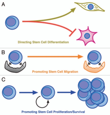
How do the Snail family proteins participate in stem cell functions? Although the role of the individual family members remains largely unaddressed in adult stem cells, we already have some pieces of knowledge (Fig. 1). Slug levels decrease upon stem cell differentiation and regulate the stem cell differentiation itself (Fig. 1A) (ref. 1) Now, Yang and colleagues4 extend these new functions to neuronal stem cell differentiation. Moreover, Slug promotes stem cell migration (ref. 1) (Fig. 1B). Finally, both proteins, Snail and Slug, mediate DNA damage response in mice, suggesting a role in stem cell survival and proliferation (Fig. 1C).8,9 These observations underlie the fact that the cellular context is of critical importance for correctly interpreting Snail/Slug functions. This cellular context specificity is also underscored by the fact that many Slug-expressing lineages showed no obvious phenotypes in Slug mutant mice. Thus, these data indicate that Snail family proteins are not only late-acting pro-disseminating factors, but also key proteins involved in early stages of both physiological development and cancer.

Figure 1.

Figure 1

New functions for Snail-family members in stem cell differentiation and proliferation. Although the importance of every particular member for these functions has yet to be fully defined, their main roles in controlling stem cell properties are: A) Directing stem cell differentiation. Slug ans Snail are involved in stem cell differentiation in several systems like haematopoietic (Slug), adipose (Slug), chondrocytes (Snail), osteoblasts (Snail) and neuroblasts (Slug and Snail) tissue and may act by controlling expression of the self-renewal machinery. B) Promoting stem cell migration. Slug favours migration of normal uncommitted progenitor to their physiological environments. If this is not achieved in a specific period of time, they would undergo apoptosis as they have been deprived of required external signals. C) Promoting stem cell survival/proliferation. Slug is essential for stem cell viability in several systems and thus important for proliferation

There are many challenges ahead before translating this new information into clinical benefits for patients. The evidence discussed above shows that Snail family members seem to play a critical role in determining stem cell function. In turn, several developmental and physiological processes depend on these cellular functions. The best proof of this principle is the existence of human diseases caused by mutations in Snail family genes. Two critical and reciprocal questions still pending to clarify are: (1) how could the degree of differentiation affect the function of Snail factors, and (2) how Snail factors could affect cell function independently of the differentiation process. Finally, genetic studies will be essential in uncovering the physiological functions of Snail members. Such genetics studies will in turn provide tools to clarify the biochemical interactions of these Snail family proteins and their impact in the genesis and development, not only in cancer, but also in other emerging stem cell-derived diseases.

Acknowledgements

Research in our groups is partially supported by FEDER and by MICINN (SAF2009-08803 to ISG, and PLE2009-O119 to JPL), by FIS (PI070057) to JPL, by Junta de Castilla y León (CSI13A08 to ISG, SA078A09 and SAN126/ SA66/09 to JPL, and Proyecto Biomedicina 2009-2010 to JPL and ISG), by MEC OncoBIO Consolider-Ingenio 2010 (Ref. CSD2007-0017) to ISG, by NIH grant (R01 CA109335-04A1) to ISG, by Sandra Ibarra Foundation to JPL and ISG, by CSIC (200920I137) to JPL, and by Group of Excellence Grant (GR15) from Junta de Castilla y Leon to ISG.

References

Cell Cycle. 2010 Jul 15;9(14):2704–2712. doi: 10.4161/cc.9.14.12380

Kinases involved in Rec8 phosphorylation revealed

Comment on: Rumpf C, et al. Cell Cycle 2010; 9:2657–62

Martin Anger 1,

During meiosis, the number of chromosomes is reduced by half in two nuclear divisions following only a single DNA replication in premeiotic S phase.1 Successful segregation of chromosomes during this process requires stepwise removal of cohesion between sister chromatids, which is mediated by cohesin complex.2 Cohesion between chromosome arms, which in meiosis is essential for holding homologous chromosomes together after chiasmata formation, is removed during meiosis I, whereas centromeric cohesion, mediating connection between sister chromatids, has to be maintained until meiosis II. It was demonstrated that the removal of cohesin during meiosis requires proteolytic cleavage of its α-kleisin subunit Rec8 by Separase.3 How is it ensured that the centromeric cohesion is preserved, while the cohesion along chromosome arms is removed, when the same proteins are presumably participating on both? It was shown that the protection of cohesin at the centromere requires complex containing shugoshin and phosphatase PP2A.4 Because of the participation of phosphatase in this process, it was considered that phoshorylation might play a role in cohesin cleavage. Rec8 was among the targets, which might be potentially dephosphorylated by shugoshinPP2A complex, and therefore several laboratories recently focused their attention on identification of the kinase, which phosphorylates Rec8 in meiosis. Considering multiple roles of Rec8 during meiosis, and particularly in chromosome segregation, it is crucial to understand how the stability and potentially other functions of Rec8 are regulated by its phoshorylation.

Previously it was suggested that Rec8 phosphorylation in meiosis might be mediated by Polo kinase.5 However, replacing the identified Polo kinase phosphorylation sites by alanines did not prevent cells from cleaving Rec8 and entering anaphase. In this issue of Cell Cycle 6 Rumpf and collaborators used series of methods to identify the kinase responsible for Rec8 phoshorylation in meiosis in fission yeast. They first used mass spectrometry to identify residues phosphorylated in Rec8, which was purified from cells harvested after induction of meiosis. Residues identified in two rounds of mass spectrometry showed only mild nondisjunction defect, when they were all replaced by alanines. This effect was, however, much stronger when these mutations were combined with mutation in one of the Separase cleavage sites. The authors realized that some of the identified residues matched the casein kinase I (CK1) phosphorylation consensus site. To confirm this, they used CK1 inhibitor, which blocked phosphorylation of Rec8 fragment by Hhp1 (one of CK1 isoforms) purified from cells in meiosis. Absence of Rec8 phosphorylation, together with the retention of Rec8-GFP signal in anaphase cells, was observed in cells, in which CK1 δ/ε isoforms Hhp1 and Hhp2 were mutated. The role of Hhp1 and Hhp2 in Rec8 phosphorylation was further supported by chromatin immunoprecipitation, which showed enrichment of Hhp1 and Hhp2 at the Rec8 binding sites. Results of Rumpf and collaborators are consistent with recently published reports showing that in budding and fission yeast, Rec8 phosphorylation, essential for segregation of homologous chromosomes, requires CK1 rather than Polo kinase.7,8

It is obvious that regulation of Rec8 phosphorylation is essential for successful chromosome segregation in meiosis and it seems that the role of shugoshin-PP2A in this process is to overcome the effect of CK1 and CDC78 kinases and to keep Rec8 at the centromere unphosphorylated. In human female gametes, chromosome segregation is frequently incorrect, which has severe consequences, including pregnancy loss and developmental abnormalities.9 Whether a failure in regulation of Rec8 phosphorylation plays any role in etiology of oocyte aneuploidy, needs to be tested. However, this process could also be sensitive to the long prophase arrest, which might take even decades.

References

Cell Cycle. 2010 Jul 15;9(14):2704–2712. doi: 10.4161/cc.9.14.12591

Regulation of vitamin metabolism by p53 and p63 in development and cancer

Comment on: Kirschner R, et al. Cell Cycle 2010; 9:2177–88

Huiqing Zhou 1, Hans van Bokhoven 1,

Vitamin A and its derivatives have been long implicated in development and tumor suppression. The major active derivative of vitamin A, retinoic acid (RA), has been shown to regulate major embryonic growth and patterning decision in vertebrates. Pioneering studies by Josef Warkany and others in the 1940's and 50's established the key role of vitamin A during fetal development in rats. By now vitamin A metabolites have been implicated in a plethora of cellular processes, such as cell differentiation, apoptosis and proliferative potential of progenitor cells in organ development and in the maintenance of various epithelia.1 In addition, there are several lines of evidence that have revealed an important role of vitamin A in tumor prevention or suppression. These effects of vitamin A in development and tumor suppression show some striking parallels with the function of p53 gene family members. In the work of Kirschner and colleagues, a link between vitamin A metabolism and transcriptional regulation by p53 and p63 has been established and their results suggest that vitamin A and its derivatives can play a role in tumor suppression and development in a p53/p63 dependent manner.

In vertebrates, the p53 family contains the transcription factors p53, p63 and p73 that share highly conserved protein architecture. The p53 protein is well known as the “guardian of the genome” for its tumor suppression activities. Activated by various stress signals, p53 regulates its target genes and initiates several cellular programs such as cell cycle arrest, senescence and apoptosis to prevent proliferation of damaged cells that could lead to cancer.2 Although a high level of p63 expression has been associated with several tumor types,3 the role of p63 in cancer is still unclear. In contrast, p63 has been established as a master regulator in ectodermal development, which is supported by animal models4 as well as human developmental diseases.5 In the highly homologous DNA-binding domains (DBD) of p53 and p63 (∼65%), a number of hotspot mutations in p53 have been found in various tumors,2 and their corresponding amino acid residues in p63 have been associated with developmental disorders that manifest with limb defects, orofacial clefting and ectodermal dysplasias.5 Some of these defects are also seen upon prenatal vitamin A depletion or in mouse models in which key players of retinoic acid metabolism have been disrupted.1 Alternative transcription start sites result in two different N-terminal isoforms in both p53 and p63, the TA isoform that contains a transcription activation (TA) domain and the ΔN isoform that lacks the TA domain and is generally considered as a repressor towards the TA isoform.6 However, the ΔN isoform is also found to have transactivation activity at promoters of some epidermal genes.3 Different splicing routes of the p63 gene give rise to three isoforms (α, β and γ) of the p63 protein at the C-terminal end.6 The shortest γ isoform of p63 is most similar to the p53 protein at the C-terminus.

Because TAp63γ is the p63 isoform that structurally resembles the p53 isoform with tumor suppression activity, a lot of efforts have been made to test TAp63γ in comparison to p53. Consistent with the similarity in structure, TAp63γ has been shown to have similar transactivation activity on a number of p53-responsive promoters in overexpression settings.6 Recently, TAp63γ has been shown to cause senescence in a p53-independent manner and repress tumor progression in vivo in p53−/− background,7 indicating that TAp63γ does function as a tumor suppressor similarly to p53. In the present work, Kirschner and co-workers identified the retinal dehydrogenase/reductase retSDR1/DHRS3 gene that is involved in vitamin A metabolism as a novel potential target gene of p53 and p63. The promoter region of retSDR1/DHRS3 can be bound directly by p53 and p63. The expression of retSDR1/DHRS3 can be activated by p53 and TAp63γ, and the activation is abolished by mutations in the DNA-binding domain of p53 and p63. In addition, there is increased binding of p53 and p63γ to the retSDR1 promoter following DNA damage and a correlation between the increased expression of TAp63 and retSDR1 in colon carcinomas was observed. Therefore, this work suggests an interesting model that vitamin A may exert its effect in development and tumor suppression through p53- and p63- gene regulation.

The present observations raise several new questions. First, it is possible that the cross talk between p53/p63 and vitamin A and derivatives is more extensive than appreciated. There are a number of studies showing that retinoic acid regulates the expression of ΔNp63α during differentiation of various epithelial cell lineages.8 Further dissection of possible coregulatory pathways is therefore warranted. Second, it would be interesting to investigate in a more physiological cellular system such as primary keratinocytes how retSDR1/DHRS3 is regulated by ΔNp63α, which is the most abundant isoform in most tissues. Third, as the work of Kirschner et al. shows a differential deregulation of retSDR1/DHRS3 expression by several p63 mutations associated with different developmental defects in humans, it would be interesting to analyze the effects on retSDR1/ DHRS3 expression in vivo in fetal material from p63 mutation knockin mouse models.9

References

Cell Cycle. 2010 Jul 15;9(14):2704–2712. doi: 10.4161/cc.9.14.12592

ΔNp73β is oncogenic in hepatocellular carcinoma by blocking apoptosis signaling via death receptors and mitochondria

Comment on: Schuster A, et al. Cell Cycle 2010; 9:2629–39

Heike Bantel 1,, Hans-Uwe Simon 2,

The observation that p53 function is lost in most cancers makes it a unique molecular target for new cancer therapies. Reconstitution of p53 activity in p53-deficient tumor cells has been demonstrated to be feasible and practical. Beyond p53, the discovery of the p53-related genes p63 and p73 raised the possibility that they may be cancer-associated genes and, as a consequence, that p53 is not the only component in predicting prognosis and response to therapy, but instead part of a network containing p53, p63 and p73. All three p53 family members, each use multiple promoters and alternative splicing to generate an array of isoforms, including full-length isoforms with a transactivation (TA-) domain homologous to that of full-length p53, and amino-terminally truncated (ΔN-) isoforms. Whereas the full length TA isoforms of p63 and p73 can activate downstream target genes and induce apoptosis, the ΔN isoforms, which lack the transactivation domain, can act as dominant inhibitors of the full-length forms of p53, p63 and p73, inhibiting the transactivation of target genes and apoptosis.1 TAp73 −/− and TAp73 +/− mice are tumor-prone, and the wild-type allele is typically lost in the heterozygotes.2 This has established TA p73 as a tumor suppressor. In contrast, a predisposition for malignant tumors has not been reported for ΔNp73 −/− mice.35 On the contrary, ΔNp73 seems to function as an oncogene. Furthermore, the finding that a significant percentage of tumors select for dominant negative p63- and/or p73-isoforms argues for their oncogenic role.

In their comprehensive study Schuster et al. provide insight into the molecular mechanisms accounting for the oncogenic role of ΔNp73 in hepatocellular carcinoma (HCC).6 The authors have previously shown that an altered ratio of TAp73/ΔNp73 isoforms is often present in HCC rather than a total loss or mutation in p73 isoforms.7 Furthermore, transgenic expression of ΔNp73α in mice from the liver-selective albumin-promoter caused early-onset hepatocellular adenomas with progression to carcinomas.8 Schuster et al. now describe three novel findings accounting for the oncogenic role of ΔNp73 in HCC.6

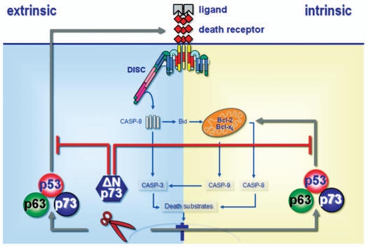
  1. The authors have identified new target genes of TAp73/ΔNp73-mediated apoptosis: TAp73, like p53, engages the major apoptosis pathways in the cell, death receptors and mitochondria, via transcription-dependent mechanisms. On the contrary, ΔNp73 confers apoptosis- and drug-resistance by inhibition of both pathways.

  2. Of clinical relevance, the authors show that the ratio TAp73/ΔNp73 regulates the apoptotic response of cancer cells, thereby playing a decisive role for treatment sensitivity versus drug resistance of HCC.

  3. Most interestingly, the authors further demonstrate that ΔNp73 is a prognostic marker in HCC and that the ΔNp73 target gene signature is a predictor of adverse outcome in patients suffering from hepatocellular carcinoma.

In order to assess the prognostic relevance of p73-dependent apoptosis signaling pathways in HCC the authors have compared their in vitro data set with the data set of Lee and Thorgeirsson9 who had previously performed gene expression profiling of human HCC tumors and identified two distinctive subclasses that are highly associated with survival. This human data set was clustered on the basis of the gene expression data set of ΔNp73-regulated target genes. Thus, the authors could show that clustering HCC genetic profiles on the basis of ΔNp73 target genes is of prognostic relevance. This finding was confirmed by Kaplan Meier analyses, which demonstrated that the ΔNp73 target gene signature can predict the prognosis of patients suffering from HCC.

In conclusion, in their elegant study, Schuster et al. propose a model (Fig. 1) which links the regulation of apoptosis by the relative expression of TAp73 and ΔNp73 to therapy response and clinical outcome in hepatocellular carcinoma. This study provides relevant findings for a central role of the p53/p63/p73 network in tumor development, treatment response and prognosis of HCC.

Figure 1.

Figure 1

Model summarizing the work of Schuster el al. which argues for an oncogenic role of ΔNp73 in hepatocellular carcinoma. Upon DNA damage, TAp73, like p53, engages the major apoptosis pathways in the cell, death receptors and mitochondria, via transcription-dependent mechanisms.6 On the contrary, ΔNp73 confers apoptosis- and drug-resistance by inhibition of both pathways. These data suggest that the ratio TAp73/ΔNp73 regulates the apoptotic response of cancer cells following treatment with DNA damaging agents thereby playing a decisive role for treatment sensitivity versus drug resistance. Of clinical relevance, the authors show that the ΔNp73 target gene signature is a predictor of adverse outcome in patients with HCC and establish ΔNp73 as a prognostic marker in hepatocellular carcinoma.

The study of Schuster et al.6 is further strengthened by a novel mouse model specifically lacking ΔNp73 engineered by Wilhelm et al.4,5 Wilhelm et al. found that ΔNp73−/− MEFs were indistinguishable from wild-type controls. However, treatment with DNA-damaging agents that induce double strand breaks revealed that cells lacking ΔNp73 displayed enhanced chemosensitivity and show an increase in p53-dependent apoptosis. When analyzing the DNA damage response (DDR) in ΔNp73−/− cells, Wilhelm et al. found that ΔNp73 localizes directly to the site of DNA damage, interacts with the DNA damage sensor protein p53BP1, and inhibits ATM activation and subsequent p53 phosphorylation. These findings establish ΔNp73 as a negative regulator of the DDR in vivo and together with the findings of Schuster et al.6 provide novel explanations why tumors with high levels of ΔNp73 show increased resistance to chemotherapy.

Thus, interfering with the expression or function of ΔNp73 in malignant tumors may render such tumors more responsive to therapy and reduce their aggressiveness and metastatic capacity.

References

Cell Cycle. 2010 Jul 15;9(14):2704–2712. doi: 10.4161/cc.9.14.12593

A novel neuronal differentiation mechanism in which Slug and Escargot are major players

Comment on: Yang D, et al. Cell Cycle 2010; 9:2789–802

Masamitsu Yamaguchi 1,

The Drosophila Escargot (Esg) and its family gene snail (sna) encode transcriptional regulators with similar C2H2 type zinc finger domains showing 76% amino acid identity. The Drosophila pan neural gene scratch also has a similar zinc finger domain, however it is more distantly related to esg and sna. In mammals, two members of this family, Snail and Slug have been reported.1 These mammalian members show a slightly greater degree of identity to the product of Drosophila esg than to that of Drosophila sna. The essential role of the Esg protein in the development of the tracheal system, wing and compound eye is well known.2,3,4 Although many lines of evidence suggest that Esg also plays a role in the nervous system, it has not been clearly demonstrated that Esg is involved in neural development or neural differentiation.

Neuro-stem cells can either differentiate into neurons or into glia—supporting non-neural cells such as astrocytes and oligodendrocytes. The process by which this happens is an important issue which remains to be addressed. In Drosophila, mechanosensory bristles are developed from neural stem cells.5 The sensory organ precursor (SOP) I (bristle stem cells) can be differentiated into SOP IIb (neural cells) and SOP IIa (supporting cells) that are further differentiated into socket cells and hair cells. Therefore Drosophila bristles provide invaluable system to examine genetic effects on neural differentiation by simply monitoring morphological or numeric changes of bristles.

In this issue of Cell Cycle Yang et al. showed that proneural precursor cells can be differentiated into neurons by Esg or Slug in both the Drosophila model system described above and also in cultured mammalian proneural N2A cells.6 The data indicate that Esg can activate not only the Drosophila but also the mammalian neural differentiation machinery. Similarly Slug can activate both the mammalian and the Drosophila neural differentiation machinery. Therefore neural differentiation appears to be highly conserved between mammals and Drosophila. Daughterless (Da) plays an essential role in bristle formation and neural differentiation in Drosophila,7 and its mammalian homologue HEB also called as TCF12/HTF4 has been suggested to be critical for the maintenance of neural stem cells. Interestingly Esg and Slug directly interact with Da and HEB, and then the complex between these proteins can be eliminated by protein degradation mediated by Drosophila sina (E3 ubiquitin ligase) and its mammalian homologue siah-1. These mechanisms are reminiscent of the Snail-p53 complex degradation in mammalian cells that was previously described by the same group.8 Since the Snail-p53 complex is secreted in human cancer,9 secretion of the HEB complex in addition to the notch/delta signaling system may be responsible for paracrine propagation of neural differentiation. More work needs to be done to address this point.

Mutations in the slug gene have been found in the human genetic disease Waardenberg syndrome (WS), a congenital disorder caused by defective functioning of the embryonic neural crest.10 WS type 1 is caused by mutations in the PAX3 gene, while WS type 2 is more heterogenous. Some WS type 2 families have mutations in the microphthalmia (MITF) gene and others have homozygous deletions in Slug. MITF is likely a trans-activator of the Slug gene and therefore defects in the Slug gene itself or its expression appear to be responsible for WS type 2. Dissection of the novel neuronal differentiation mechanism in which Slug plays a major role should therefore contribute to a deeper understanding of the pathological features of Waardenberg syndrome.

References

Cell Cycle. 2010 Jul 15;9(14):2704–2712. doi: 10.4161/cc.9.14.12594

Dancing with p53: The role of p38MAPK in mitosis of p53-deficient tetraploid cells

Comment on: Vitale I, et al. Cell Cycle 2010; 9:2823–9

Sheau-Yann Shieh 1,

p38MAPK, a member of the mitogen-activated protein kinase superfamily, plays a role in the stress response induced by ultraviolet (UV) radiation, osmotic shock, and proinflammatory stimuli such as lipopolysaccharide. Of particular interest is its involvement in mitotic progression. In the Xenopus oocyte, p38 is required for meiotic transition from G2 to M and for metaphase arrest.1,2 p38 has also been shown to be essential for the spindle assembly checkpoint (SAC) in somatic cells such as NIH3T3 and HeLa cells.3 Interestingly, work by Vitale et al.4 reveals yet another role of p38 in mitotic progression of p53-null but not p53 wild-type tetraploid cells. In their experimental setting (tetraploid HCT116 p53−/−), p38 facilitated multipolar division in the absence of p53; upon depletion or pharmacological inhibition of p38, the SAC was activated and multinucleated cells increased. Intriguingly, they found that p38 was activated in mitotic p53−/− tetraploid but not in p53+/+ tetraploid or p53−/− diploid cells. Phosphorylated (activated) p38 colocalized with spindle poles and later with the midbody. Inactivation of p38 in p53−/− tetraploid cells by either siRNA-mediated protein depletion or the chemical inhibitor SB203580 led to persistent SAC activation and marked reduction in multipolar division. These and previous observations of the seemingly conflicting roles of p38 in the SAC illustrate the multifaceted nature of the molecule and the complexity of signaling cross talk in the control of the cell cycle.

Tetraploidy represents a genomically unstable state that, upon abnormal mono-polar or multipolar cell division, creates aneuploidy that often leads to the development of cancers.5 Multipolar division of tetraploid cells is inhibited by the tumor suppressor p53.6 However, in the absence of p53, the asymmetrically dividing cells are allowed to survive. Vitale et al. demonstrated that, in the cellular context, p38 is required to permit multipolar mitosis, suggesting that p38 plays a role in suppressing or bypassing the SAC induced in p53-deficient tetraploid cells.4 In this regard, p38 appears to act similarly to the oncoprotein Mos, which has been shown recently by the same group to facilitate multipolar division of tetraploid cells.6 Although the mechanism by which p53 safeguards bipolar cell division is not understood fully, p38 appears to have a contrasting role in promoting multipolar division. The relationship between p53 and p38 contrasts with their previously characterized partnership, in which they work together to promote the expression of apoptosis-related genes.7 In this case, UV-activated p38 phosphorylates p53 at Ser33 and Ser46, thus activating p53.7 How these different scenarios are entailed remains to be explored. One possibility is that the negative feedback regulation of p38 mediated by p53-inducible wip1/PPM1D phosphatase8 is impaired in p53-null tetraploid cells, therefore allowing the constitutive activation of p38. Whether this is the case awaits further investigation. Nevertheless, the study by Vitale et al.4 raises the possibility that inhibition of p38 can suppress the growth and expansion of p53-null tumor cells, an approach that warrants further consideration.

References


Articles from Cell Cycle are provided here courtesy of Taylor & Francis

RESOURCES