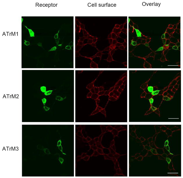
Fig. 2. Membrane localization of AeATr in HEK293/Gα16gust44 cells.
Fluorescence images of HEK293/Gα16gust44 cells transfected with the three different AeATr-pEYFP-N1 DNAs. ATrM1 (top), ATrM2 (middle) and ATrM3 (bottom). The cell surface location of the AT receptor in transfected cells was recognized by expressing them fused to YFP (Yellow Fluorescent Protein) (first row) and marking the plasma membrane glycoproteins with biotin-conjugated concanavalin A and streptavidin-conjugated Alexa Fluor 633 (second row). Yellow color in the overlay indicates AT receptor expression at the cell membrane (third row). Scale bars, 10 μm. Similar results were obtained in two independent transfection experiments.

