Abstract
Right hepatectomy for hepatocellular carcinoma is the most common major operation in liver surgery; therefore, liver surgeons should know the fundamental surgical concept and techniques of the Glissonean pedicle approach. A J-shaped or reversed T laparotomy is performed in the right subcostal area. The Glissonean pedicle approach is performed at the hepatic hilus. In this approach, the right anterior and posterior Glissonean pedicles are encircled and ligated without liver dissection. The right hepatic artery, right portal vein and right hepatic duct in the hepatoduodenal ligament should be divided if the Glissonean pedicles cannot be approached easily. After confirming the border between the right and left liver, the liver parenchyma is dissected from the anterior surface of the liver to the anterior surface of the inferior vena cava (IVC). The V5 and V8 middle hepatic veins are divided and liver dissection is performed along the main middle hepatic vein. Finally, the anterior surface of the IVC and the trunk of the right hepatic vein are identified in the liver. This approach is widely known as the anterior approach described by Lai and Fan (World J Surg 20:314–8, 1996). However, this procedure had already been reported by Takasaki et al. (Shoukakigeka 7:1545–51, 1984) but since they did not report this procedure in English, their pioneering work on the anterior approach has not been recognized. The liver hanging maneuver described by Belghiti et al. (J Am Coll Surg 193:109–111, 2001) is also useful in right hepatectomy. Among the techniques used in right hepatectomy, the Glissonean pedicle approach, the anterior approach and the liver hanging maneuver are considered to be the most important.
Electronic supplementary material
The online version of this article (doi:10.1007/s00534-011-0445-y) contains supplementary material, which is available to authorized users.
Keywords: Hepatic resection, Hepatocellular carcinoma, Huge liver tumor, Liver mobilization, Liver hanging maneuver
Introduction
Bismuth has previously described two basic procedures in right hepatectomy [1]. One is the so-called controlled hepatectomy method and the other is approaching the right Glissonean pedicle in the liver after hepatic dissection. The controlled hepatectomy method was reported by Lortat-Jacob [2]; however, Foster and Berman mentioned in their book entitled Solid Liver Tumors that Honjo in Japan performed anatomical right hepatectomy in 1949, which involved dividing the right hepatic artery, right portal vein and right hepatic duct at the hepatic hilus [3, 4]. Another procedure was reported by Lin et al. [5] from Taiwan and Tung and Quang [6] from Vietnam who dissected the liver parenchyma first and then approached the right Glissonean pedicle in the liver. Bismuth [1] combined these two procedures into his own right hepatectomy procedure. The procedure reported by Lin et al. and Tung and Quang might be considered as one of the anterior approaches in right hepatectomy. Takasaki et al. [7] first reported an anterior approach for a huge tumor in the right liver in 1984. They mentioned that the inflow vessels were ligated and divided. Liver dissection should be performed before mobilization of the right liver to avoid tumor manipulation. However, Takasaki et al. failed to report this procedure in English. Ozawa [8] described an anterior approach to avoid prolonged rotation and displacement of the hepatic lobes, which result in impairment of afferent and efferent circulation. Lai et al. [9] reported this anterior approach in English in 1996. Mobilization of the right liver is sometimes difficult and unsafe in right hepatectomy for a huge tumor due to massive bleeding or iatrogenic tumor rupture. If the tumor is manipulated during the operation, intraoperative dissemination of tumor cells occurs. Liu et al. [10, 11] showed that the anterior approach improved the surgical outcome, and demonstrated the usefulness of the anterior approach in right hepatectomy. This procedure is a safe and effective treatment for a huge tumor in the right liver compared with the conventional approach. Liver surgeons should know how to perform this technique in right hepatectomy.
Operation techniques
A reversed T-shaped incision or a J-shaped incision is made in the right subcostal area. The J-shaped incision is preferred because of the low risk of incisional herniation after surgery. If the operative field is not sufficient, thoraco-abdominal incision should be performed to obtain a good surgical view. In carrying out right hepatectomy, the Kent retractor (Fig. 1) manufactured by Takasago Medical Industry Co., Ltd., Tokyo, Japan is useful. Three adapters in the cranial side of a patient are commonly set at the 10, 11 and 2 o’clock positions.
Fig. 1.
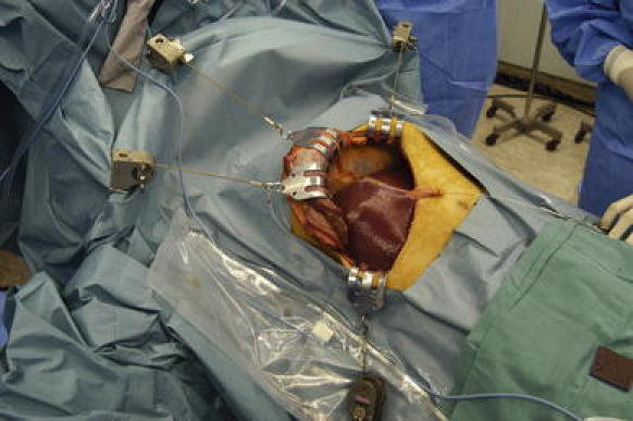
The Kent retractor manufactured by Takasago Medical Industry Co., Tokyo, Japan is useful for carrying out right hepatectomy. Three adapters in the cranial side of a patient are set at the 10, 11 and 2 o’clock positions in right hepatectomy
After laparotomy, the ligament of teres hepatis is ligated and divided. Thereafter, the falciform ligament from the ligament of teres hepatis is divided and the ventral side of the suprahepatic inferior vena cava (IVC) is confirmed. After cholecystectomy, the cystic plate is dissected. Behind the cystic plate, the right anterior Glissonean pedicle is identified. The hilar plate is detached from the liver parenchyma and the right anterior Glissonean pedicle is approached carefully. The right anterior pedicle is dissected all around the pedicle and the posterior surface of the pedicle should be confirmed. The pedicle is taped and clamped to confirm the area that is fed by the pedicle. The right posterior pedicle can be taped following the right main pedicle. The tape is pulled at the ramification point between the right anterior pedicle and the right posterior pedicle. During pedicle clamping, the color of the area changes and the tumor location is confirmed by ultrasonography. When the pedicle is ligated, the tape should be extracted strongly and the ligation should be near the liver parenchyma to avoid injuring the elements that go to another section. The pedicle should be double ligated by a transfixing suture to avoid slippage of the ligated string from the pedicle stump. The pedicle can be divided before or during liver dissection. The Glissonean pedicle approach at the hepatic hilus without liver dissection was previously reported by Couinaud [12, 13] and Takasaki et al. [14–17].
The Pringle maneuver and clamping of the infrahepatic IVC are used to control bleeding during liver dissection. The Pringle forceps is gently used to interrupt blood flow at the portal pedicle. On the other hand, intraoperative bleeding from the outflow system readily occurs when the central venous pressure is higher than 5 cm H2O. Clamping of the infrahepatic IVC is effective in reducing blood loss during hepatectomy [18] (Fig. 2). After confirming the demarcated line between the left liver and the right liver, liver dissection is performed from the anterior surface of the liver to the anterior surface of the IVC (Fig. 3). If the plane is perfectly dissected, a branch of the peripheral portal tracts will not be seen. The branches of middle hepatic veins originate from the anterior section; for example, V5 and V8 are divided and the liver parenchyma is dissected along the main middle hepatic vein. After this, the right side of the IVC and the cranial side of the caudate process are dissected. The small Glissonean pedicle to the right caudate process is preserved and the liver parenchyma is preserved behind the right main Glissonean pedicle (Fig. 4).
Fig. 2.
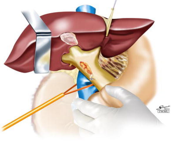
The clamping of the infrahepatic IVC is used to control bleeding during liver dissection
Fig. 3.
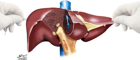
After confirming the demarcated line between the left liver and the right liver, liver dissection is performed from the anterior surface of the liver to the anterior surface of the IVC
Fig. 4.
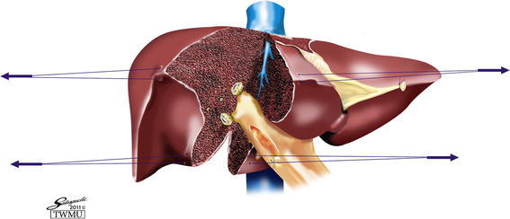
The small Glissonean pedicle to the right caudate process is preserved and the liver parenchyma is maintained behind the right main Glissonean pedicle
While dissecting the liver parenchyma of the caudate area, short hepatic veins are ligated and divided. Finally, the root of the right hepatic vein is identified (Fig. 5). Another useful anterior approach in right hepatectomy is the liver hanging maneuver reported by Belghiti et al. [19].
Fig. 5.
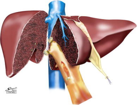
While dissecting the liver parenchyma of the caudate area, short hepatic veins are ligated and divided. The root of the right hepatic vein is indentified
In the anterior approach technique, the ventral side of the right hepatic vein and the IVC can be confirmed in the liver; however, the dorsal side of the right hepatic vein is a blind spot. After dissecting the cranial side of the right hepatic vein, the surgeon’s right pointing finger is slowly and carefully inserted behind the right hepatic vein by blunt dissection. The finger technique is useful for confirming the width of the right hepatic vein (Fig. 6). The right hepatic vein is encircled and clamped using vascular forceps. The right hepatic vein is then divided and continuously sutured using 2–0 nonabsorbent braided polyester.
Fig. 6.
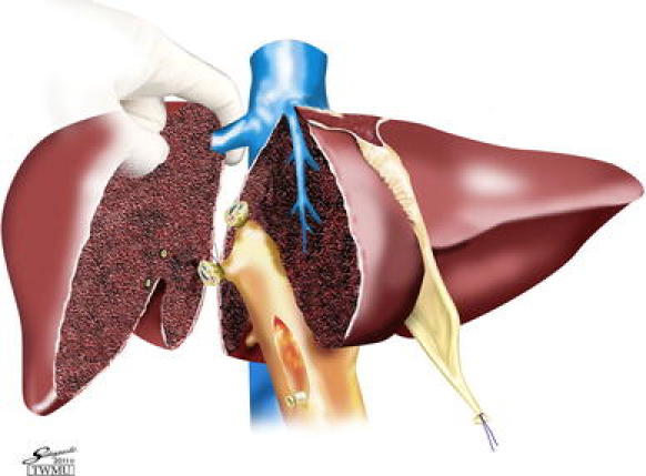
The surgeon’s right pointing finger is slowly and carefully inserted behind the right hepatic vein with blunt dissection. The finger technique is useful for confirming the width of the right hepatic vein
Thereafter, the resected right liver is dissected from the retroperitoneum, upper and lower coronary ligaments and right triangular ligament. Finally, the right adrenal gland is carefully dissected from the resected right liver. After removing the specimen, bleeding is carefully checked at the root of the Glissonean pedicle, the stump of the right hepatic vein and the resected surface of the liver parenchyma (Fig. 7).
Fig. 7.
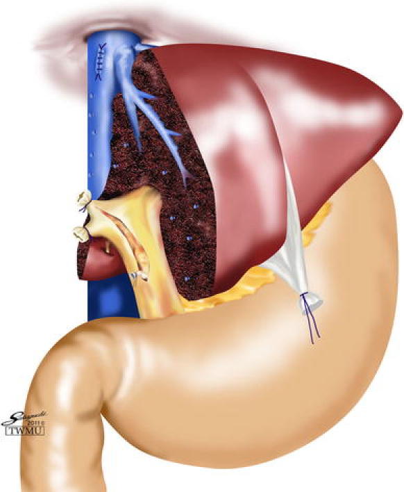
Postoperative view. Bleeding is carefully checked at the root of the Glissonean pedicle, the stump of the right hepatic vein and the resected surface of the liver parenchyma
Discussion
The Glissonean pedicle transection method was classified as either the extra-fascial approach or the extra-fascial and transfissural approach by Couinaud [13]. The extra-fascial approach to the right Glissonean pedicle starts by dissecting the hilar plate from the liver parenchyma without liver dissection, and continues by encircling the anterior and posterior Glissonean pedicles. The right main Glissonean pedicle must not be ligated en masse because of the risk of injuring the elements of the left liver such as the left hepatic duct and the left portal vein. Therefore, the right anterior and right posterior Glissonean pedicles should be ligated and divided separately.
When each pedicle is ligated, the ligation point must be close to the liver parenchyma. This procedure avoids injuring the elements that feed another section [14–17]. If a huge tumor is located near the hepatic hilus, it is difficult to perform the extra-fascial Glissonean pedicle approach. The extra-fascial and transfissural approach reported by Lin et al. [5] and Tung and Quang [6] should be considered instead. In this procedure, the right anterior and posterior pedicles can be easily found after liver dissection. Another important point regarding this procedure is that surgeons do not have to consider variation in the elements of the portal pedicle. Liver surgeons should be able to select either of these two approaches in right hepatectomy.
The liver hanging maneuver is also a useful and safe technique for right hepatectomy without liver mobilization [19]. After dividing the right anterior and posterior Glissonean pedicles, the caudate process is dissected from the IVC. Following dissection of the caudate process, the length of the blind approach behind the liver becomes <3 cm. Moreover, taping behind the liver is feasible and safe. It is possible to perform right hepatectomy with minimally invasive laparotomy using the liver hanging maneuver and the Glissonean pedicle transection method with the anterior approach. These combined methods will be the standard operative techniques for right hepatectomy.
In conclusion, the Glissonean pedicle approach, liver hanging maneuver and anterior approach are the most and important standard techniques that liver surgeons should learn and master.
Electronic supplementary material
Below is the link to the electronic supplementary material.
Conflict of interest
The authors declare that they have no conflict of interest.
Open Access
This article is distributed under the terms of the Creative Commons Attribution Noncommercial License which permits any noncommercial use, distribution, and reproduction in any medium, provided the original author(s) and source are credited.
Footnotes
This article is based on studies first reported in Highly Advanced Surgery for Hepato-Biliary-Pancreatic Field (in Japanese). Tokyo: Igaku-Shoin, 2010.
References
- 1.Bismuth H. Surgical anatomy and anatomical surgery of the liver. World J Surg. 1982;6:3–9. doi: 10.1007/BF01656368. [DOI] [PubMed] [Google Scholar]
- 2.Lortat-Jacob JL, Robert HG. Hepatectomie droitereglee. Presse Med. 1952;60:539–551. [PubMed] [Google Scholar]
- 3.Foster JH, Berman MM. Highlights in the history of liver tumors and their resection. Solid liver tumors. WB Saunders Company, Philadelphia; 1977.
- 4.Honjo I, Araki C. Total resection of the right lobe of the liver. J Int Coll Surg. 1955;23:23–28. [PubMed] [Google Scholar]
- 5.Lin TY, Chem KM, Liu TK. Total right hepatic lobectomy for primary hepatoma. Surgery. 1960;48:1048–1060. [PubMed] [Google Scholar]
- 6.Tung TT, Quang ND. A new technique for operating on the liver. Lancet. 1963;26:192–193. doi: 10.1016/S0140-6736(63)91210-8. [DOI] [Google Scholar]
- 7.Takasaki K, Kobayashi S, Muto H, Saito A, Akimoto S, Watayou T, et al. Extended right hepatectomy for huge liver cancer with non-touch isolation. Shoukakigeka. 1984;7:1545–1551. [Google Scholar]
- 8.Ozawa K. Hepatic function and liver resection. J Gastroenterol Hepatol. 1990;5:296–309. doi: 10.1111/j.1440-1746.1990.tb01632.x. [DOI] [PubMed] [Google Scholar]
- 9.Lai EC, Fan ST, Lo CM, Ghu KM, Liu CL. Anterior approach for difficult major right hepatectomy. World J Surg. 1996;20:314–318. doi: 10.1007/s002689900050. [DOI] [PubMed] [Google Scholar]
- 10.Liu CL, Fan ST, Lo CM, Tung-Ping Poon R, Wong J. Anterior approach for major right hepatic resection for large hepatocellular carcinoma. Ann Surg. 2000;232:25–31. doi: 10.1097/00000658-200007000-00004. [DOI] [PMC free article] [PubMed] [Google Scholar]
- 11.Liu CL, Fan ST, Cheung ST, Lo CM, Ng IO, Wong J. Anterior approach versus conventional approach right hepatic resection for large hepatocellular carcinoma. A prospective randomized controlled study. Ann Surg. 2006;244:194–203. doi: 10.1097/01.sla.0000225095.18754.45. [DOI] [PMC free article] [PubMed] [Google Scholar]
- 12.Couinaud C. A simplified method for controlled left hepatectomy. Surgery. 1985;97:358–361. [PubMed] [Google Scholar]
- 13.Couinaud C. Surgical anatomy of the liver revisited. Paris: Self-printed; 1989.
- 14.Takasaki K, Kobayashi K, Tanaka S, Muto H, Watayo K, Saito A, et al. Newly developed systematized hepatectomy by Glissonean pedicle transection method. Shujutsu. 1986;40:7–14. [Google Scholar]
- 15.Takasaki K. Glissonean pedicle transection method for hepatic resection: a new concept of liver segmentation. J Hepatobiliary Pancreat Surg 1998; 5:286–291. [DOI] [PubMed]
- 16.Takasaki K. Glissonean pedicle transection method for hepatic resection. Tokyo: Springer; 2007. [DOI] [PubMed] [Google Scholar]
- 17.Takasaki K. Procedures for hepatic resection. Anterior approach technique. Glissonean pedicle transection method for hepatic resection. Tokyo: Springer; 2007. pp. 76–82. [Google Scholar]
- 18.Otsubo T, Takasaki K, Yamamoto M, Katsuragawa H, Katagiri S, Yoshitoshi K, et al. Bleeding during hepatectomy can be reduced by clamping the inferior vena cava below the liver. Surgery. 2004;135:67–73. doi: 10.1016/S0039-6060(03)00343-X. [DOI] [PubMed] [Google Scholar]
- 19.Belghiti J, Guevara OA, Noun R, Saldinger PF, Kianmanesh R. Liver hanging maneuver: a safe approach to right hepatectomy without liver mobilization. J Am Coll Surg. 2001;193:109–111. doi: 10.1016/S1072-7515(01)00909-7. [DOI] [PubMed] [Google Scholar]
Associated Data
This section collects any data citations, data availability statements, or supplementary materials included in this article.


