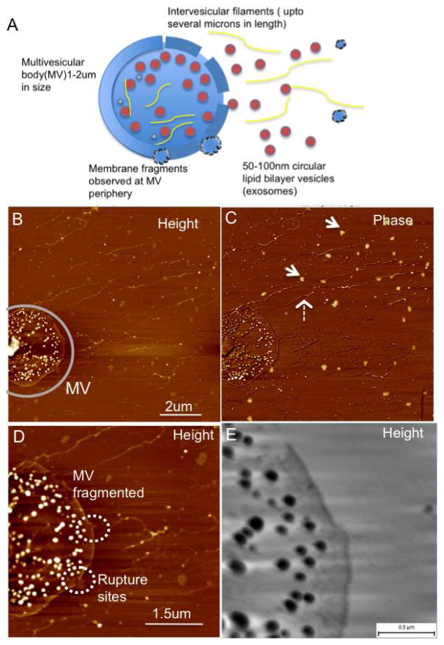
Figure 3.
Release of exosomes from multivesicular bodies (MVs) seen in oral cancer patient salivary exosomes (A) Schematics of a single MV membrane rupturing at multiple sites and release of several nanoscale vesicles, exosomes along with intervesicular filaments from the MV lumen (B) AFM topographic and (C) Phase image of a single multi-vesicular body filled with several exosome vesicles. Inter-vesicular filaments (broken arrow) without the characteristic exosome-like phase contrast (arrows) are observed. (D) At higher resolution the ruptures in the multivesicular body are seen clearly with membrane fragments appearing in the vicinity of the membrane breaks (short broken circles). Additionally the inter-vesicular filaments are also seen within the lumen of the MVB. A large rupture of the limiting membrane is seen in the top region at higher resolution in (E). Samples were imaged under ambient conditions.

