

Abstract
Renal dysplasia is a hereditary disease characterized by abnormal differentiation of renal tissue. The ultrasonographic appearance of dysplastic canine kidneys has been reported in the late stage of the disease where inflammatory and degenerative changes are already present and the dogs are in chronic renal failure. In this study, we describe the ultrasonographic appearance of the kidneys of five related Cairn Terriers affected with renal dysplasia before the onset of clinical or laboratory evidence of renal failure. Common findings included poor corticomedullary definition and multifocal hyperechoic speckles in the renal medulla, or a diffusely hyperechoic medulla. Severity of ultrasonographic changes was related to the severity of histopathologic findings. The ability to detect dysplastic changes before clinical signs develop makes ultrasound a potentially useful screening method for canine renal dysplasia.
Keywords: dysplasia, renal, ultrasound
Introduction
Renal dysplasia is a hereditary condition observed in many canine breeds including the Cairn Terrier.1–8 It is characterized by abnormal or asynchronous differentiation of renal tissue, leading to disorganized development of the renal parenchyma. Immature or fetal glomeruli persist past 6 months of age and mesenchymal tissue may be present in the medulla.9 Other findings may include persistent metanephric ducts, atypical epithelium, and dysontogenic metaplasia.10,11 As the disease progresses, degenerative and inflammatory components are superimposed and may obscure the primary lesions. Severity and rate of progression are variable among individuals; onset of clinical signs ranges from 4 weeks to 5 years or more.1,5 Diagnosis currently requires renal biopsy, as no mutation-based DNA test is available. A surgical wedge biopsy is the only reliable diagnostic method, as approximately 100 glomeruli have to be evaluated, and the architecture of the renal cortex has to be assessed.12 The prolonged time until clinical signs develop and the invasiveness of the diagnostic procedure pose a problem for breeders who have to determine if they want to use a specific dog for breeding. Availability of a noninvasive diagnostic tool to screen dogs before breeding would contribute to making sound breeding decisions and the understanding of disease development and progression.
Ultrasound is a sensitive tool to evaluate the kidney, and has been used for detection of feline polycystic kidney disease at an early age.13,14 The ultrasonographic appearance of dysplastic canine kidneys has been described2–8 but all subjects already had clinical signs of chronic renal failure.
Our purpose was to describe the ultrasonographic appearance of the kidneys of five related Cairn Terriers affected with renal dysplasia before the onset of clinical or laboratory evidence of renal failure, and to compare the severity of ultrasonographic and histopathologic changes.
Materials and Methods
Five Cairn Terriers were evaluated, four were female and one was male. The dogs underwent renal ultrasound examination as a part of a screening program for renal dysplasia performed in a group of dogs from, or related to, a line known to produce dogs affected with juvenile renal disease. Two dogs (Dogs 2 and 3) were siblings. The kidneys in these five dogs were diagnosed as ultrasonographically abnormal and the dogs subsequently underwent repeated ultrasonographic examination and surgical wedge biopsy of the kidney. Four dogs had an initial ultrasound examination at the age of 4 months, one dog at the age of 11 months. Repeated ultrasonographic examinations were performed between the age of 9 and 32 months (Table 1). Ultrasonographic examinations were performed using a GE logiq 9 ultrasound system* with either an 8 MHz microcurved or a 10 MHz linear matrix array transducer. Still images were recorded electronically.
Table 1.
Renal Length, Severity of Ultrasonographic Findings, and Histopathologic Changes
| Dog | Age (months) | Renal Length
|
Severity of Ultrasonographic Findings
|
Severity of Histopathologic Changes Left Kidney* | ||
|---|---|---|---|---|---|---|
| Left (mm) | Right (mm) | Left Kidney | Right Kidney | |||
| 1 | 4 | 38 | 38 | Mild | Mild | |
| 8 | Mild | |||||
| 18 | 37 | 38 | Mild | Mild | ||
| 2 | 4 | 33 | 36 | Severe | Moderate | |
| 9 | 37 | 40 | Severe | Moderate | Severe | |
| 24 | 34 | 35 | Severe | Severe | ||
| 3 | 4 | 33 | 33 | Severe | Severe | |
| 9 | 35 | 34 | Severe | Severe | Severe | |
| 24 | 30 | 30 | Severe | Moderate | ||
| 4 | 4 | 35 | 39 | Moderate | Moderate | |
| 11 | Moderate | |||||
| 16 | 37 | 41 | Severe | Severe | ||
| 5 | 11 | 40 | 38 | Moderate | Moderate | |
| 15 | 40 | 39 | Moderate | Moderate | Moderate | |
| 32 | 39 | 41 | Moderate | Moderate | ||
Biopsy samples were only obtained from the left kidney. Mild, <10% fetal glomeruli; moderate, 11–15% fetal glomeruli; and severe, >25% fetal glomeruli.
All dogs had surgical wedge biopsies of their left kidney cortex performed at the age of 9 months (three dogs), 11 and 15 months (one dog each), followed by histopathologic examination.
Representative ultrasound images of both kidneys were collected and patient information and examination dates removed. The images were reviewed retrospectively by a board certified radiologist (G.S.), who was unaware of the patient data and examination date. Presence of the following ultrasonographic abnormalities was recorded: renal surface irregularity, decreased corticomedullary definition, presence of hyperechoic speckles within the medulla, hyperechogenicity of the renal cortex or medulla, and presence of a hyperechoic corticomedullary rim. The severity of the changes was subjectively graded as mild, moderate, or severe for each kidney at each time point. Resistive index (RI) as well as renal length was recorded for each examination, if available.
Histopathologic features were classified retrospectively by a board certified pathologist (R.C.) who was unaware of the ultrasonographic findings. Because biopsy samples were taken from cortical tissue only, histopathologic findings were limited to the persistence of fetal glomeruli after 6 months of age. Lesions were qualified as mild if <10% of glomeruli were fetal, moderate when 11–25% of glomeruli were fetal and severe if >25% of glomeruli were fetal. Additional changes such as dilation of Bowman’s capsules and presence of eosinophilic fluid within Bowman’s space were noted.
Descriptive statistics were performed for RI values. RI values were compared between different visits of the same dog using a Wilcoxon’s rank test for nonparametric data. All statistical tests were performed using commercially available software† and the level of significance was set at P<0.05.
Results
All dogs were clinically healthy and had normal hematologic and serum chemistry parameters, urinalyses and urine protein to creatinine ratios at the time of presentation as well as at all follow up examinations. None of the dogs had developed clinical signs of renal disease at the time of submission of this manuscript.
Based on the ultrasonographic appearance of the kidneys, the changes were mild at all time points in one dog, one dog had moderate changes at all time points, one dog had moderate changes at the first examination and severe changes at the second, and two dogs had severe changes in the left kidney at all time points and moderate to severe changes in the right kidney (Fig. 1, Table 1).
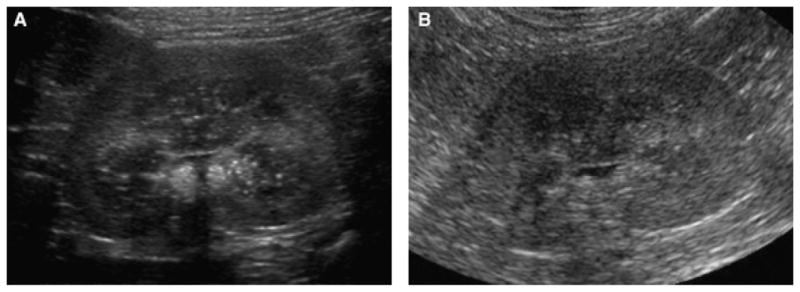
Fig. 1.

(A–C): Dorsal plane ultrasound images of the left kidney of three dogs at the age of 4 months with different degrees of ultrasonographic changes. (A) Dog 1 with mild hyperechoic speckling of the medulla and slightly reduced corticomedullary definition. (B) Dog 4 had moderate ultrasonographic changes. The predominant abnormality is the hyperechoic speckling of the medulla. (C) Dog 3 with severe ultrasonographic changes: there is poor corticomedullary definition and a hyperechoic medulla. Renal length measured between 3.3 cm (Dog 3) and 3.8 (Dog 1).
The most consistent abnormality, seen in all dogs was a decrease in corticomedullary definition and presence of multifocal hyperechoic speckles within the renal medulla or generalized medullary hyperechogenicity (Figs. 2 and 3). Mild to moderate cortical hyperechogenicity was seen in four dogs, the cortices of Dog 1 were considered to be normal in all images. A hyperechoic corticomedullary rim was discerned in one dog (Dog 5). At the time of the last examination three dogs had a wedge-shaped defect in one left renal pole; this was considered to be caused by the surgical wedge biopsy, as it corresponded to the biopsy site as described in the surgery report.
Fig. 2.
Dorsal plane ultrasound images of the left kidney of Dog 4 at the age of 4 months (A) and 16 months (B): The ultrasonographic changes were more severe at 16 months with persistent hyperechoic medullary speckles but a decreased corticomedullary definition. Mild pyelectasia is also present.
Fig. 3.
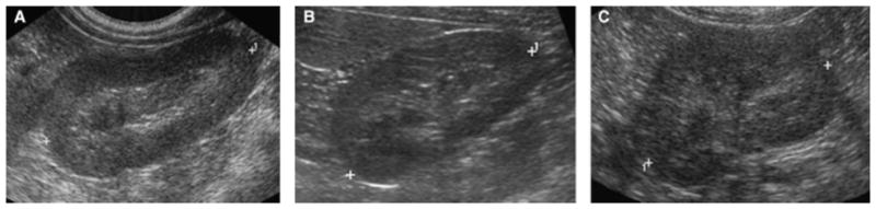
Dorsal plane ultrasound images of the left kidney of Dog 2 at the age of 4 months (A), 9 months (B), and 24 months (C). The ultrasonographic changes include poor corticomedullary definition and medullary hyperechogenicity, the changes were severe at all time points.
At the time of the initial ultrasound examination, all kidneys were normal in size for the dog’s body weight. All dogs were over 12 months of age at the last examination, at this time point renal length was at the lower limit of normal for the dogs’ weight (Table 1).15
Resistive indices were available for all dogs at time of the last ultrasound examination and four dogs had an RI measurement performed at a previous examination. The median RI (range) for the left kidney was 0.61 (0.58–0.69) at the first time of measurement and 0.64 (0.56–0.7) at the last time point. Median (range) RI for the right kidney was 0.6 (0.59–0.61) at the first and 0.59 (0.53–0.66) at the last time of measurement. There was no significant difference between the first and last RI value (P = 0.8).
Histopathologic lesions were limited to glomeruli, as all biopsy specimens consisted of cortical tissue only (Table 1). Because the size of each biopsy varied (from 1 × 5 × 2 mm to 5 × 10 × 3 mm), the number of glomeruli available for evaluation also varied. Lesions were mild in Dog 1, in which two of 28 glomeruli (11%) were fetal. Moderate lesions were present in two dogs: in Dog 4, 10 of 50 glomeruli (20%) were fetal (Fig. 4) and in Dog 5, 14 of 86 glomeruli (16%) were fetal. The remaining two dogs were diagnosed with severe renal dysplasia. Forty-two of 88 glomeruli (48%) were fetal in Dog 2 and 37 of 80 glomeruli (46%) were fetal in the Dog 3. The biopsy sample from the latter dog contained additional histopathologic lesions, namely occasional dilation of Bowman’s space by eosinophilic fluid. Cortical tubules and interstitium were normal in all biopsies.
Fig. 4.
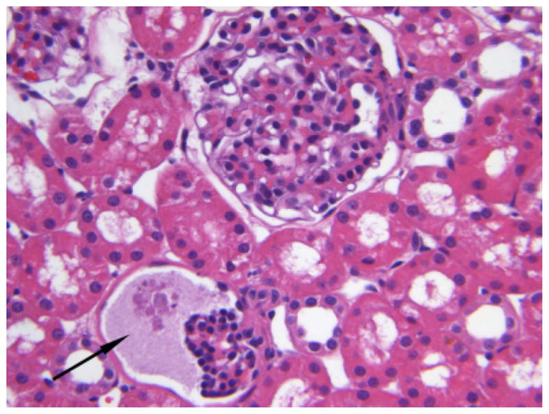
H&E staining of the renal biopsy sample of Dog 4, ×40 magnification. Two glomeruli are included, a large, normally developed glomerulus, and a small fetal glomerulus with a dilated Bowman’s capsule (arrow).
Ultrasonographic findings matched the histopathologic grade of disease severity in each dog (Table 1).
Discussion
Dogs affected with renal dysplasia usually present in advanced stages when clinical signs of renal insufficiency become apparent. At this time, small irregular hyperechoic kidneys with poor corticomedullary demarcation are observed commonly.3,6 Irregular kidney surface and decreased medullary thickness was found in one puppy in renal failure.5 These changes are similar to ultrasonographic findings in other types of chronic renal disease, highlighting the difficulty in distinguishing imaging findings related to the primary dysplasia vs. those related to secondary inflammatory and degenerative changes. Histopathologically, embryonic renal tissue was seen in chronic renal dysplasia, but secondary changes such as mineralization, and cellular proliferation as well as tubular necrosis were also present.4
In comparison, the dogs in our study had no evidence of renal failure at the time of examination and no histopathologic evidence of secondary degenerative changes. Common abnormalities in our study included decreased corticomedullary definition and a hyperechoic cortex. In addition, we observed pronounced hyperechoic speckling of the medulla or general medullary hyperechogenicity. It is difficult to determine the origin of these changes, as even surgical wedge biopsies do not usually include the renal medulla due to risk of hemorrhage. In necropsy reports of dysplastic kidneys, presence of mesenchymal tissue in the renal medulla is described, it remains to be determined if this tissue could lead to the speckled appearance of the medulla. Medullary hyperechogenicity has been described in human neonates and is considered a normal variant if it disappears within 10 days.16 Other conditions in which hyperechogenicity can be seen in the medulla include diffuse glomerulosclerosis and hypernatremic dehydration where the hyperechoic foci are thought to be crystal depositions.17,18
The RI is a unitless expression of the resistance to blood flow within an artery obtained by pulsed wave Doppler ultrasound. It was elevated in a group of 12 dogs with renal dysplasia.19 In our study, however, the median RI was within the normal range of 0.56–0.67 and there was no significant increase during the study period. This may be another reflection of the fact that we examined the dogs during an early phase of the disease. The RI may be elevated in dogs with renal dysplasia due to the degenerative lesions associated with a late stage of the disease such as interstitial fibrosis, inflammatory cell infiltrates, and cystic glomerular atrophy, which may cause swelling, vascular compression, or vasculitis.19 These degenerative changes were minimal or absent in our dogs. Serial measurements of the RI in dogs with renal dysplasia may be helpful in determining the onset of degenerative changes within the renal parenchyma, however, this has yet to be determined in longitudinal studies. Renal length did not differ much between the five dogs, nor did it increase or decrease over time beyond a few millimeters, which is likely within the range of variation between different operators and the accuracy of the method. Interpretation of renal size was complicated by the fact that the dogs were growing during the study period, and normal values for puppies of that age are not established. Dog 3 had the smallest kidneys and also had severe ultrasonographic and histopathologic changes.
Even though the number of dogs is small, the severity of the ultrasonographic findings was comparable to the severity of the histopathologic findings. Some inconsistency in assessment of severity of ultrasonographic changes may be associated with the retrospective analysis of still images, with an inherent variability of image quality. The fact that ultrasonographic changes could be detected in the dogs before development of secondary inflammatory or degenerative lesions is encouraging, and ultrasound may be a useful method for screening dogs in breeding programs for renal dysplasia.
Footnotes
GE healthcare Inc., Milwaukee, WI.
MedCalc Software, Mariakerke, Belgium.
References
- 1.Greco DS. Congenital and inherited renal disease of small animals. Vet Clin North Am Small Anim Pract. 2001;31:393–399. doi: 10.1016/s0195-5616(01)50211-9. [DOI] [PubMed] [Google Scholar]
- 2.Abraham LA, Beck C, Slocombe RF. Renal dysplasia and urinary tract infection in a Bull Mastiff puppy. Aust Vet J. 2003;81:336–339. doi: 10.1111/j.1751-0813.2003.tb11507.x. [DOI] [PubMed] [Google Scholar]
- 3.De Morais HS, DiBartola SP, Chew DJ. Juvenile renal disease in Golden Retrievers: 12 cases (1984–1994) J Am Vet Med Assoc. 1996;209:792–797. [PubMed] [Google Scholar]
- 4.Felkai CS, Voros K, Vrabely T, Vetesi F, Karsai F, Papp L. Ultrasonographic findings of renal dysplasia in Cocker Spaniels: eight cases. Acta Veterinaria Hungarica. 1997;45:397–408. [PubMed] [Google Scholar]
- 5.Hoppe A, Karlstam E. Renal dysplasia in Boxers and Finnish Harriers. J Small Anim Pract. 2000;41:422–426. doi: 10.1111/j.1748-5827.2000.tb03237.x. [DOI] [PubMed] [Google Scholar]
- 6.Lobetti RG, Pearson J, Jimenez M. Renal dysplasia in a Rhodesian Ridgeback dog. J Small Anim Pract. 1996;37:552–555. doi: 10.1111/j.1748-5827.1996.tb02319.x. [DOI] [PubMed] [Google Scholar]
- 7.Ohara K, Kobayashi Y, Tsuchiya N, Furuoka H, Matsui T. Renal dysplasia in a Shih Tzu dog in Japan. J Vet Med Sci. 2001;63:1127–1130. doi: 10.1292/jvms.63.1127. [DOI] [PubMed] [Google Scholar]
- 8.Olenick CL. Congenital renal dysplasia and psychogenic polydipsia in a Bernese Mountain dog. Can Vet J. 1999;40:425–526. [PMC free article] [PubMed] [Google Scholar]
- 9.Khan KNM, Alden CL. Kidney. In: Haschek WM, Rousseaux CG, Wallig MA, editors. Handbook of toxicologic pathology. Vol. 2. San Diego: Elsevier Science; 2002. pp. 255–330. [Google Scholar]
- 10.Picut CA, Lewis RM. Microscopic features of canine renal dysplasia. Vet Pathol. 1987;24:156–163. doi: 10.1177/030098588702400209. [DOI] [PubMed] [Google Scholar]
- 11.Picut CA, Lewis RM. Comparative pathology of canine hereditary nephropathies: an interpretive review. Vet Res Commun. 1987;11:561–581. doi: 10.1007/BF00396371. [DOI] [PubMed] [Google Scholar]
- 12.Vaden SL. Renal biopsy of dogs and cats. Clin Techn Small Anim Pract. 2005;20:11–22. doi: 10.1053/j.ctsap.2004.12.003. [DOI] [PubMed] [Google Scholar]
- 13.Bonazzi M, Volta A, Gnudi G, et al. Comparison between ultrasound and genetic testing for the early diagnosis of polycystic kidney disease in Persian and Exotic Shorthair cats. J Feline Med Surg. 2009;11:430–434. doi: 10.1016/j.jfms.2008.10.003. [DOI] [PMC free article] [PubMed] [Google Scholar]
- 14.Domanjko-Petric A, Cernec D, Cotman M. Polycystic kidney disease: a review and occurrence in Slovenia with comparison between ultrasound and genetic testing. J Feline Med Surg. 2008;10:115–119. doi: 10.1016/j.jfms.2007.07.004. [DOI] [PMC free article] [PubMed] [Google Scholar]
- 15.Barr FJ, Holt PE, Gibbs C. Ultrasonographic measurement of normal renal parameters. J Small Anim Pract. 1990;31:180–184. [Google Scholar]
- 16.Howlett DC, Greenwood KL, Jarosz JM, MacDonald LM, Saunders AJS. The incidence of transient renal medullary hyperechogenicity in neonatal ultrasound examination. Br J Radiol. 1997;70:140–143. doi: 10.1259/bjr.70.830.9135439. [DOI] [PubMed] [Google Scholar]
- 17.Salame H, Damry N, Vandenhout K, Hall M, Avni FE. The contribution of ultrasound for the differential diagnosis of congenital and infantile nephrotic syndrome. Eur Radiol. 2003;13:2674–2679. doi: 10.1007/s00330-003-1920-x. [DOI] [PubMed] [Google Scholar]
- 18.Ali US, Sengupta K, Andankar O, Saraf S, Chawla A, Deshpande S. Reversible renal medullary hyperechogenicity in neonatal hypernatremic dehydration. Pediatr Nephrol. 2004;19:1050–1052. doi: 10.1007/s00467-004-1510-4. [DOI] [PubMed] [Google Scholar]
- 19.Morrow KL, Salman MD, Lappin MR, Wrigley R. Comparison of the resistive index to clinical parameters in dogs with renal disease. Vet Radiol Ultrasound. 1996;37:193–199. [Google Scholar]