Introduction
Epidermal inclusion cyst (EIC) presenting as a lump in the breast is a rare and benign condition. Usually, it arises from the skin. It is also called as keratinous cyst or epidermoid cyst. Less than forty cases have been reported in the English literature [1, 2]. It is not uncommon that breast epidermal cysts are misdiagnosed clinically. We report here a confirmed case of epidermal inclusion cyst in the male patient. The present case of epidermal inclusion cyst was a surprise cytohistopathological diagnosis. EIC presenting as a lump in the male breast is extremely rare entity. As per our knowledge only three cases have been reported in the English literature [3].
Case report
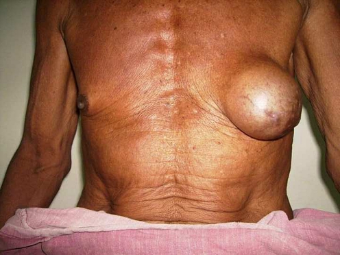
We encountered 65 years old male patient with enlargement of left breast since 5 years. Swelling was insidious in onset and slowly and gradually increased in the size. There was no history of pain, fever, cough or any trauma. Patient did not reveal history of any chronic liver disease, mumps, leprosy, pulmonary tuberculosis or prolonged use of any medicine. Local examination revealed a single, oval swelling in the left breast region measuring 10 × 8 cm. Nipple and areola were deviated inferolaterally (Fig. 1). It was nontender, smooth, soft, noncompressible and attached to the skin but free from pectoralis major muscle and chest wall. There was no discharge from the nipple.
Fig. 1.
Preoperative photograph showing left breast swelling
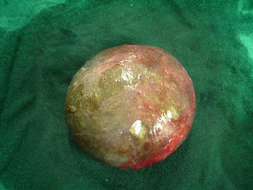
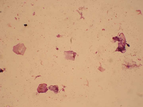
Ipsilateral axilla was normal. Systemic examination was within normal limit. Clinically, it was diagnosed as a cystic lesion arising either from the skin or the breast tissue. Fine needle aspiration cytology (FNAC) showed plenty of anucleate squamous cells without any breast tissue, suggestive of a keratinous cyst. The patient was subjected for excision of the swelling. Brownish black and very thin walled cyst was excised completely with preservation of nipple and areola (Fig. 2). Histopathology confirmed the diagnosis of EIC (Fig. 3). Patient is asymptomatic with 18 months follow up after the surgery.
Fig. 2.
Gross specimen of the epidermal inclusion cyst of the breast
Fig. 3.
Microphotograph showing anucleate squamous cells
Discussion
Various types of cystic swellings arise from the skin epithelium. The epidermal cysts account for 80% of cystic swelling. Epidermal inclusion cysts occur in the scalp, back and the neck region [2]. It is also reported on face, palm, sole, extremities and female external genitalia. EIC occurring in the breast is very rare entity and less than 40 cases have been reported in the English literature [3]. It can be congenital or acquired. Most often it occurs due to obstruction of the hair follicle [4, 5]. It may be preceded by trauma, reduction mammoplasty or breast augmentation [5]. Another described possible origin is the squamous metaplasia of the columnar epithelium of the ducts [4, 5]. Even inflammatory down growth of the epidermis is blamed for the formation of EIC [5]. It occurs during third or fourth decades of the life. EIC is well defined, smooth and rounded swelling attached to the skin but free from the breast and deeper tissues [3].It is firm, nontender and noncompressible swelling. It appears as a solid and well circumscribed on sonography and mammography respectively [1, 6]. Pavel Crystal et al described the onion ring pattern of epidermal cysts on the sonography [1]. Aspiration from the epidermal cyst yields cheesy and flaky material [4].Possible complications of EIC are spontaneous rupture as a result of inflammation, infection, hemorrhage and malignant transformation [4–6]. Complete surgical excision of the cyst is the treatment of choice since it has a tendency to recur [4].
Conclusion
Epidermal inclusion cysts (keratinous cysts) growing inside the breast tissue need to be considered as the differentional diagnosis for the benign breast tumor.
References
- 1.Pavel Crystal, Ruthy Shaco-Levy (2005) Concentric rings within a breast mass on sonography: lamillated keratin in an epidermal inclusion cyst AJR; 184:547–48 [DOI] [PubMed]
- 2.Beromann-Koester CU, Kolberg HC, Rudolf I, Krueger S, Gellisen J, Stoeckelhuber BM. Epidermal cyst of the breast mimicking malignancy: clinical, radiological and histological correlation. Arch Gynecol Obstet. 2006;273(5):312–314. doi: 10.1007/s00404-005-0019-9. [DOI] [PubMed] [Google Scholar]
- 3.Cooper RA, Ramamurthy L. Epidermal inclusion cyst in the male breast. Can Assoc Radiol J. 1996;47:92–94. [PubMed] [Google Scholar]
- 4.Chantra PK, Tang JT, Stanley TM, Bassett LW. Circumscribed fibrocystic mastopathy with formation of an epidermal cyst. AJR. 1994;163:831–832. doi: 10.2214/ajr.163.4.8092018. [DOI] [PubMed] [Google Scholar]
- 5.Whang IY, Lee JH, Kim JS, Kim KT, Shin OR. Ruptured epidermal inclusion cysts in the subareolar area: sonographic findings in two cases. Korean J Radiol. 2007;8(4):356–359. doi: 10.3348/kjr.2007.8.4.356. [DOI] [PMC free article] [PubMed] [Google Scholar]
- 6.Kwack JY, Park HL, Kim JY, Chung SY, Kwon TH, Hong HS, et al. Imaging finding in a case of epidermal inclusion cyst arising within the breast parenchyma. J Clin Ultrasound. 2004;32(3):141–143. doi: 10.1002/jcu.20005. [DOI] [PubMed] [Google Scholar]


