Abstract
An unusual anamoly of the extrahepatic bitiary system is reported in which the common hepatic duct was found to enter the gallbladder, whereas the cystic duct drained the entire biliarysystem into the duodenum. Excision of the gallbladder and cystic duct and a roux-en-Y hepaticojejunostomy was performed. Identification and treatment options of this rare anomaly are briefly discussed.
Keywords: Extrahepatic biliary tract anamoly, gallbladder interposition, hepaticocystic duct
Case Report
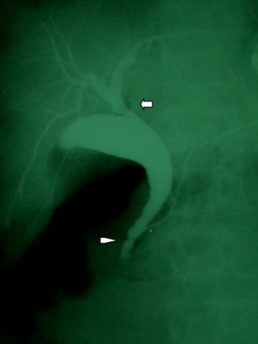
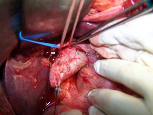
A 55 year old lady presented with history of recurrent episodes of upper abdominal pain of four years duration. Ultrasonography revealed a distended gallbladder with a 2 cm polyp at the fundus and normal common bile duct (CBD). Her liver function tests were normal. CT scan revealed a 2 cm intraluminal soft tissue mass in fundus of gallbladder. Midportion of CBD appeared dilated (2 cm in size) with gradual tapering of intrapancreatic portion. In view of the isolated CBD dilatation with no intrahepatic dilatation, a possibility of associated choledochal cyst was considered. At laparotomy there was a thin walled distended gallbladder with a soft polyp at the fundus of the gallbladder. After dissecting the calot’s triangle the cystic duct appeared markedly dilated and unusually long. To be sure of the exact anatomy gallbladder was dissected off its bed by fundus first method. It was transected at the neck and an intraoperative cholangiogram was obtained. Intraoperative cholangiogram (Fig. 1) revealed a normally forming confluence. Common hepatic duct was emptying into neck of gallbladder. Dilated cystic duct joined the pancreatic duct and opened into the duodenum through a long common channel. Further dissection isolated a normal sized common hepatic duct (6 mm) opening into the neck of gallbladder (Fig. 2). Excision of the gallbladder and cystic duct was performed and a Roux-en-Y hepaticojejunostomy was performed. A 2 cm wide anastomosis was created by lowering hilar plate and extending opening on to the left hepatic duct. Postoperative recovery was normal. Histology of the gallbladder polyp revealed an adenoma .Patient remains symptom free at 1 year follow up.
Fig. 1.
Intraoperative cholangiogram (after partial cholecystectomy) shows common hepatic duct opening into neck of gallbladder (arrow). Cystic duct joins the pancreatic duct with a ABPU and provides the only drainage route (arrowhead)
Fig. 2.
Operative image showing dissected normal sized common hepatic duct (arrow) draining into the gallbladder neck with wide cystic duct passing towards duodenum
Discussion
Upto half the patients undergoing a cholecystectomy have variations of the classically described biliary anatomy. Aberrant anatomy has been reported in upto 17% of cases with bile duct injury [1]. The biliary anatomy described in our case is extremely rare with few published reports in literature [2–9]. The anomaly has been described as ‘interposition of gall bladder’, ‘hepaticocystic duct’ and also by some as ‘cholecystohepatic duct’ [3–7]. The term ‘hepaticocystic’ is preferred to ‘cholecystohepatic’ ducts as direction of flow of bile is from the hepatic ducts into the cystic duct [7] which provides the sole route of drainage of bile into duodenum. Losanoff et al described four variants, type I in which right and left ducts open separately into gallbladder, type II where the ducts join upon entering gallbladder and type III seen in our case where the common hepatic ducts opens into the gallbladder [7]. Losanoff further subdivided Type III into four types based on entry of CHD into the gall bladder wall. CHD may enter gallbladder wall superiorly (III A), at its neck (III B) as seen in our pateint, posteriorly (III C),or at the fundus (III D). A type IV variant has also been reported in which multiple segments of liver drain to the gallbladder directly.
Differential diagnosis include Mirrizzi’s syndrome and choledochal cyst. A similar appearance of the CHD opening into the gallbladder may be seen in type III - IV Mirrizzi’s, however absence of gallstones and any fibrosis/ fistulization at the gallbladder neck rules out Mirrizzi’s syndrome. Choledochal cyst was considered in the preoperative diagnosis in this patient, cholangiogram revealed a fusiform dilated cystic duct with abnormal pancreaticobiliary union (ABPU). This association of interposition of gallbladder with ABPU has not been reported previously. Awareness of this anomaly is important to avoid bile duct injury during cholecystectomy. Ultrasound and CT scan may miss this anomaly . The anomaly is revealed best by a preoperative magnetic resonance cholangiogram and/or intraoperative cholangiogram but rarity of such anomalies does not justify routine usage of these investigations. The presence of a impacted cystic duct has been reported to suggest a major hepatocystic duct [8] but similar picture may be seen in periampullary pathology. Intraoperative awareness of this anomaly, dissection of gallbladder by fundus first method, performing a cholecystocholangioram at the slightest suspicion (such as a wide cystic duct/ absence of cystic duct) will allow timely recognition of the anomaly and repair. During laparoscopic cholecystectomy clear visualization of gallbladder neck-cystic duct junction is needed to ensure that there are no tubular structures opening above it. Once the anomaly is detected surgical options include partial cholecystectomy or hepaticojejunostomy. In our patient due to presence of abnormal dilated cystic duct, presence of adenomatous change in gallbladder mucosa and associated ABPU we performed excision of cystic duct portion and hepaticojejunostomy. Some reports describe partial cholecystectomy with closure of residual gallbladder over T -tube [3, 9]. However long term follow up is unavailable and there is always a risk of recurrent stone formation in the residual gallbladder leading to biliary obstruction. Therefore we believe that hepaticojejunostomy should be the preferred surgical option if the lesion is encountered during cholecystectomy.
References
- 1.Suhochi PV, Meyers WC. Injury to aberrant bile ducts during cholecystectomy: a common cause of diagnostic error and treatment delay. AJR. 1989;172:955–959. doi: 10.2214/ajr.172.4.10587128. [DOI] [PubMed] [Google Scholar]
- 2.Hashmonai M, Kopelman D. An anomaly of extrahepatic biliary system. Arch Surg. 1995;130:673–675. doi: 10.1001/archsurg.1995.01430060111023. [DOI] [PubMed] [Google Scholar]
- 3.Losanoff JE, Kjossev KT, Kalrov E. Hepaticocystic duct - A case report. Surg Radiol Anat. 1996;8:339–341. doi: 10.1007/BF01627614. [DOI] [PubMed] [Google Scholar]
- 4.Walia HS, Abraham TK, Baraka A. Gall-bladder interposition: a rare anomaly of the extrahepatic ducts. Int Surg. 1986;71:117–121. [PubMed] [Google Scholar]
- 5.Redkar RG, Rivkin LA, Myers N. Association of esophageal atresia and cholecystohepatic duct. Pediatr Surg Int. 1999;15:21–23. doi: 10.1007/s003830050503. [DOI] [PubMed] [Google Scholar]
- 6.Lamah M, Karanjia ND, Dickson GH. Anatomical variations of extrahepatic biliary tree: a review of world literature. Clin Anat. 2001;14:167–172. doi: 10.1002/ca.1028. [DOI] [PubMed] [Google Scholar]
- 7.Losanoff JE, Jones JW, Richman BW. Hepaticocystic duct: a rare anomaly of extrahepatic biliary system. Clin Anat. 2002;15:314–315. doi: 10.1002/ca.10032. [DOI] [PubMed] [Google Scholar]
- 8.Pottakat B, Sikora SS. Aberrant right hepatic duct presenting as empyema of the gall bladder. Australas Radiol. 2007;51:303–305. doi: 10.1111/j.1440-1673.2007.01826.x. [DOI] [PubMed] [Google Scholar]
- 9.Shah O. The missing common bile duct (hepaticocystic duct) Surgery. 2007;142:424–425. doi: 10.1016/j.surg.2006.11.004. [DOI] [PubMed] [Google Scholar]


