Abstract
Background
In recent years, research efforts exploring the possibility of using biomaterial nanoparticles for intravitreous drug delivery has increased significantly. However, little is known about the effect of material properties on intravitreous tissue responses.
Principal Findings
To find the answer, nanoparticles made of hyaluronic acid (HA), poly (l-lactic acid) (PLLA), polystyrene (PS), and Poly N-isopropyl acrylamide (PNIPAM) were tested using intravitreous rabbit implantation model. Shortly after implantation, we found that most of the implants accumulated in the trabecular meshwork area followed by clearance from the vitreous. Interestingly, substantial reduction of intraocular pressure (IOP) was observed in eyes implanted with particles made of PS, PNIPAM and PLLA, but not HA nanoparticles and buffered salt solution control. On the other hand, based on histology, we found that the particle implantation had no influence on cornea, iris and even retina. Surprisingly, substantial CD11b+ inflammatory cells were found to accumulate in the trabecular meshwork area in some animals. In addition, there was a good relationship between recruited CD11b+ cells and IOP reduction.
Conclusions
Overall, the results reveal the potential influence of nanoparticle material properties on IOP reduction and inflammatory responses in trabecular meshwork. Such interactions may be critical for the development of future ocular nanodevices with improved safety and perhaps efficacy.
Introduction
Posterior ocular diseases, including glaucoma, macular degeneration, uveal melanoma and retinoblastoma are often hard to be treated due to ocular tissue barriers [1]–[3]. While topical administration is effective in the treatment of anterior chamber diseases, it is ineffective in the treatment of diseases afflicting the posterior segments of the eye [2]. Major problems include washing away of the drug by tears and the inefficient diffusion of drug from the corneal side to the posterior [4], [5]. Systemic injection does deliver drugs to the posterior of the eye but is also associated with non-specific accumulation of drug in other organs. In addition the blood retinal barrier also hinders the diffusion of drug into the posterior chamber [2]. In light of this information, intraocular drug injections have gained in importance. However, although they achieve therapeutic drug levels, they are associated with high vitreal clearance which necessitates multiple injections. This in turn leads to complications of endophthalmitis and retinal detachment [2], [3], [6]. There is a need for the development of alternative treatments for posterior ocular diseases.
Many therapeutic strategies have been developed in recent years. One such method is the use of biomaterial drug delivery devices either in the form of implants or as micro or nanoparticles [2], [7], [8]. Despite of their ability to release therapeutic agents for a prolonged period of time, ocular rod implants have been found to be responsible for causing retinal detachment and endophthalmitis [6]. With the expansion of nanotechnology in medicine, a wide variety of nanoparticle drug releasing devices have been fabricated and tested for their ability to treat a wide range of diseases [1], [9]–[12]. Many studies have been done to explore the possibility of using polymeric micro and nanoparticles for anterior and posterior chamber drug delivery [1], [9]–[15]. Although microparticles have better drug loading capacity than nanoparticles, the latter is recognized as favorable drug carrier due to its low risk on hampering normal vision [16], [17]. Although different types of nanoparticles have been investigated for their ability to target different cells, tissues and to cure different ocular diseases [9]–[12], [14], [15], [18]–[22]. very limited studies have been done to systematically evaluate the effect of material physical and chemical properties on their ocular tissue and cell compatibility.
It is well established that the physical and chemical properties of materials affect their cell and tissue compatibility [23]–[27]. We thus assumed that nanoparticles made of different materials are likely to cause different extents of acute tissue responses in the eye. To test this hypothesis, nanoparticles made of different materials were included in this study. Specifically, nanoparticles were made out of degradable polymers like poly (l-lactic acid) (PLLA), hydrogels like poly N-isopropyl acrylamide (PNIPAM), non-degradable materials like polystyrene (PS), and biological materials like hyaluronic acid (HA). The ocular compatibility of these nanoparticles was evaluated using rabbit intravitreous implantation model. After implantation for different periods of time, we measured the changes in intraocular pressure (IOP). At the end of the studies, animals were sacrificed and ocular tissues were histologically evaluated. The effect of material properties on the ocular tissue responses was then determined to show that it can play a key role in determining the fate of nanoparticles in the eye.
Methods
Ethics Statement
The animal use protocols (A06-028, A09-028) were reviewed and approved by the Institutional Animal Care and Use Committee of the University of Texas at Arlington.
Materials
N-Isopropylacrylamide was purchased from Polysciences. Sodium acrylate (NaAc), N,N′-Methylene-bis-acrylamide (BIS) and potassium persulfate (KPS) were purchased from Bio-Rad (Hercules, CA, USA). Poly (L-lactic acid) (PLLA, MW 1.37×10⁶ kDa) was purchased from Birmingham Polymers (Birmingham, AL, USA). N-(3 Dimethylaminopropyl)-N′-ethylcarbodiimide hydrochloride (EDAC), Fluorescein isothiocyanate (FITC), Polystyrene and hyaluronic acid were purchased from Sigma, St. Louis, USA. Balanced Salt Solution (BSS) was obtained from Alcon Inc (Fort Worth, TX, USA).
Nanoparticle synthesis
Both poly N-isopropylacrylamide (PNIPAM) and PS nanoparticles were synthesized based on established techniques [28], [29], [30]–[32]. To study the distribution of the intravitreous injected nanoparticles, some PNIPAM nanoparticles were conjugated with FITC using carbodiimide chemistry [33], [34]. HA nanoparticles were synthesized as described earlier [35]. Briefly, acetone was added in a weight ratio of 100∶80 to a 0.5 wt% HA solution and the HA/water/acetone mixture was stirred for 2 hours. EDAC was added to the mixture in a weight ratio of 0.05∶100 to form a crosslinked mixture. This mixture was then stirred at 20 to 22°C for approximately 24 hours after which acetone in a weight ratio of approximately 60∶100 was added to form the final mixture that was stirred for 20 hours and dialyzed against distilled water to form HA nanoparticles.
Animal implantation
Dutch rabbits (4–5 lb) were purchased from Myrtle's Rabbitry Inc (Thompsons Station, TN). All experimental procedures were approved by the Institutional Animal Care and Use Committee (IACUC) at the University of Texas at Arlington and carried out under veterinary supervision. Prior to the procedures, animals were sedated by subcutaneous injection of a 1∶5 mixture of 100 mg/ml xylazine (Rompun; Miles Laboratories, Shawnee Mission, KS) and 100 mg/ml Ketamine HCl (Ketaset; Bristol laboratories. Syracuse, NY). One drop of topical anesthetic Proparacaine HCl (0.5%; Alcon Laboratories, Fort Worth, TX) was administered to each eye before injection. One hundred µl (20 g/l) of each type of particle was injected in to intravitreal space of the right eye via 30 gauge needle on a 1 ml tuberculin syringe. The intravitreal space of the left eye was injected with 100 µl of balanced salt solution (BSS) and served as control. The points where the intravitreal injections were made were approximately 2–3 mm from the corneal limbus as suggested in early publications [36], [37]. All injections were performed by the same researcher to avoid individual operational variations.
Intraocular pressure measurement
Intraocular pressure (IOP) has often been used as an indicator for various ocular diseases [38], [39]. The IOP was measured using a calibrated pneumatonometer (Model 30 Classic; Mentor Co., Norwell, MA, U.S.A.) one day before and daily for 3 days after nanoparticle injection as described earlier [40]. The results of the IOP reading were taken in the confidence interval greater than or equal to 95%. Measurements were taken at the same hour in order to avoid circadian changes. The changes in IOP were calculated by subtracting the IOPs at the end of 3 days from that before particle injection.
Ocular imaging and histological Analyses
After intravitreous implantation of nanoparticles for different periods of time (2 hrs, 4 hrs and 1 day), rabbits were euthanized and eyes were recovered and frozen sectioned. To image intravitreous distribution of nanoparticles, ocular tissue sections from animals implanted with PNIPAM nanoparticles were scanned using a Genepix Microarray analyzer (Molecular Devices, Sunnyvale, CA). The fluorescence intensities in different areas of the ocular tissues were analyzed and then compared. Furthermore, some tissue sections were H&E stained and the extent of implant-associated ocular tissue responses was quantified by measuring the thickness of various ocular tissues like cornea, iris and retina using a Leica microscope (Leica Microsystems, Wetzlar, GmbH) equipped with a Nikon E500 Camera (8.4 V, 0.9 A, Nikon Corp., Japan). To assess the extent of acute inflammatory responses, some tissue sections underwent immunohistological staining for CD11b+ inflammatory cells in which images were captured with a CCD camera (Retiga EXi, Qimaging, Surrey BC, Canada) and cell densities were analyzed using NIH ImageJ program as described in our previous publications [41], [42].
Statistical Analyses
All results were expressed as mean ± SD. All statistical comparisons were made with BSS controls using t-tests. Differences were considered statistically significant at p<0.05 Linear regression analyses was conducted with the Intraocular pressure change represented by mm of Hg was used as the dependent variable, while the CD11b+ cell numbers were the explanatory variables.
Results
Changes in intraocular pressure after intravitreous particle injection
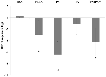
Particles made of different materials were used in this investigation (Table 1). These particles had diameter between 100–200 nm. Both PLLA and PS particles are hydrophobic while HA and PNIPAM particles were hydrophilic. It should be noted that PLLA and HA particles are biodegradable whereas PS and PNIPAM particles are non-degradable. Following intravitreous implantation, nanoparticle implants did not trigger apparent adverse response or abnormality. Rather surprisingly, we found that the intravitreous implantation of particles affected IOP significantly. Specifically, the injection of PLLA, PS and PNIPAM particles caused substantial reduction of IOP (Figure 1). On the other hand, the injection of HA nanoparticles and BSS (as control) had no effect on IOP. Such pressure changes lasted for less than 3 days. Although the cause of such particle implant-mediated IOP reduction was not known, it is likely that the material property of particle implants affects their acute ocular compatibility.
Table 1. Physical properties of nanoparticles used for intravitreous implantation.
| Material | Type | Wettability | Degradable | Size nm |
| Buffered Salt Solution | Solution | Aqueous solution | NA | NA |
| Poly (l-lactic) Acid | Polymeric | Hydrophobic | Yes | 143 |
| Polystyrene | Polymeric | Hydrophobic | No | 100 |
| Hyaluronic Acid | Polymeric | Hydrophilic | Yes | 200 |
| Poly N-isopropyl acrylamide | Polymeric Hydrogel | Hydrophilic | No | 100 |
Figure 1. Assessment of mean intraocular pressure variations after administration of particles made of poly-L lactic acid (PLLA), polystyrene (PS), hyaluronic acid (HA), and poly N-isopropyl acrylamide (PNIPAM) and Balanced Salt Solution (BSS) in the vitreous.
Data are mean ± standard deviation. Significance of PLLA, PS, HA, PNIPAM vs. BSS control; * p<0.05.
Distribution of intra-vitreous injected nanoparticles
To determine the potential interaction between injected particles with ocular tissues, we first monitored the particle distribution following intravitreous implantation using FITC-labeled PNIPAM particles. As shown in the whole ocular section images, we found that, at 2 hours, implanted particles were only found in the posterior, but not anterior segment of eye. We also found uniform fluorescence along the wall of the posterior segments indicating the even spread of particles over the retinal tissue (Figure 2A). Interestingly, we found that substantially more particles accumulated in the trabecular meshwork area even at an early time point – 2 hours. With increasing amount of time (4 and 24 hours) following implantation, we found that the particle-associated fluorescence intensities reduced substantially in the posterior chamber (Figure 2A and B). Interestingly, by 4 hours the fluorescence intensity at the trabecular meshwork was significantly higher than the rest of the eye and substantial fluorescent signals were also found outside the ocular tissue nearby the trabecular outflow region. Based on the fluorescent intensity measurements and distribution, noticeably, most of the particles cleared from the central portion of posterior chambers while majority of the residual fluorescence intensity was seen in the area of trabecular meshwork prior to clearance from the posterior cavities shortly after 24 hours. Quantification of the distribution of fluorescent particles throughout the ocular tissues further confirmed the presence of particles in the trabecular meshwork area (Figure 2C). These results suggest that the particle implants have little or no contact with corneal and iris tissues. On the other hand, based on the fluorescence distribution, it is likely that many posterior ocular tissues, including retina and trabecular meshwork, were exposed to particle implants.
Figure 2. Distribution of FITC-labeled PNIPAM nanoparticles after intravitreous administration over a day.
Localization of fluorescence in the posterior segments of the eye at various time points (2, 4 and 24 hours) was observed (A) and quantified (B). Quantification of the normalized fluorescence intensity at various locations (TB: Trabecular Meshwork; RA: Retina; ON: Optic Nerve) at various time points (C).
Effect of material properties on ocular tissue responses
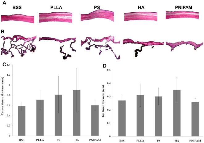
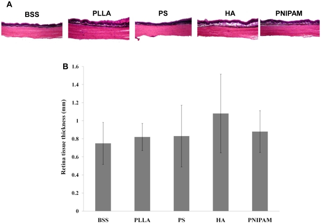
To assess the potential ocular compatibility of particle implants, various ocular tissues in both anterior and posterior segments were histologically analyzed. As expected, we found that the intravitreous injection of particles have no apparent influence on the anatomical structure of cornea (Figure 3A) and iris (Figure 3B) tissue based on morphological assessment of tissue thickness (Figures 3C & D). Although PNIPAM particle-implanted animals showed the lowest corneal and iris tissue thickness, the differences were not statistically significant as compared with those in animals injected with BSS control. Rather surprisingly, despite of the apparent interaction between particles and retinal tissue, we found that particle implants have no influence on the anatomical structure and thickness of retinal tissues (Figure 4A & B). How the injection of particle implants reduced IOP, was yet to be answered.
Figure 3. Histological assessment of corneal and iris tissue after intravitreous implantation.
The representative H&E images of the cornea (A) and iris (B) tissue were shown here. Based on H&E staining images, the influence of particle property on the thickness of the corneal (C) and iris (D) thickness were quantified. Data are mean ± standard deviation. Significance of PLLA, PS, HA, PNIPAM vs. BSS control; * p<0.05.
Figure 4. Evaluation of retinal tissue morphology following intravitreal injection.
Representative H&E stained retinal tissues were shown here (A). The thickness of the retinal tissue was also calculated and then compared (B). Data are mean ± standard deviation. Significance of PLLA, PS, HA, PNIPAM vs. BSS control; * p<0.05.
CD11b+ Inflammatory cell accumulation in trabecular meshwork tissue
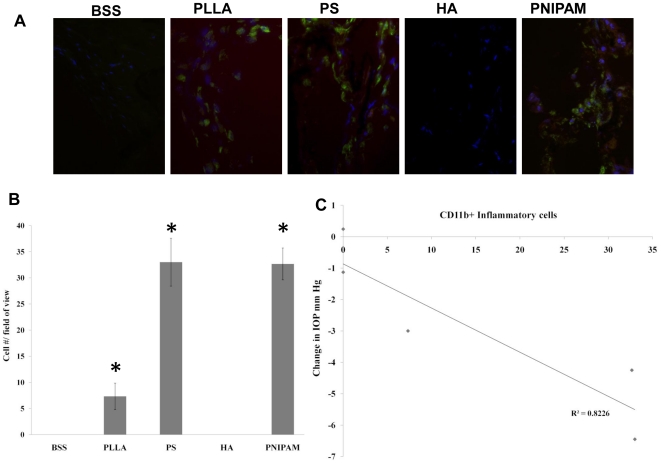
It is well established that trabecular meshwork is responsible for controlling IOP [43], and increased inflammatory responses in trabecular meshwork have been shown to cause IOP reduction in human patients [44]. Since our results have shown that substantial portion of the injected particles accumulated in trabecular meshwork prior to clearance from the posterior chamber, we assumed that some particle implants may trigger such inflammatory responses in trabecular meshwork tissue and then lead to the reduction of IOP. To test this hypothesis, immunohistological analysis of ocular tissues was performed by examining the presence or absence of CD11b+ inflammatory cells in the trabecular meshwork area around the ciliary body. Indeed, we found that both PS and PNIPAM particle groups were associated with very high CD11b+ cell accumulation (labeled green) (Figure 5A) while PLLA particles prompt less (∼20%) CD11b+ cell accumulation as compared with PS and PNIPAM particle groups. It should be noted that there were almost no CD11b+ cells in the HA nanoparticle group as well as BSS control. The average CD11b+ cell numbers from numerous sections were quantified to substantiate the visual observations (Figure 5B). To investigate the relationship between inflammatory cell accumulation and IOP reduction, then numbers of CD11b+ cells from different sample groups were then correlated with averages of IOP from respective sample groups. As expected, there was a very good relationship between CD11b+ cell numbers found in the trabecular meshwork tissues and overall IOP reduction (Figure 5C).
Figure 5. The accumulation of CD11b+ inflammatory cells in the trabecular meshwork.
The representative immunohistochemically stained images showed the accumulation of CD11b+ inflammatory cells (labeled green; DAPI staining to locate cell nucleus) in the trabecular meshwork following particle injection (A). The extent of CD11b+ cell accumulation in the trabecular meshwork was quantified (B). Data are mean ± standard deviation. Significance of PLLA, PS, HA, PNIPAM vs. BSS control; * p<0.05 Correlation between CD11b+ inflammatory cells and average IOP changes in different test groups was also determined (C).
Discussion
Drug delivery to the back of the eye, especially the posterior segments, is a key research area and important considering numerous ocular diseases that afflict that region. However, most drug administration techniques like topical administration or even implantable rods have their own drawbacks as has been reviewed in our earlier publication [3]. Administration of drug directly into the vitreous is also associated with a lot of problems; clearance of the drug being one of the main drawbacks [2], [45]. In light of this fact, nanotechnology has come to the forefront of ocular drug delivery and as such, understanding ocular tissue response to implanted nano-biomaterials is of paramount significance [2], [3], [8], [46]. Our selection of materials to synthesize various nanoparticles was based on the following facts. First, a vast majority of drug releasing nanodevices are made out of FDA approved polymers like PLLA and PLGA and are usually in the range of 10 nm to 1000 nm [2], [8], [47]. In fact, previous studies have shown that particles in the range of 20 nm to 200 nm have the highest affinity to tissue [13], [48]. Second, hydrogels like PNIPAM have been extensively researched for drug delivery applications [14], [49]–[51]. Thirdly, HA is a major component of the vitreous and as such, their presence in the eye should be tolerable. HA makes up a sizeable proportion in the retinal pigment epithelium and interphotoreceptor matrix [52]. Considering this information, we selected nanoparticles made out of PLLA, PS, HA and PNIPAM in the size range of 100 to 200 nm.
It is well established that material properties trigger different extent of soft tissue responses [25], [53]. We thus assumed that material properties would exert some influence on ocular tissue reactions. Very few studies have been done to assess the effect of material properties on ocular compatibility of particles. Nevertheless, studies have found that PLLA and PLGA nanoparticles can be used for delivery of high molecular weight drugs to the retina [54], and poly (ε-caprolactone) is well tolerated by retinal tissue for at least 4 weeks [55]. Non-toxic chitosan and hyaluronic acid have been found to be good drug carriers [56], [57], and carbodiimide crosslinked hyaluronic acid has been shown to have good ocular compatibility in the anterior chamber [58]. PNIPAM hydrogel grafted with chitosan has also been applied as a thermally responsive ophthalmic drug delivery device [51]. Also most of the studies until now have mainly focused on visual signs of inflammation to suggest lack of biocompatibility.
To determine the ocular tissue responses to particle implants, we first measured the IOP changes following intravitreous implantation of particle. The fluctuation of IOP indicates the balance between production and drainage of aqueous humor and hence it was measured to determine the impact of various nanoparticle injections on aqueous humor drainage [43], [59]. In addition, it has been documented that ocular inflammation strongly influences the IOP [60]–[63]. Diseases like glaucoma have been shown to increase IOP while inflammatory conditions produced by anterior uveitis and iritis were found to reduce IOP [44], [64]–[67]. Substantial studies in glaucoma research have focused on using various pharmacological approaches to reduce IOP for prolonged period of time [64], [68]. Interestingly, our studies have found that the intravitreous implantation of particles prompted different extent of IOP reduction. Specifically, we found that PNIPAM and PS particles triggered the maximum reduction in IOP, while PLLA particles caused a rather mild reduction in IOP. Most interestingly, our results show that the implantation of HA particles trigger minimal or no IOP reduction. In fact, many studies have shown that HA particles have superb tissue compatibility in other body parts [56], [58], [69], [70], in different animal models [70], [71]. In fact a study evaluating the biocompatibility of intravitreal hyaluronic acid implants found that there were no evident signs of inflammation following implantation [71]. These results suggest that HA particles are good nanocarriers for posterior drug delivery.
To find the cause for particle implant-mediated IOP reduction, we first observed the distribution of the implants following intravitreous implantation. Although extensive research efforts have been placed on the development of nanocarriers for anterior and posterior ocular drug delivery, little has been done to study the fate of particle implants following injection. Briefly, these studies have revealed that nano and microparticles can reach the intraocular tissues when administered systemically or through periocular administration routes [48], [72]. However, it has also been reported that systemic administration of drug requires high doses to offset loss due to non-specific targeting and systemic side effects [73]. Our studies revealed that, rather surprisingly, majority of the particle implants injected intravitreally, only stay in the posterior segments for a very short period of time (∼24 hours). These results support that that, differing from common assumption that intravitreous administration will lead to better distribution, humor flow actively pushed particle implants out of the posterior. In addition, particle implants may reach retinal tissues shortly after intravitreous injection. Although the fate of the particles is yet to be determined, it is plausible that most of the particle implants exit the posterior chamber via the trabecular meshwork based on the fluorescent intensity distribution. This observation is supported by several earlier observations that particles and drugs may leave vitreous compartment via the trabecular meshwork [43], [74].
Particle implants have been shown to trigger immune reactions in the surrounding tissues [23], [25], and it is likely that similar particle-mediated tissue responses are also found in ocular tissues. To the best of our knowledge, very few studies have been done in this regard. A few studies have tested poly(ortho esters) following intravitreal injection and found no evident signs of inflammatory reaction up to 3 months [75]. Studies involving PLGA microspheres [76], and porous silicon microparticles [77], found that they were well tolerated with no clinical signs of inflammation based on visual examination even four days to four months after implantation. A recent study has also evaluated the ocular compatibility of gluteraldehyde crosslinked and EDAC crosslinked hyaluronic acid implants in the anterior chamber and found that EDAC crosslinked implants were more compatible [58]. However, it is mostly unclear whether the intravitreous implantation of particles would trigger immune reactions in different ocular tissues, including cornea, iris, retina and trabecular meshwork. To our surprise, we found that all the tested particle implants had no significant influence on the morphology and anatomical structure of cornea, iris, retina tissue despite of apparent short term accumulation of particle implants in nearby retinal tissue.
It is well established that trabecular meshwork and surrounding ciliary body is responsible for maintaining the IOP [78]–[81]. To search for the cause of IOP changes following particle implantation, we examined the tissue responses in the trabecular meshwork. It should be noted that the potential influence of intravitreous particle implants on trabecular meshwork function have not been evaluated prior to this work. The trabecular meshwork, upon examination showed signs of apparent acute inflammatory responses with accumulation of CD11b+ cells in some groups of animals, especially animals with PS particle or PNIPAM implants. Mild accumulation of CD11b+ cells in trabecular meshwork was also found in animals implanted with PLLA particles. Interestingly, no CD11b+ cells were found in the tissues isolated from animals implanted with either HA particles or BSS controls. Equally importantly, we found a good relationship between the numbers of CD11b+ cells in trabecular meshwork and the average IOP reduction in different groups of implants. These results suggest that particle implant-associated inflammatory responses in trabecular meshwork are responsible for IOP reduction and are supported by many earlier works in which inflammatory responses inside trabecular meshwork have been linked to the reduction of IOP [44], [65], [82].
The results from this study have emphasized the fact that IOP should be measured as part of the evaluation of tissue compatibility of ocular implants, specifically in the case of nanoparticle and microparticle implants. Furthermore, the “normal” anatomical structure of retinal, corneal and iris tissue does not guarantee the safety of ocular particle implants. Rather, the histological evaluation of inflammatory responses in trabecular meshwork should be done as an indicator of ocular compatibility of intravitreous implants. Finally, further studies are needed to investigate the influence of material physical and chemical properties on IOP changes and on trabecular tissue responses.
Footnotes
Competing Interests: The authors have declared that no competing interests exist.
Funding: This work was supported by National Institutes of Health grant EB007271. The funder had no role in study design, data collection and analysis, decision to publish, or preparation of the manuscript.
References
- 1.Bourges JL, Gautier SE, Delie F, Bejjani RA, Jeanny JC, et al. Ocular drug delivery targeting the retina and retinal pigment epithelium using polylactide nanoparticles. Invest Ophthalmol Vis Sci. 2003;44:3562–3569. doi: 10.1167/iovs.02-1068. [DOI] [PubMed] [Google Scholar]
- 2.Janoria KG, Gunda S, Boddu SH, Mitra AK. Novel approaches to retinal drug delivery. Expert Opin Drug Deliv. 2007;4:371–388. doi: 10.1517/17425247.4.4.371. [DOI] [PubMed] [Google Scholar]
- 3.Nair A, Thevenot P, Hu W, Tang L. Nanotechnology in the Treatment and Detection of Intraocular Cancers. J Biomed Nanotechnol. 2008;4:410–418. doi: 10.1166/jbn.2008.004. [DOI] [PMC free article] [PubMed] [Google Scholar]
- 4.Kaur IP, Kanwar M. Ocular preparations: the formulation approach. Drug Dev Ind Pharm. 2002;28:473–493. doi: 10.1081/ddc-120003445. [DOI] [PubMed] [Google Scholar]
- 5.Sakurai E, Ozeki H, Kunou N, Ogura Y. Effect of particle size of polymeric nanospheres on intravitreal kinetics. Ophthalmic Res. 2001;33:31–36. doi: 10.1159/000055638. [DOI] [PubMed] [Google Scholar]
- 6.Choonara YE, Pillay V, Danckwerts MP, Carmichael TR, du Toit LC. A review of implantable intravitreal drug delivery technologies for the treatment of posterior segment eye diseases. J Pharm Sci. 2010;99:2219–2239. doi: 10.1002/jps.21987. [DOI] [PubMed] [Google Scholar]
- 7.Hsu J. Drug delivery methods for posterior segment disease. Curr Opin Ophthalmol. 2007;18:235–239. doi: 10.1097/ICU.0b013e3281108000. [DOI] [PubMed] [Google Scholar]
- 8.Vandervoort J, Ludwig A. Ocular drug delivery: nanomedicine applications. Nanomedicine (Lond) 2007;2:11–21. doi: 10.2217/17435889.2.1.11. [DOI] [PubMed] [Google Scholar]
- 9.Li H, Tran VV, Hu Y, Saltzman WM, Barnstable CJ, et al. A PEDF N-terminal peptide protects the retina from ischemic injury when delivered in PLGA nanospheres. Exp Eye Res. 2006;83:824–833. doi: 10.1016/j.exer.2006.04.014. [DOI] [PubMed] [Google Scholar]
- 10.Merodio M, Irache JM, Valamanesh F, Mirshahi M. Ocular disposition and tolerance of ganciclovir-loaded albumin nanoparticles after intravitreal injection in rats. Biomaterials. 2002;23:1587–1594. doi: 10.1016/s0142-9612(01)00284-8. [DOI] [PubMed] [Google Scholar]
- 11.Padilla De Jesus OL, Ihre HR, Gagne L, Frechet JM, Szoka FC., Jr Polyester dendritic systems for drug delivery applications: in vitro and in vivo evaluation. Bioconjug Chem. 2002;13:453–461. doi: 10.1021/bc010103m. [DOI] [PubMed] [Google Scholar]
- 12.Vega E, Egea MA, Valls O, Espina M, Garcia ML. Flurbiprofen loaded biodegradable nanoparticles for ophtalmic administration. J Pharm Sci. 2006;95:2393–2405. doi: 10.1002/jps.20685. [DOI] [PubMed] [Google Scholar]
- 13.Amrite AC, Kompella UB. Size-dependent disposition of nanoparticles and microparticles following subconjunctival administration. J Pharm Pharmacol. 2005;57:1555–1563. doi: 10.1211/jpp.57.12.0005. [DOI] [PubMed] [Google Scholar]
- 14.Hsiue GH, Hsu SH, Yang CC, Lee SH, Yang IK. Preparation of controlled release ophthalmic drops, for glaucoma therapy using thermosensitive poly-N-isopropylacrylamide. Biomaterials. 2002;23:457–462. doi: 10.1016/s0142-9612(01)00127-2. [DOI] [PubMed] [Google Scholar]
- 15.Lim JI, Wolitz RA, Dowling AH, Bloom HR, Irvine AR, et al. Visual and anatomic outcomes associated with posterior segment complications after ganciclovir implant procedures in patients with AIDS and cytomegalovirus retinitis. Am J Ophthalmol. 1999;127:288–293. doi: 10.1016/s0002-9394(98)00443-7. [DOI] [PubMed] [Google Scholar]
- 16.Sahoo SK, Dilnawaz F, Krishnakumar S. Nanotechnology in ocular drug delivery. Drug Discov Today. 2008;13:144–151. doi: 10.1016/j.drudis.2007.10.021. [DOI] [PubMed] [Google Scholar]
- 17.Irache JM, Merodio M, Arnedo A, Camapanero MA, Mirshahi M, et al. Albumin nanoparticles for the intravitreal delivery of anticytomegaloviral drugs. Mini Rev Med Chem. 2005;5:293–305. doi: 10.2174/1389557053175335. [DOI] [PubMed] [Google Scholar]
- 18.Huhtala A, Pohjonen T, Salminen L, Salminen A, Kaarniranta K, et al. In vitro biocompatibility of degradable biopolymers in cell line cultures from various ocular tissues: extraction studies. J Mater Sci Mater Med. 2008;19:645–649. doi: 10.1007/s10856-007-3192-5. [DOI] [PubMed] [Google Scholar]
- 19.Kawakami S, Harada A, Sakanaka K, Nishida K, Nakamura J, et al. In vivo gene transfection via intravitreal injection of cationic liposome/plasmid DNA complexes in rabbits. Int J Pharm. 2004;278:255–262. doi: 10.1016/j.ijpharm.2004.03.013. [DOI] [PubMed] [Google Scholar]
- 20.Kawashima Y, Niwa T, Handa T, Takeuchi H, Iwamoto T, et al. Preparation of controlled-release microspheres of ibuprofen with acrylic polymers by a novel quasi-emulsion solvent diffusion method. J Pharm Sci. 1989;78:68–72. doi: 10.1002/jps.2600780118. [DOI] [PubMed] [Google Scholar]
- 21.Pignatello R, Bucolo C, Spedalieri G, Maltese A, Puglisi G. Flurbiprofen-loaded acrylate polymer nanosuspensions for ophthalmic application. Biomaterials. 2002;23:3247–3255. doi: 10.1016/s0142-9612(02)00080-7. [DOI] [PubMed] [Google Scholar]
- 22.Quintana A, Raczka E, Piehler L, Lee I, Myc A, et al. Design and function of a dendrimer-based therapeutic nanodevice targeted to tumor cells through the folate receptor. Pharm Res. 2002;19:1310–1316. doi: 10.1023/a:1020398624602. [DOI] [PubMed] [Google Scholar]
- 23.Kamath S, Bhattacharyya D, Padukudru C, Timmons RB, Tang L. Surface chemistry influences implant-mediated host tissue responses. J Biomed Mater Res A. 2008;86:617–626. doi: 10.1002/jbm.a.31649. [DOI] [PMC free article] [PubMed] [Google Scholar]
- 24.Liu H, Webster TJ. Nanomedicine for implants: A review of studies and necessary experimental tools. Biomaterials. 2006;28:354–369. doi: 10.1016/j.biomaterials.2006.08.049. [DOI] [PubMed] [Google Scholar]
- 25.Nair A, Zou L, Bhattacharyya D, Timmons RB, Tang L. Species and density of implant surface chemistry affect the extent of foreign body reactions. Langmuir. 2008;24:2015–2024. doi: 10.1021/la7025973. [DOI] [PMC free article] [PubMed] [Google Scholar]
- 26.Roach P, Eglin D, Rohde K, Perry CC. Modern biomaterials: a review - bulk properties and implications of surface modifications. J Mater Sci Mater Med. 2007;18:1263–1277. doi: 10.1007/s10856-006-0064-3. [DOI] [PubMed] [Google Scholar]
- 27.Thevenot P, Hu W, Tang L. Surface chemistry influences implant biocompatibility. Curr Top Med Chem. 2008;8:270–280. doi: 10.2174/156802608783790901. [DOI] [PMC free article] [PubMed] [Google Scholar]
- 28.Nguyen KT, Shukla KP, Moctezuma M, Braden AR, Zhou J, et al. Studies of the cellular uptake of hydrogel nanospheres and microspheres by phagocytes, vascular endothelial cells, and smooth muscle cells. J Biomed Mater Res A. 2009;88:1022–1030. doi: 10.1002/jbm.a.31734. [DOI] [PMC free article] [PubMed] [Google Scholar]
- 29.Sakuma S, Sudo R, Suzuki N, Kikuchi H, Akashi M, et al. Mucoadhesion of polystyrene nanoparticles having surface hydrophilic polymeric chains in the gastrointestinal tract. Int J Pharm. 1999;177:161–172. doi: 10.1016/s0378-5173(98)00346-9. [DOI] [PubMed] [Google Scholar]
- 30.Fessi H, Devissaguet JP, Puisieus F, Thies C. Proédé de préparation de systèmes colloidaux dispersibles d'une substance, sous forme de nanoparticles. 1986. In: French Patent 2, 988, editor. France.
- 31.Fessi H, Puisieus F, Devissaguet JP, Ammoury N, Benita S. Nanocapsule formation by interfacial polymer deposition following solvent displacement. International Journal of Pharmaceutics. 1989;55:R1–R4. [Google Scholar]
- 32.Riley T, Govender T, Stolnik S, Xiong CD, Garnett MC, et al. Colloidal stability and drug incorporation aspects of micellar-like PLA–PEG nanoparticles. Colloids and Surfaces B: Biointerfaces. 1999;16:147–159. [Google Scholar]
- 33.Cejkova J, Hanus J, Stepanek F. Investigation of internal microstructure and thermo-responsive properties of composite PNIPAM/silica microcapsules. J Colloid Interface Sci. 2010;346:352–360. doi: 10.1016/j.jcis.2010.02.060. [DOI] [PubMed] [Google Scholar]
- 34.Gao Q, Wang C, Liu H, Wang C, Liu X, et al. Suspension polymerization based on inverse Pickering emulsion droplets for thermo-sensitive hybrid microcapsules with tunable supracolloidal structures. Polymer. 2009;50:2587–2594. [Google Scholar]
- 35.Hu Z, Xia X, Tang L. Process for synthesizing oil and surfactant-free hyaluronic acid nanoparticles and microparticles. United States of America: University of North Texas (Denton, TX, US); 2009. [Google Scholar]
- 36.Ma WJ, Li XR, Li YX, Xue ZX, Yin HJ, et al. Antiinflammatory effect of low-level laser therapy on Staphylococcus epidermidis endophthalmitis in rabbits. Lasers Med Sci. 2011 doi: 10.1007/s10103-011-0991-1. [DOI] [PubMed] [Google Scholar]
- 37.Nishida K, Kamei M, Du ZJ, Xie P, Yamamoto T, et al. Safety threshold of intravitreal activated protein-C. Graefes Arch Clin Exp Ophthalmol. 2011;249:833–838. doi: 10.1007/s00417-010-1566-8. [DOI] [PubMed] [Google Scholar]
- 38.Johnson M, Kass MA, Moses RA, Grodzki WJ. Increased corneal thickness simulating elevated intraocular pressure. Arch Ophthalmol. 1978;96:664–665. doi: 10.1001/archopht.1978.03910050360012. [DOI] [PubMed] [Google Scholar]
- 39.Ponte F, Giuffre G, Giammanco R, Dardanoni G. Risk factors of ocular hypertension and glaucoma. The Casteldaccia Eye Study. Doc Ophthalmol. 1994;85:203–210. doi: 10.1007/BF01664928. [DOI] [PubMed] [Google Scholar]
- 40.Urcola JH, Hernandez M, Vecino E. Three experimental glaucoma models in rats: comparison of the effects of intraocular pressure elevation on retinal ganglion cell size and death. Exp Eye Res. 2006;83:429–437. doi: 10.1016/j.exer.2006.01.025. [DOI] [PubMed] [Google Scholar]
- 41.Nair A, Thevenot P, Dey J, Shen J, Sun MW, et al. Novel polymeric scaffolds using protein microbubbles as porogen and growth factor carriers. Tissue Eng Part C Methods. 2010;16:23–32. doi: 10.1089/ten.tec.2009.0094. [DOI] [PMC free article] [PubMed] [Google Scholar]
- 42.Thevenot PT, Nair AM, Shen J, Lotfi P, Ko CY, et al. The effect of incorporation of SDF-1alpha into PLGA scaffolds on stem cell recruitment and the inflammatory response. Biomaterials. 2010;31:3997–4008. doi: 10.1016/j.biomaterials.2010.01.144. [DOI] [PMC free article] [PubMed] [Google Scholar]
- 43.Raju HB, Hu Y, Vedula A, Dubovy SR, Goldberg JL. Evaluation of magnetic micro- and nanoparticle toxicity to ocular tissues. PLoS One. 2011;6:e17452. doi: 10.1371/journal.pone.0017452. [DOI] [PMC free article] [PubMed] [Google Scholar]
- 44.Akler ME, Johnson DW, Burman WJ, Johnson SC. Anterior uveitis and hypotony after intravenous cidofovir for the treatment of cytomegalovirus retinitis. Ophthalmology. 1998;105:651–657. doi: 10.1016/S0161-6420(98)94019-2. [DOI] [PubMed] [Google Scholar]
- 45.Khoobehi B, Stradtmann MO, Peyman GA, Aly OM. Clearance of sodium fluorescein incorporated into microspheres from the vitreous after intravitreal injection. Ophthalmic Surg. 1991;22:175–180. [PubMed] [Google Scholar]
- 46.Lloyd AW, Faragher RG, Denyer SP. Ocular biomaterials and implants. Biomaterials. 2001;22:769–785. doi: 10.1016/s0142-9612(00)00237-4. [DOI] [PubMed] [Google Scholar]
- 47.Lu JM, Wang X, Marin-Muller C, Wang H, Lin PH, et al. Current advances in research and clinical applications of PLGA-based nanotechnology. Expert Rev Mol Diagn. 2009;9:325–341. doi: 10.1586/erm.09.15. [DOI] [PMC free article] [PubMed] [Google Scholar]
- 48.Amrite AC, Edelhauser HF, Singh SR, Kompella UB. Effect of circulation on the disposition and ocular tissue distribution of 20 nm nanoparticles after periocular administration. Mol Vis. 2008;14:150–160. [PMC free article] [PubMed] [Google Scholar]
- 49.Fitzpatrick SD, Jafar Mazumder MA, Lasowski F, Fitzpatrick LE, Sheardown H. PNIPAAm-grafted-collagen as an injectable, in situ gelling, bioactive cell delivery scaffold. Biomacromolecules. 2010;11:2261–2267. doi: 10.1021/bm100299j. [DOI] [PubMed] [Google Scholar]
- 50.Nair A, Shen J, Thevenot P, Zou L, Cai T, et al. Enhanced intratumoral uptake of quantum dots concealed within hydrogel nanoparticles. Nanotechnology. 2008;19:485102. doi: 10.1088/0957-4484/19/48/485102. [DOI] [PubMed] [Google Scholar]
- 51.Cao Y, Zhang C, Shen W, Cheng Z, Yu LL, et al. Poly(N-isopropylacrylamide)-chitosan as thermosensitive in situ gel-forming system for ocular drug delivery. J Control Release. 2007;120:186–194. doi: 10.1016/j.jconrel.2007.05.009. [DOI] [PubMed] [Google Scholar]
- 52.Kaneko M. Interphotoreceptor matrix glycosaminoglycans in bovine eye. Ophthalmic Res. 1987;19:330–337. doi: 10.1159/000265517. [DOI] [PubMed] [Google Scholar]
- 53.Weng H, Zhou J, Tang L, Hu Z. Tissue responses to thermally-responsive hydrogel nanoparticles. J Biomater Sci Polym Ed. 2004;15:1167–1180. doi: 10.1163/1568562041753106. [DOI] [PubMed] [Google Scholar]
- 54.Yasukawa T, Ogura Y, Sakurai E, Tabata Y, Kimura H. Intraocular sustained drug delivery using implantable polymeric devices. Adv Drug Deliv Rev. 2005;57:2033–2046. doi: 10.1016/j.addr.2005.09.005. [DOI] [PubMed] [Google Scholar]
- 55.Beeley NR, Rossi JV, Mello-Filho PA, Mahmoud MI, Fujii GY, et al. Fabrication, implantation, elution, and retrieval of a steroid-loaded polycaprolactone subretinal implant. J Biomed Mater Res A. 2005;73:437–444. doi: 10.1002/jbm.a.30294. [DOI] [PubMed] [Google Scholar]
- 56.Liao YH, Jones SA, Forbes B, Martin GP, Brown MB. Hyaluronan: pharmaceutical characterization and drug delivery. Drug Deliv. 2005;12:327–342. doi: 10.1080/10717540590952555. [DOI] [PubMed] [Google Scholar]
- 57.Prabaharan M, Mano JF. Chitosan-based particles as controlled drug delivery systems. Drug Delivery. 2005;12:41–57. doi: 10.1080/10717540590889781. [DOI] [PubMed] [Google Scholar]
- 58.Lai JY, Ma DH, Cheng HY, Sun CC, Huang SJ, et al. Ocular biocompatibility of carbodiimide cross-linked hyaluronic acid hydrogels for cell sheet delivery carriers. J Biomater Sci Polym Ed. 2010;21:359–376. doi: 10.1163/156856209X416980. [DOI] [PubMed] [Google Scholar]
- 59.Lu PL, Lai JY, Tabata Y, Hsiue GH. A methodology based on the “anterior chamber of rabbit eyes” model for noninvasively determining the biocompatibility of biomaterials in an immune privileged site. J Biomed Mater Res A. 2008;86:108–116. doi: 10.1002/jbm.a.31619. [DOI] [PubMed] [Google Scholar]
- 60.Binkhorst CD. Inflammation and intraocular pressure after the use of Healon in intraocular lens surgery. J Am Intraocul Implant Soc. 1980;6:340–341. doi: 10.1016/s0146-2776(80)80031-0. [DOI] [PubMed] [Google Scholar]
- 61.Musa F, Srinivasan S, King CM, Kamal A. Raised intraocular pressure and orbital inflammation: a rare IgE-mediated allergic reaction to sub-Tenon's hyaluronidase. J Cataract Refract Surg. 2006;32:177–178. doi: 10.1016/j.jcrs.2005.11.024. [DOI] [PubMed] [Google Scholar]
- 62.Taniguchi T, Kawakami H, Sawada A, Iwaki M, Tsuji A, et al. Effects of nitric oxide synthase inhibitor on intraocular pressure and ocular inflammation following laser irradiation in rabbits. Curr Eye Res. 1998;17:308–315. doi: 10.1076/ceyr.17.3.308.5225. [DOI] [PubMed] [Google Scholar]
- 63.Williamson J, Eilon LA, Walker SR. Clobetasone butyrate eye drops. Effect on ocular inflammation and intraocular pressure. Trans Ophthalmol Soc U K. 1981;101:27–29. [PubMed] [Google Scholar]
- 64.Dietlein TS. [Glaucoma and uveitis. Causes of and treatment options for increased intraocular pressure in cases of inflammatory ophthalmology]. Ophthalmologe. 2003;100:991–1006; quiz 1007–1008. doi: 10.1007/s00347-003-0831-1. [DOI] [PubMed] [Google Scholar]
- 65.Davis JL, Taskintuna I, Freeman WR, Weinberg DV, Feuer WJ, et al. Iritis and hypotony after treatment with intravenous cidofovir for cytomegalovirus retinitis. Arch Ophthalmol. 1997;115:733–737. doi: 10.1001/archopht.1997.01100150735008. [DOI] [PubMed] [Google Scholar]
- 66.Herbert HM, Viswanathan A, Jackson H, Lightman SL. Risk factors for elevated intraocular pressure in uveitis. J Glaucoma. 2004;13:96–99. doi: 10.1097/00061198-200404000-00003. [DOI] [PubMed] [Google Scholar]
- 67.Kinshuck D. Glauline (metipranolol) induced uveitis and increase in intraocular pressure. Br J Ophthalmol. 1991;75:575. doi: 10.1136/bjo.75.9.575. [DOI] [PMC free article] [PubMed] [Google Scholar]
- 68.Ohno S, Ichiishi A, Matsuda H. Hypotensive effect of carteolol on intraocular pressure elevation and secondary glaucoma associated with endogenous uveitis. Ophthalmologica. 1989;199:41–45. doi: 10.1159/000310013. [DOI] [PubMed] [Google Scholar]
- 69.Fernandez-Cossio S, Castano-Oreja MT. Biocompatibility of two novel dermal fillers: histological evaluation of implants of a hyaluronic acid filler and a polyacrylamide filler. Plast Reconstr Surg. 2006;117:1789–1796. doi: 10.1097/01.prs.0000214656.07273.b0. [DOI] [PubMed] [Google Scholar]
- 70.Kamelger FS, Marksteiner R, Margreiter E, Klima G, Wechselberger G, et al. A comparative study of three different biomaterials in the engineering of skeletal muscle using a rat animal model. Biomaterials. 2004;25:1649–1655. doi: 10.1016/s0142-9612(03)00520-9. [DOI] [PubMed] [Google Scholar]
- 71.Avitabile T, Marano F, Castiglione F, Bucolo C, Cro M, et al. Biocompatibility and biodegradation of intravitreal hyaluronan implants in rabbits. Biomaterials. 2001;22:195–200. doi: 10.1016/s0142-9612(00)00169-1. [DOI] [PubMed] [Google Scholar]
- 72.Amrite AC, Ayalasomayajula SP, Kompella UB. Ocular Distribution of Intact Nano- & MicroParticles Following Subconjunctival & Systemic Routes of Administration. Drug Development and Delivery. 2003;3 [Google Scholar]
- 73.Insler MS, Helm CJ, George WJ. Topical vs systemic gentamicin penetration into the human cornea and aqueous humor. Arch Ophthalmol. 1987;105:922–924. doi: 10.1001/archopht.1987.01060070058028. [DOI] [PubMed] [Google Scholar]
- 74.Ueda J, Sawaguchi S, Hanyu T, Yaoeda K, Fukuchi T, et al. Experimental glaucoma model in the rat induced by laser trabecular photocoagulation after an intracameral injection of India ink. Jpn J Ophthalmol. 1998;42:337–344. doi: 10.1016/s0021-5155(98)00026-4. [DOI] [PubMed] [Google Scholar]
- 75.Einmahl S, Ponsart S, Bejjani RA, D'Hermies F, Savoldelli M, et al. Ocular biocompatibility of a poly(ortho ester) characterized by autocatalyzed degradation. J Biomed Mater Res A. 2003;67:44–53. doi: 10.1002/jbm.a.10597. [DOI] [PubMed] [Google Scholar]
- 76.Giordano GG, Chevez-Barrios P, Refojo MF, Garcia CA. Biodegradation and tissue reaction to intravitreous biodegradable poly(D,L-lactic-co-glycolic)acid microspheres. Curr Eye Res. 1995;14:761–768. doi: 10.3109/02713689508995797. [DOI] [PubMed] [Google Scholar]
- 77.Cheng L, Anglin E, Cunin F, Kim D, Sailor MJ, et al. Intravitreal properties of porous silicon photonic crystals: a potential self-reporting intraocular drug-delivery vehicle. Br J Ophthalmol. 2008;92:705–711. doi: 10.1136/bjo.2007.133587. [DOI] [PMC free article] [PubMed] [Google Scholar]
- 78.Fleenor DL, Shepard AR, Hellberg PE, Jacobson N, Pang IH, et al. TGFbeta2-induced changes in human trabecular meshwork: implications for intraocular pressure. Invest Ophthalmol Vis Sci. 2006;47:226–234. doi: 10.1167/iovs.05-1060. [DOI] [PubMed] [Google Scholar]
- 79.Kymionis GD, Diakonis VF, Charisis S, Pallikaris AI, Bouzoukis DI, et al. Effects of topical mitomycin C on the ciliary body and intraocular pressure after PRK: an experimental study. J Refract Surg. 2008;24:633–638. doi: 10.3928/1081597X-20080601-14. [DOI] [PubMed] [Google Scholar]
- 80.Rosa N, Lanza M, Borrelli M, De Bernardo M, Palladino A, et al. Low intraocular pressure resulting from ciliary body detachment in patients with myotonic dystrophy. Ophthalmology. 2011;118:260–264. doi: 10.1016/j.ophtha.2010.06.020. [DOI] [PubMed] [Google Scholar]
- 81.Stoiber J, Fernandez V, Lamar PD, Decker SJ, Dubovy S, et al. Trabecular meshwork alteration and intraocular pressure change following pulsed near-infrared laser trabeculoplasty in cats. Ophthalmic Surg Lasers Imaging. 2005;36:471–481. [PubMed] [Google Scholar]
- 82.Ambati J, Wynne KB, Angerame MC, Robinson MR. Anterior uveitis associated with intravenous cidofovir use in patients with cytomegalovirus retinitis. Br J Ophthalmol. 1999;83:1153–1158. doi: 10.1136/bjo.83.10.1153. [DOI] [PMC free article] [PubMed] [Google Scholar]


