Abstract
A 56-year-old patient presented with shock and severe abdominal pain. Initial blood tests, erect chest x-ray and focused ultrasound were not diagnostic. CT imaging demonstrated bleeding from a superior mesenteric artery aneurysm. Operative treatment with ligation of the bleeding point and packing of the abdomen successfully controlled the bleeding. He eventually made a full recovery.
Background
The great majority of clinicians will recognise abdominal aortic aneurysm (AAA) as an important cause of shock and abdominal pain. Visceral artery aneurysm (VAA) is much more rarely encountered, and may be overlooked as an initial differential diagnosis. Focused emergency ultrasound is becoming widely used to look for evidence of AAA, but will not identify VAA.
Case presentation
A 56-year-old male presented to the emergency department with a recent history of sudden onset, severe abdominal pain. The pain was localised to the epigastric and periumbilical regions of the abdomen. There was no radiation, and no associated symptoms. There was no history of recent trauma.
The patient was noted to have undergone an aortic valve replacement (for a congenital abnormality) 20 years previously, and remained anticoagulated with sinthrome. He had also had a partial colectomy 18 years previously owing to the presence of a non-malignant tumour. He was otherwise healthy.
On examination, the patient was pale and clammy. His blood pressure was 88/62 with barely palpable radial pulse. He was tender in the epigastric and periumbilical regions with guarding, but with no palpable masses. Bowel sounds were normal.
Investigations
Baseline urea and electrolytes, liver function tests, amylase and full blood count were normal. International normalised ratio was therapeutic at 3.5.
Erect chest x-ray did not reveal any free air under the diaphragm. Focused emergency ultrasound demonstrated a normal calibre abdominal aorta.
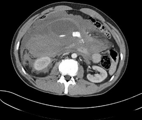
As the diagnosis remained unclear, CT abdomen was performed. This demonstrated active intra-abdominal bleeding with an origin in the bowel mesentery (figure 1).
Figure 1.
CT image demonstrating mesenteric bleed with contrast extravasation into the peritoneal cavity. Aorta is of normal calibre, and the inferior vena cava is collapsed commensurate with hypovolaemia. The actual aneurysm is not visible.
Differential diagnosis
Prior to imaging, our differential diagnosis included AAA, pancreatitis and perforated hollow viscus. VAA was not considered until the CT result was available.
Treatment
Initial resuscitation involved vitamin K and prothrombin compound concentrate to reverse his anticoagulation, and fluid resuscitation with 1.5 l of crystalloid. At this point he was stabilised sufficiently to obtain the CT.
The patient was transferred to an operating theatre, where 4.5–5 l of fresh and clotted blood were evacuated. Active bleeding was identified at the origin of the superior mesenteric artery. The bleeding point was ligated, and ongoing ooze controlled with packing. Forty units of blood products were transfused in the perioperative period.
Outcome and follow-up
The patient was ventilated on intensive therapy unit for 10 days. The packs were removed and the abdomen closed on day 3, when a section of ischaemic bowel was resected with a defunctioning ileostomy performed. He made steady progress, and was transferred to a surgical ward on day 14 and discharged home on day 33.
Discussion
Aneurysms of the visceral arteries are a recognised but rare problem, with an estimated prevalence of 0.1–2.0%.1 Aneurysms of the superior mesenteric artery represent an estimated 5.5% of this group; less than 0.5% of all intra-abdominal aneurysms.2 Their potential for life-threatening haemorrhage must be recognised; 38–50% of patients with an aneurysm of the superior mesenteric artery will present with rupture, carrying a 30% risk of mortality.3 4 The majority of patients have some history of intermittent abdominal pain, although this is frequently not recognised as relating to an aneurysm until after rupture has occurred.5 Unusually, our patient was entirely asymptomatic until the presenting episode.
VAAs have been successfully treated with surgical and interventional radiology techniques.6–8 As patients frequently attend with collapse after rupture of the aneurysm, surgical intervention has been the more widely used approach.
Diagnosis of VAA has been made by ultrasound, MRI and CT imaging. It is worth noting that focused bedside ultrasound to assess for AAA, as performed in the UK, will not reliably detect aneurysms of branches of the aorta.
Learning points.
-
▶
VAA is much rarer than AAA, but does rarely present to the emergency department.
-
▶
Focused emergency ultrasound for AAA will not detect VAA.
-
▶
Early consideration of this differential, formal imaging, and involvement of a vascular surgeon are required to achieve the best outcomes.
Acknowledgments
Thanks to Dr M Dobson for assistance with obtaining CT images.
Footnotes
Competing interests None.
Patient consent Obtained.
References
- 1.Tulsyan N, Kashyap VS, Greenberg RK, et al. The endovascular management of visceral artery aneurysms and pseudoaneurysms. J Vasc Surg 2007;45:276–83; discussion 283 [DOI] [PubMed] [Google Scholar]
- 2.Zimmerman-Klima PM, Wixon CL, Bogey WM, Jr, et al. Considerations in the management of aneurysms of the superior mesenteric artery. Ann Vasc Surg 2000;14:410–14 [DOI] [PubMed] [Google Scholar]
- 3.Shanley CJ, Shah NL, Messina LM. Uncommon splanchnic artery aneurysms: pancreaticoduodenal, gastroduodenal, superior mesenteric, inferior mesenteric, and colic. Ann Vasc Surg 1996;10:506–15 [DOI] [PubMed] [Google Scholar]
- 4.Lindberg CG, Stridbeck H. Aneurysms of the superior mesenteric artery and its branches. Gastrointest Radiol 1992;17:132–4 [DOI] [PubMed] [Google Scholar]
- 5.Akkary E, Cramer T, Patel M, et al. Superior mesenteric artery aneurysm: an uncommon disease with potentially serious complications. W V Med J 2010;106:10–14 [PubMed] [Google Scholar]
- 6.Marone EM, Mascia D, Kahlberg A, et al. Is open repair still the gold standard in visceral artery aneurysm management? Ann Vasc Surg 2011;25:936–46 [DOI] [PubMed] [Google Scholar]
- 7.Ferrero E, Ferri M, Viazzo A, et al. Visceral artery aneurysms, an experience on 32 cases in a single center: treatment from surgery to multilayer stent. Ann Vasc Surg 2011;25:923–35 [DOI] [PubMed] [Google Scholar]
- 8.Piffaretti G, Lomazzi C, Carrafiello G, et al. Visceral artery: management of 48 cases. J Cardiovasc Surg (Torino) 2011;52:557–65 [PubMed] [Google Scholar]


