Abstract
We developed two types of chimeric Sindbis virus (SINV)/western equine encephalitis virus (WEEV) alphaviruses to investigate their potential use as live virus vaccines against WEE. The first-generation vaccine candidate, SIN/CO92, was derived from structural protein genes of WEEV strain CO92-1356, and two second-generation candidates were derived from WEEV strain McMillan. For both first- and second-generation vaccine candidates, the nonstructural protein genes were derived from SINV strain AR339. Second-generation vaccine candidates SIN/SIN/McM and SIN/EEE/McM included the envelope glycoprotein genes from WEEV strain McMillan; however, the amino-terminal half of the capsid, which encodes the RNA-binding domain, was derived from either SINV or eastern equine encephalitis virus (EEEV) strain FL93-939. All chimeric viruses replicated efficiently in mammalian and mosquito cell cultures and were highly attenuated in 6-week-old mice. Vaccinated mice developed little or no detectable disease and showed little or no evidence of challenge virus replication; however, all developed high titers of neutralizing antibodies. Upon intranasal challenge with high doses of virulent WEEV strains, mice vaccinated with ≥105 PFU of SIN/CO92 or ≥104 PFU of SIN/SIN/McM or SIN/EEE/McM were completely protected from disease. These findings support the potential use of these live-attenuated vaccine candidates as safe and effective vaccines against WEE.
Keywords: alphavirus, western equine encephalitis virus, vaccine, virulence
INTRODUCTION
Western equine encephalitis virus (WEEV), a member of the family Togaviridae, genus Alphavirus, is an important mosquito-borne human and veterinary pathogen in the Americas [1]. It is also a potential biological weapon and an NIAID category B Priority Pathogen (www3.niaid.nih.gov/topics/BiodefenseRelated/Biodefense/research/CatA.htm). The virus contains a single stranded, positive-sense RNA genome of approximately 11.5 kb. Four nonstructural proteins (nsP1-4), encoded by an open reading frame in the 5’ two-thirds of the genome, are required for viral replication and polyprotein processing. A structural polyprotein is translated from a subgenomic (26S) RNA and cleaved into three major structural proteins (capsid and envelope glycoproteins, E2 and E1) that are involved in receptor recognition, virus attachment and penetration, membrane fusion and virion assembly [2].
First isolated in 1930 in California, WEEV is endemic in western North America and several strains have also been isolated from South America and Cuba [3]. In nature, WEEV is transmitted enzootically in western North America among passerine birds by mosquito vectors, primarily Culex tarsalis. Humans and equids become infected through mosquito bites that can result in periodic, extensive equine epizootics and epidemics of encephalitis. In humans, WEEV causes signs and symptoms ranging from fever and headache to severe encephalitis that can result in the development of neurologic sequelae. Overall, the estimated case fatality rate in humans is 3–4%, but these rates have been reported to be as high as 8–15% during epidemics [1, 4].
When aerosolized, WEEV causes high primate mortality [5] and is therefore considered a potential bioterrorism agent [6]. Unfortunately, neither a human vaccine nor antiviral drugs are available for the prevention and treatment of WEE during a natural outbreak or a bioterrorism attack. A DNA vaccine candidate completely protects mice against WEEV challenge; however, 3 immunizations by ballistic (intradermal) delivery were required to achieve protection [7].
Recent work has focused on recombinant WEEV vaccines using human adenovirus as a delivery vector [8, 9]. In mice, a single-dose injection of an adenovirus-vectored WEEV vaccine provides complete protection against both homologous and heterologous strains of WEEV at intranasal doses of ≤4 x 10⁴ PFU. However, the use of an adenovirus-vectored vaccine in humans is not without risk. Preexisting immunity against adenoviruses in the general population [10, 11] and acute toxicity to adenovirus vectors [12] are relevant concerns with this vaccine strategy.
Live-attenuated viral vaccines generally provide rapid and long-term protection after a single dose. However, there is a potential for live arbovirus vaccines to be transmitted to secondary hosts [13] and possibly to revert to a more virulent phenotype. To overcome this problem, one of the least human-pathogenic alphaviruses, Sindbis virus (SINV), has been successfully used as a vector for the expression of structural proteins derived from more pathogenic, encephalitic alphaviruses, such as Venezuelan (VEEV) and eastern equine encephalitis (EEEV) viruses [14–16].
In the present study, 3 recombinant alphavirus vaccine candidates were developed and assessed as live vaccine candidates for protection against WEE. The nonstructural protein genes and cis-acting RNA genome elements were derived from SINV strain AR339 [17], and the structural protein genes were derived from either WEEV strain CO92-1356 or ON41-McMillan. The recombinant vaccine candidates were evaluated in mice to assess attenuation, immunogenicity, and protection against WEEV challenge. Our findings suggest that the recombinant SIN/WEEV vaccines are safe and efficacious.
MATERIALS AND METHODS
Cells
Baby hamster kidney (BHK-21) and African green monkey (Vero) cells were purchased from the American Type Culture Collection (Bethesda, MD) and grown at 37°C in Eagles minimal essential medium (MEM) with 10% fetal bovine serum (FBS) and 0.05 mg/ml of gentamicin sulfate (Invitrogen, Carlsbad, CA). The Aedes albopictus mosquito cell line C710, a gift from Henry Huang at Washington University, was maintained in MEM at 32°C with 10% FBS and 10% tryptose phosphate broth.
Viruses
Three WEEV strains were included to study their pathogenesis in mice: strain CO92-1356 was isolated in Colorado from Culex tarsalis mosquitoes in 1992; strain ON41-McMillan, a 1941 Ontario, Canada strain, was kindly provided by Charles Calisher (Centers for Disease Control and Prevention, Fort Collins, CO); and strain TX71-TBT235, a 1971 Texas isolate, was provided by Robert Tesh (University of Texas Medical Branch, Galveston, TX). Details of the passage histories for each WEEV are summarized in Table 1. All viruses were passaged once in C6/36 mosquito cell cultures from stock virus before use in the mouse infectivity studies.
Table 1.
Strains of WEEV used in this study
| Virus strain | Year of isolation | Location of isolation | Host | Passage historya |
|---|---|---|---|---|
| CO92-1356 | 1992 | Larimer City, CO, United States | Culex tarsalis | V(1), BHK(1), C6/36(1) |
| ON41-McMillan | 1941 | Ontario, Canada | Human | M(2), SM(2), V(2), C6/36(1) |
| TX71-TBT235 | 1971 | Texas, United States | Gopherus berland | WC(1), DE(1), SM(1), BHK(1), C6/36(1) |
V, Vero cells; SM, suckling mouse; C6/36, mosquito cells; BHK, baby hamster kidney cells; M, mouse; WC, wet chick; DE, duck embryo.
RT-PCR and sequencing
WEEV RNAs were extracted from cell culture supernatants using a QIAamp Viral RNA Mini Kit (Qiagen, Valencia, CA) according to the manufacturer’s protocol. Coding regions of WEEV strains CO92, McMillan, and TBT235 were amplified by the Titan One Tube RT-PCR kit (Roche, Indianapolis, IN) and sequenced using the Applied Biosystems BigDye Terminator v3.1 Cycle Sequencing Kit (Foster City, CA) and ABI3100 Genetic Analyzer. All primers were designed based on the genomic sequence of WEEV strain 71V-1658 (GenBank accession no. AF214040) and are available upon request.
Construction of recombinant plasmids
A recombinant plasmid, pSIN/CO92/1, encoded the genome of a chimeric virus, in which the 5’ and 3’ untranslated genome regions (UTRs), subgenomic promoter, and nonstructural protein genes were derived from the SINV strain AR339 genome. The 5’UTR of the subgenomic RNA and structural protein genes were derived from the WEEV strain CO92-1356 genome. Recombinant plasmid pSIN/McM encoded the genome of chimeric virus, in which the 5’ and 3’ UTRs, subgenomic promoter and the nonstructural protein genes were derived from the SINV strain AR339 genome. The structural genes and the 5’UTR of the subgenomic RNA were derived from the WEEV strain McMillan. Junction between the SINV subgenomic promoter and 5’UTR of the WEEV subgenomic RNA was made by ligating PCR. The WEEV McMillan structural polyprotein-coding sequence was synthesized by RT-PCR as 3 fragments. WEEV-infected BHK-21 cells were used as a source of genetic material. To avoid introduction of PCR-mediated mutations, before assembly into chimeric virus genome, all of the PCR-synthesized fragments were initially cloned into pRS2 plasmid and sequenced. Then they were assembled into the subgenomic RNA-coding sequence of the chimeric virus genome using BglII, BsrGI, SphI and HindIII restriction sites. Recombinant plasmid, pSIN/SIN/McM, encoded the genome of a chimeric virus, in which the 5’ and 3’ UTRs, subgenomic promoter, the nonstructural protein genes, and the 5’UTR of the subgenomic RNA were derived from the SINV strain AR339 genome. The structural genes were derived from the WEEV strain McMillan genome, but the fragment encoding 102 amino terminal amino acids of the WEEV capsid was replaced by the corresponding fragment coding 106 amino terminal amino acids of the SINV capsid gene. The chimeric capsid gene was constructed by ligating PCR, and cloned into pSIN/McM using BamHI restriction site located upstream of the subgenomic promoter, and BsiWI restriction site in the WEEV capsid gene. Recombinant plasmid, pSIN/EEE/McM, encoded the genome of a chimeric virus, in which the 5’ and 3’ UTRs, subgenomic promoter, and the nonstructural protein genes were derived from the SINV strain AR339 genome. DNA fragment encoding 102 amino terminal amino acids of WEEV capsid were replaced by the corresponding 104 codons derived from the EEEV strain FL93 capsid gene [18]. The 5’ UTR of the subgenomic RNA was also derived from this EEEV genome. This DNA fragment was assembled by ligating PCR, and cloned into pSIN/McM genome as described above. Sequences of the recombinant genomes can be provided upon request.
In vitro transcription, transfection, and production of infectious virus
Plasmids were purified by centrifugation in CsCl gradients. Before the transcription reaction, the viral and replicon genome-coding plasmids were linearized by NotI digestion. RNAs were synthesized by SP6 RNA polymerase in the presence of cap analog. The yield and integrity of the transcripts were analyzed by gel electrophoresis under non-denaturing conditions. Aliquots of transcription reactions were used for electroporation without additional purification.
Electroporation of BHK-21 cells was performed under previously described conditions [19]. To rescue the viruses, 1 μg of in vitro-synthesized viral genome RNA was electroporated into cells, which were then seeded into 100-mm dishes and incubated until cytopathic effects (CPE) were observed. Virus titers were determined using a standard plaque assay on BHK-21 or Vero cells [20]. To assess RNA infectivity, an infectious center assay was performed in which 10-fold dilutions of electroporated BHK-21 cells were seeded in 6-well Costar plates containing subconfluent, naïve cells. Electroporated BHK-21 cells with in vitro-synthesized RNA from SINV strain Toto1101 was used as a control. After 1 h incubation at 37°C, cells were overlaid with 2 ml 0.5% agarose supplemented with MEM and 3% FBS. Plaques were stained with crystal violet after 48 h of incubation at 37°C, and infectivity was determined as plaque-forming units (PFU) per μg of transfected RNA.
Virus replication in cell culture
Replication kinetics of parental strains (WEEV strain CO92, WEEV strain McMillan, and SINV strain AR339) and chimeras (SIN/CO92, SIN/SIN/McM, and SIN/EEE/McM) were compared in Vero and C710 cells. Confluent monolayers grown in 12-well plates were infected in triplicate at a multiplicity of infection (MOI) of 1 PFU/cell at 37°C for Vero cells and 32°C for C710 cells. After 1 h of incubation, the plates were washed with phosphate buffer saline (PBS) and 2 ml of MEM with 2% FBS was added to each well. At 4, 8, 12, 24 and 32 h post infection, cell culture medium was collected and replaced with fresh medium. Virus titers were determined in the harvested media by plaque assay on Vero cells (limit of detection 0.9 log10 PFU/ml).
Mouse infections
Female and pregnant NIH Swiss mice were purchased from Harlan Sprague Dawley, Inc. (Indianapolis, IN) and maintained under specific-pathogen-free conditions. Newborn mice were held for 4–6 days after birth prior to subcutaneous (SC) or intracerebral (IC) infections. To monitor body temperature, 5-week-old mice were anaesthetized with isoflurane 7 days before infection and implanted SC with a pre-programmed telemetry chip according the manufacturer’s instructions (IPTT-300; Bio Medical Data Systems, Inc., Seaford, DE). Body temperatures and weights were recorded daily or weekly without anesthesia.
Mice were infected SC in the medial thigh or intraperitoneally (IP) in a total volume of 100 μl. For intranasal infections (IN), a total volume of 20 μl was used. Blood samples were collected from the retroorbital sinus and virus titers were determined by plaque assay using Vero cells (limit of detection 0.9 log10 PFU/ml). Four-day-old mice were inoculated SC in the dorsal shoulder at a dose of 4.6 log10 PFU in a volume of 50 μl, and 6-day-old mice were inoculated IC at a dose of 5.3 log10 PFU in a volume of 20 μl.
Immunization and challenges with WEEV
Six-week-old mice were vaccinated SC in the medial thigh. Controls were sham-infected with PBS. Blood samples were collected from the retroorbital sinus to detect viremia for 3 days after vaccination. Animals were observed daily for clinical signs of infection, and body weight measurements were performed on days 1–4, 7, 14, 21 and 28 after vaccination. Sera were collected on day 28 after vaccination, then mice were challenged with either WEEV strain TBT235 (5.3 log10 PFU) or McMillan (5.0 log10 PFU) by the IN route.
Serological assays
Neutralizing antibodies were assayed using an 80% plaque reduction neutralization test (PRNT80), and IgG responses were measured using an enzyme-linked immunosorbent assay (ELISA) [21]. For the ELISA, mouse brain antigens, derived from WEEV strain Fleming, and control mouse brain antigens (Centers for Disease Control and Prevention, Fort Collins, CO) were resuspended in 0.5 ml of water and then diluted 1:800 in PBS. One hundred μl of diluted antigens were added to each well of a 96-well plate (PolySorp, Nalge Nunc International, Rochester, NY) and incubated at 4°C overnight. The plate was then washed with buffer [PBS with 0.1% Tween 20 (Sigma)] and blocked with PBS containing 0.1% Tween 20 and 5% dry milk, and incubated at 37°C for 1 h. Following the addition of experimental mouse sera at a 1:100 dilution, IgG was detected with horseradish peroxidase-conjugated goat antimouse IgG (Kirkegaard & Perry Laboratories, Inc., Gaithersburg, MD). Sera yielding absorbance values that exceeded the mean values of negative control sera by more than 2 standard deviations were considered IgG positive for WEEV.
Statistical analysis
Statistical comparisons of body temperatures and weights of mice in different groups were performed using a one-way ANOVA followed by a Tukey-Kramer multiple comparisons test. Differences were considered significant when P<0.05.
RESULTS
Mouse model for WEE
Previous studies have shown that both epizootic and enzootic WEEV strains kill young mice (3–5 weeks of age) by the IP and SC routes [22, 23]. However, mice become resistant to disease produced by some WEEV strains (e.g. Argentina strain Ag80–646) after 5 weeks of age and no signs of infection are seen after IP inoculation. Another study showed that some but not all WEEV strains (e.g. Fleming strain and others isolated in California during 1994 and 1995) are lethal by the SC route in adult outbred mice [24]. In contrast, a different study showed that all WEEV strains tested by IN route were 100% lethal in adult Balb/c mice [25]. Despite high mortality, older mice do not consistently develop viremia following WEEV infection [24].
To develop an animal model for our WEEV challenge experiments, we initially tested WEEV strain CO92-1356, used for vaccine design, in several different strains of 8–10-week-old mice using IP (6 log10 PFU) and IN (5.3 log10 PFU) inoculations (Table 2). All animals developed encephalitis and died after IN infection, but the mice exhibited no signs of disease after IP infection despite seroconversion. Among the mouse strains tested, C57BL/6 mice were more sensitive to WEE after IN infection and died at an average of 7.6 days post infection. Other strains of mice were also susceptible to WEE after IN infection and died between 8–10 days post infection (Table 2).
Table 2.
Survival of mouse strains inoculated intranasally (IN) and intraperitoneally (IP) with WEEV strain CO92
| Mouse strain | Age (weeks) | No. of animals tested | Mean survival time after IN inoculation (days ± SD)a | Mean survival time after IP inoculation (days)b |
|---|---|---|---|---|
| C57BL/6 | 8 | 3 | 7.6 ± 0.1 | > 21 |
| Balb/c | 8 | 3 | 8.5 ± 0.0 | > 21 |
| CD1 | 8 | 3 | 8.2 ± 1.5 | > 21 |
| NIH Swiss | 10 | 3 | 9.5 ± 0.9 | > 21 |
Inoculated IN with 5.3 log10 PFU in 20 μl volume.
Inoculated IP with 6 log10 PFU in 100 μl volume.
To develop an outbred mouse model, we further compared the virulence of 3 other WEEV strains in 9-week-old NIH Swiss mice, and the results indicated that WEEV strains TX71-TBT235 and ON41-McMillan were highly virulent when compared to CO92-1356 (Table 3). Both TX71-TBT235 and ON41-McMillan strains killed NIH Swiss mice within 4 days post infection by IN delivery. Therefore, high virulent strains TBT235 and McMillan were used for WEEV challenge experiments.
Table 3.
Survival of 9-week-old female NIH Swiss mice inoculated intranasally (IN) or intraperitoneally (IP) with different strains of WEEV
| Virus strain | No. of animals tested | Route of inoculationa | Mean survival time (days ± SD) | Survival (%) |
|---|---|---|---|---|
| CO92 | 3 | IN | 9.5 ± 0.9 | 0 |
| 3 | IP | >21 | 100 | |
| McMillan | 5 | IN | 3.2 ± 0.1 | 0 |
| 5 | IP | 4.6 ± 0.9 | 0 | |
| TBT235 | 5 | IN | 3.8 ± 0.2 | 0 |
| 5 | IP | >21 | 100 |
IP dose, 6 log10 PFU in 100 μl volume; IN dose, 5.3 log10 PFU in 20 μl volume.
Sequence analysis of WEEV strains
To identify potential virulence determinants at the molecular level, the coding regions of the full genomes of WEEV strains CO92-1356 (GenBank Accession Nos. FJ786260 and FJ786264), ON41-McMillan (FJ786263 and FJ786265), and TX71-TBT235 (FJ786261 and FJ786262) were sequenced and compared to a low virulence WEEV strain 71V-1658 [25, 26]. Table 4 shows the deduced amino acid differences that are unique to each high-virulence WEEV strain (TBT235 and McMillan) when compared to both low virulence WEEV strains 71V-1658 and CO92. The amino acid sequence of the McMillan strain showed a total of 29 unique amino acids at the nonstructural and structural protein gene regions when compared to WEEV strains 71V-1658, CO92, and TBT235 (Table 4). There were four amino acid differences at the same positions in both high virulence WEEV strains TBT235 and McMillan (nsP3-334 A versus T/V, nsP3-365 L versus F, nsP3-469 Y versus H, capsid-250 R versus K/W). Additionally, an Asp amino acid insertion was found in the McMillan strain at position 167 of nsP3, and a deletion of Val was found in the TBT235 strain at position 352 of nsP3.
Table 4.
Deduced amino acid differences between each high virulence WEEV strain (TBT235 and McMillan) and both low virulence WEEV strains 71V-1658 and CO92-1356
| Protein | Amino acid positiona | Low virulence phenotype | High virulence phenotypeb | ||
|---|---|---|---|---|---|
| 71V-1658 | CO92 | TBT235 | McMillan | ||
| nsP1 | 59 | Q | Q | Q | E |
| 467 | K | K | K | R | |
| 513 | P | P | S | P | |
|
| |||||
| nsP2 | 39 | A | A | A | E |
| 433 | T | T | I | T | |
| 630 | K | K | K | N | |
|
| |||||
| nsP3c | 56 | R | R | K | R |
| 289 | L | L | L | P | |
| 332 | P | P | P | S | |
| 334 | A | A | V | T | |
| 365 | L | L | F | F | |
| 418 | V | V | V | D | |
| 447 | K | K | K | R | |
| 469 | Y | Y | H | H | |
|
| |||||
| nsP4 | 24 | E | E | E | D |
| 27 | L | L | L | F | |
| 28 | D | D | D | E | |
| 147 | A | A | A | S | |
| 159 | L | L | L | I | |
| 537 | P | P | S | P | |
| 602 | S | S | S | N | |
|
| |||||
| capsid | 57 | S | S | S | A |
| 74 | K | K | K | E | |
| 250 | R | R | W | K | |
| 252 | T | T | P | T | |
|
| |||||
| E2 | 23 | T | T | T | A |
| 69 | F | F | F | Y | |
| 146 | H | H | H | Q | |
| 157 | H | H | H | R | |
| 181 | E | E | E | K | |
| 214 | R | R | R | Q | |
| 224 | K | K | K | R | |
| 231 | S | S | S | L | |
|
| |||||
| 6K | 30 | V | V | V | I |
|
| |||||
| E1 | 50 | K | K | Q | K |
| 349 | V | V | V | T | |
| 374 | S | S | S | T | |
Amino acid position number corresponds to WEEV strain 71V-1658.
Boldface type specifies amino acid residues unique to the high virulence WEEV strains when compared to both low virulence WEEV strains.
There was an amino acid insertion of D at nsP3-167 in the McMillan strain and a deletion of V at nsP3-352 in the TBT235 strain.
There were 8 amino acid differences found between the McMillan strain used in our challenge studies and its published sequence of the structural protein gene region (GenBank accession No. DQ393792): capsid-102 M/V, E2–17 P/Q, E2–74 D/V, E2–81 E/K, E2–116 T/A, E2–268 V/A, E1–95 F/S, and E1–196 K/R. However, since there were no amino acid differences at the same positions in the other 3 sequenced WEEV strains, these 8 amino acid differences may be due to different passage histories of the virus and/or the sequence methods used.
First-generation vaccine candidate
Replication of SIN/CO92 in cell culture
A SIN/CO92 infectious cDNA clone was constructed in which the nonstructural protein genes were derived from SINV strain AR339 and the structural protein genes were derived from WEEV strain CO92 (a less virulent strain when compared to the McMillan strain) (Fig. 1A). The originally designed construct SIN/CO92/1 was viable, and the in vitro-synthesized RNA demonstrated the same infectivity in the infectious center assay as did control SINV Toto1101 RNA (5–10x105 PFU/μg). However, titers of SIN/CO92/1 were unusually low (2–3x10⁶ PFU/ml), and plaques were very heterogeneous in size. This was likely an indication of adaptation of the original variant to replication in cell culture. Therefore, we further passaged SIN/CO92/1 in BHK-21 cells at an MOI of 0.1 PFU/cell, and after 4 passages, harvested viruses formed uniformly large plaques in BHK-21 cell monolayers, and titers approached 109 PFU/ml. Sequencing of the plaque-purified, BHK-21-adapted virus detected a single mutation in E2 E182→K, and cloning of this mutation into the original SIN/CO92/1 produced virus (SIN/CO92/2) that replicated efficiently in BHK-21 cells. Surprisingly, this mutation was insufficient for efficient replication in Vero cells, which are normally used in vaccine production. In this cell line, SIN/CO92/2, harvested after electroporation, continued to develop pinpoint, barely visible plaques. Therefore, 5 passages were performed in Vero cells at an MOI of ~0.1 PFU/cell, which led to the selection of another variant demonstrating a large-plaque phenotype both on Vero and BHK-21 cells. This change in Vero cell plaque size was apparently the result of a K159→N mutation in the E2 glycoprotein, leading to the addition of a putative NET glycosylation site. Consistent with this interpretation, this mutant exhibited strongly reduced mobility of the E2 glycoprotein in the SDS-PAGE (data not shown), suggesting that an additional glycosylation occurred. This variant, having both E182→K and K159→N mutations, was termed SIN/CO92 and used in the following experiments.
Fig. 1.
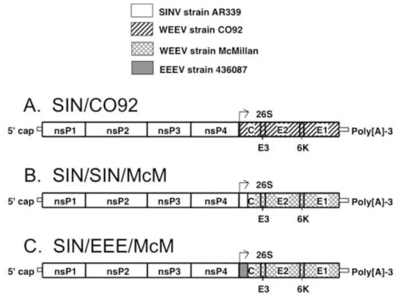
Schematic representation of SIN/CO92, SIN/SIN/McM, and SIN/EEE/McM chimeras. The SIN/CO92 chimera (A) includes the nonstructural protein gene regions of SIN strain AR339 and the structural protein gene regions of WEEV strain CO92-1356. The SIN/SIN/McM chimera (B) includes the nonstructural protein gene regions and the amino-terminal domain of the capsid of SINV strain AR339 and the carboxy-terminal domain of the capsid and envelope glycoproteins of WEEV strain McMillan. The SIN/EEE/McM chimera (C) includes the nonstructural protein gene regions of SINV strain AR339, the amino-terminal domain of the capsid of EEEV strain 436087, and the carboxy-terminal domain of the capsid and envelope glycoproteins of WEEV strain McMillan.
Replication kinetics of SIN/CO92 was compared to the parental strains at a MOI of 1 in mammalian Vero and mosquito C710 cells. Similar to the parental SINV strain AR339, there was a high yield of SIN/CO92 produced in both cell lines, with peak titers of 7.1 log10 PFU at 24 h in Vero cells (Fig. 2A) and 7.5 log10 PFU at 32 h in C710 cells (Fig. 2B).
Fig. 2.
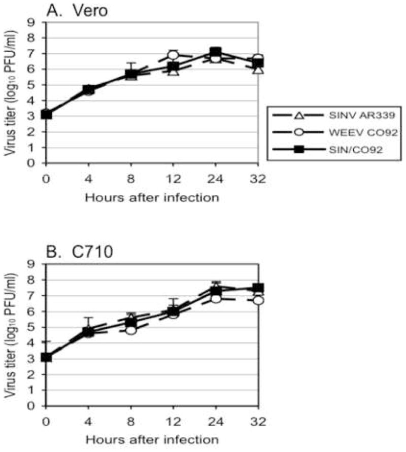
Replication of parental and chimeric SIN/CO92 strains in (A) mammalian Vero and (B) mosquito C710 cells. Cells were infected in triplicate at a MOI of 1 PFU/cell for 1 h, washed three times with PBS, and then, viral titers in the culture medium were determined by plaque assay on Vero cells at selected times after infection. Bars indicate standard deviations.
Attenuation and immunogenicity of SIN/CO92
We assessed the attenuation and safety of SIN/CO92 in 6-week-old female NIH Swiss mice. Cohorts of 10 were inoculated SC with doses of 3.5, 4.5, or 5.0 log10 PFU. Control mice (10 mice) were sham vaccinated with PBS. Viremia levels in mice were determined on days 1 to 3 post immunization. No infectious virus was detected in the sera by plaque assay (limit of detection, 8 PFU/ml). None of the vaccinated animals exhibited any signs of disease.
Four weeks after immunization, sera were collected and antibody titers were measured by PRNT and ELISA. The results showed a dose response to the SIN/CO92 inoculations, with the highest mean titer detected following immunization with 5.0 log10 PFU (Table 5). At this dose, all animals in the cohort had positive PRNT80 titers ranging from 20–640, and the mean PRNT80 titer was 190 when tested against WEEV strain CO92. When the animals received vaccine doses below 5.0 log10 PFU, the immune responses were inconsistent with only 40–50% of mice developing PRNT80 titers ≥20, but all animals had a positive IgG titer by ELISA (OD ≥ 0.20) (Table 5).
Table 5.
Neutralizing antibody and IgG response in female NIH Swiss mice 4 weeks after infection with SIN/CO92 chimera
| Vaccine dose (log10 PFU/mouse) | Mean 80% PRNT titer ± SD | No. of PRNT seropositive | Mean OD of IgG ELISA ± SD | No. of IgG seropositive |
|---|---|---|---|---|
| 3.5 | 65 ± 64 | 4/10 | 0.69 ± 0.15 | 10/10 |
| 4.5 | 60 ± 62 | 5/10 | 0.80 ± 0.23 | 10/10 |
| 5.0 | 190 ± 193 | 10/10 | 1.13 ± 0.10 | 10/10 |
Protection against WEEV challenge
On day 28 after immunization with either PBS (sham) or SIN/CO92, mice were challenged IN with virulent WEEV strain TBT235 at a dose of 5.3 log10 PFU and monitored for signs of disease and mortality for 28 days (Fig. 3). Sham-vaccinated mice showed signs of paralysis and encephalitis by days 4–5 after challenge and died on day 5. In contrast, mice vaccinated with SIN/CO92 at a dose of 5.0 log10 PFU showed no signs of disease after WEEV challenge and were completely protected. However, mice receiving vaccine doses of ≤4.5 log10 PFU were not completely protected and showed similar disease patterns to those of the sham-vaccinated mice; 50–60% of the mice died on days 5–9 after challenge. These results indicated that a dose ≥5 log10 PFU of SIN/CO92 is required for complete protection of mice from WEE. Although individual mice that died after challenge could not be matched to PRNT antibody titers, the similarity between seroconversion and protection rates suggest that the former predicted the latter outcome.
Fig. 3.
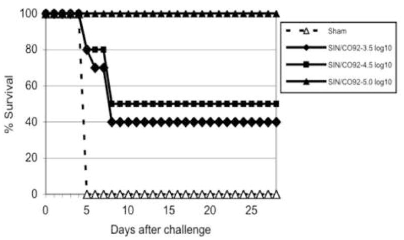
Survival following SIN/CO92 vaccination and WEEV challenge. Six-week-old female NIH Swiss mice were inoculated subcutaneously with either PBS (sham) or SIN/CO92 at the indicated doses (expressed as log10 PFU). Four weeks after immunization, mice were challenged intranasally with 5.3 log10 PFU of WEEV strain TBT235, and mortality was recorded.
Second-generation vaccine candidates
Replication of SIN/SIN/McM and SIN/EEE/McM in cell culture
Low immunogenicity of SIN/CO92 could have multiple explanations, but the most probable ones were either inefficient infectivity of the virus for mouse cells in vivo or inefficient WEEV capsid function in packaging of the genomic RNA of the chimeric virus. The data presented above demonstrated that SIN/CO92 was packaged to high titers in vitro, in highly permissive cells (Fig. 4). However, this does not necessarily mean that it was packaged with the same efficiency in mice, where SINV poorly replicates and, most likely, does not overproduce the structural components of the virions.
Fig. 4.
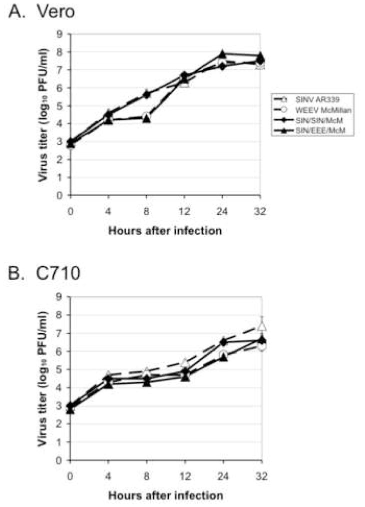
Replication of parental and chimeric SIN/SIN/McM and SIN/EEE/McM strains in (A) mammalian Vero and (B) mosquito C710 cells. Cells were infected in triplicate at a MOI of 1 PFU/cell for 1 h, washed three times with PBS, and then, viral titers in the culture medium were determined by plaque assay on Vero cells at selected times after infection. Bars indicate standard deviations.
Therefore, in an attempt to increase the immunogenicity of SIN/CO92, we designed several new chimeras, which included mouse-attenuated WEEV strain Ag80-646 [22]. However, clones containing the backbone of SINV strain AR339 and the structural protein genes of either WEEV strain Ag80-646 or McMillan did not produce high titers on Vero cells even after 10 passages (data not shown). Another chimera with the complete capsid derived from SINV and the envelope proteins derived from WEEV-McMillan also replicated poorly in Vero cells.
Two additional chimeras, SIN/SIN/McM and SIN/EEE/McM, proved more successful, in which the envelope glycoprotein genes were derived from WEEV strain McMillan. In SIN/SIN/McM, the amino-terminal domain of the capsid (including the RNA-binding domain) was derived from SINV strain AR339 (Fig. 1B). Thus, a SINV-specific RNA-binding domain was homologous to the viral genome backbone and was expected to function more efficiently in RNA packaging. The capsid gene of the second chimera, SIN/EEE/McM, contained the RNA-binding domain of EEEV strain FL93-939 (Fig. 1C). The rationale for the use of the EEEV domain was based on efficient replication of SIN/EEEV chimeric viruses [16], suggesting a high activity of the EEEV-specific capsid in RNA binding during virus assembly. No adaptive mutations in the envelope glycoproteins were required for efficient replication.
The replication kinetics of SIN/SIN/McM and SIN/EEE/McM were compared to the parental SINV and WEEV strain McMillan at a MOI of 1 in Vero and C710 cells. Similar to the parental SINV and WEEV strains, there were high yields of SIN/SIN/McM and SIN/EEE/McM produced in both cell lines (Fig. 4). In Vero cells, SIN/SIN/McM and SIN/EEE/McM produced peak titers at slightly different times, with 7.5 log10 PFU at 32 h for SIN/SIN/McM and 7.9 log10 PFU at 24 h for SIN/EEE/McM (Fig. 4A). In C710 cells, SIN/SIN/McM and SIN/EEE/McM both produced peak titers at 32 h, with titers of 6.6 and 6.7 log10 PFU at 32 h, respectively (Fig. 4B), comparable to wild-type alphavirus titers.
Attenuation and immunogenicity of SIN/SIN/McM and SIN/EEE/McM
SIN/SIN/McM and SIN/EEE/McM were passaged three times in Vero cells (to recapitulate vaccine manufacturing conditions) and then inoculated into 6-week-old NIH Swiss mice (10 mice per cohort). Cohorts were inoculated SC with PBS (sham-vaccinated), SIN/SIN/McM (at doses of either 4.8 or 5.8 log10 PFU), or SIN/EEE/McM (at doses of either 4.6 or 5.6 log10 PFU), and viremia levels in mice were determined on days 1–3 post immunization. No infectious virus was detected in the sera by plaque assay (limit of detection, 8 PFU/ml). However, on day 8 after infection, 3 mice immunized with 5.8 log10 PFU of SIN/SIN/McM became sick and 2 of these died on days 9 or 12 post infection. Also, 2 mice became sick on day 9 post infection with 4.8 log10 PFU of SIN/SIN/McM and recovered 2–3 days later. There were no signs of disease observed in mice immunized with SIN/EEE/McM.
Four weeks after immunization, sera were collected to measure antibody responses by PRNT. Compared to vaccine candidate SIN/CO92 (Table 5), both SIN/SIN/McM and SIN/EEE/McM were significantly more immunogenic regardless of dose (p<0.05, 2-way ANOVA; Table 6). When tested against WEEV strain McMillan, the mean PRNT80 titers after 5.6–5.8 log10 doses were 600 ± 113 for SIN/SIN/McM and 420 ± 252 for SIN/EEE/McM; this difference was not significant (p>0.05, 2-way ANOVA). When the animals received SIN/EEE/McM at a dose of 4.6 log10 PFU, the immune responses were less consistent with 80% of mice developing PRNT80 titers ≥ 20.
Table 6.
Neutralizing antibody titers in female 10-week-old NIH Swiss mice 4 weeks after immunization with either SIN/SIN/McM or SIN/EEE/McM chimera
| Vaccine | Vaccine dose (log10 PFU/mouse) | Mean 80% PRNT titer ± SD | No. of PRNT seropositive |
|---|---|---|---|
| SIN/SIN/McM | 4.8 | 600 ± 113 | 8/8* |
| 5.8 | 604 ± 106 | 10/10 | |
| SIN/EEE/McM | 4.6 | 420 ± 252 | 8/10 |
| 5.6 | 416 ± 215 | 10/10 |
two mice died before testing of sera for antibodies
Protection against WEEV challenge
On day 28 after immunization with PBS (sham), SIN/SIN/McM, or SIN/EEE/McM, mice were challenged IN with WEEV strain McMillan at a dose of 5.0 log10 PFU and monitored for signs of disease and mortality for 28 days (Fig. 5). Sham-vaccinated mice developed signs of paralysis and encephalitis, and all died on day 4 post challenge. In contrast, mice vaccinated with either SIN/SIN/McM or SIN/EEE/McM were completely protected, even when vaccine doses were <5 log10 PFU. Two mice with PRNT titers of <20 following SIN/EEE/McM vaccination (Table 6) were also protected. These results suggest that a dose <5 log10 PFU of either SIN/SIN/McM or SIN/EEE/McM provides complete protection from WEE.
Fig. 5.
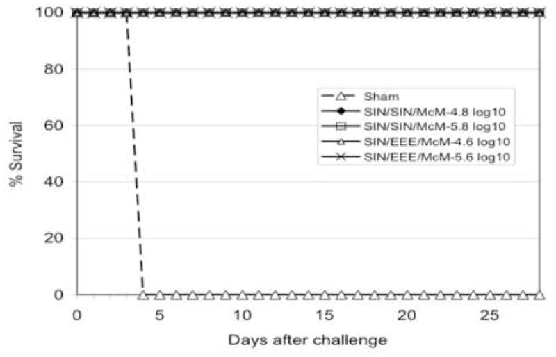
Survival following either SIN/SIN/McM or SIN/EEE/McM vaccination and WEEV challenge. Six-week-old female NIH Swiss mice were inoculated subcutaneously with either SIN/SIN/McM or SIN/EEE/McM at the indicated doses (expressed as log10 PFU). Four weeks after immunization, mice were challenged intranasally with 5.0 log10 PFU of WEEV strain McMillan, and mortality was recorded.
Body temperature, weight changes associated with vaccination and challenge
In a separate experiment, cohorts of 6-week-old female NIH Swiss mice were implanted SC with a telemetry chip to monitor body temperature before and after immunization (Fig. 6A) and challenge (Fig. 7A). Body weights were also measured before and after immunization (Fig. 6B) and challenge (Fig. 7B). Cohorts were inoculated SC with one of the three vaccine candidates (SIN/CO92, SIN/SIN/McM, SIN/EEE/McM) at 5.0 log10 PFU, or PBS as a sham control. Positive virulence control mice were infected IN with WEEV strain McMillan at a dose of 5.0 log10 PFU. No significant febrile response or change in body weight was measured in the vaccinated or sham-vaccinated mice, although some mice had slight elevations in body temperature (38–38.5°C) after infection (Fig. 6A). In contrast, mice infected with WEEV exhibited encephalitis, a significant increase in mean body temperature at day 2 post infection (39.3°C±0.3, P<0.01, ANOVA with Tukey-Kramer multiple comparisons test), and a significant loss in body weight on day 3 (P<0.01). Vaccinated- and sham-vaccinated mice gained weight throughout the 28-day period (Fig. 6B; data not shown), and there were no significant differences in body weight measurements between groups.
Fig. 6.
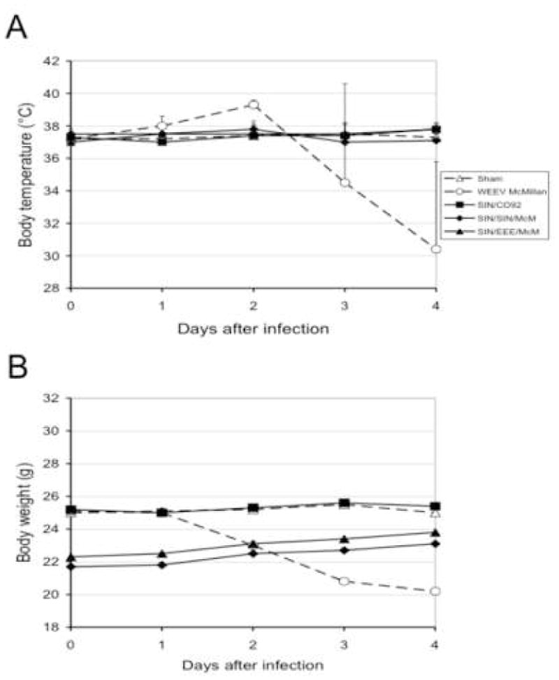
Body temperatures (A) and weights (B) in cohorts of ten 6-week-old female NIH Swiss mice after subcutaneous immunization with WEE vaccine candidates (SIN/CO92, SIN/SIN/McM, SIN/EEE/McM) or after sham-vaccination with PBS. Intranasal infection with WEEV strain McMillan was included as a positive virulence control. Bars indicate standard deviations.
Fig. 7.
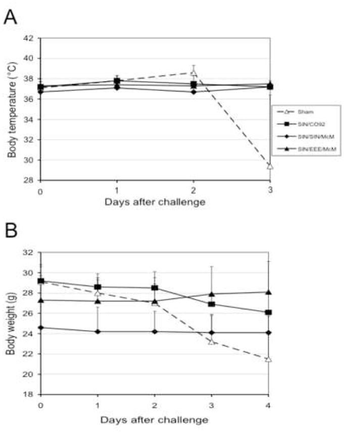
Body temperatures (A) and weights (B) of vaccinated (SIN/CO92, SIN/SIN/McM, or SIN/EEE/McM) or sham-vaccinated (PBS) female NIH Swiss mice following intranasal challenge with WEEV strain McMillan. Bars indicate standard deviations.
On day 28 post vaccination, mice were challenged IN with WEEV strain McMillan at a dose of 5.0 log10 PFU. All sham-vaccinated mice developed a febrile response (mean body temperature, 38.5°C; P>0.05) with signs of encephalitis (Fig. 7A). The sham-vaccinated mice had significant losses in body weight (Fig. 7B) and died within 4 days post-infection (mean survival time, 3.1±0.1 days). All vaccinated mice were completely protected from WEE. For the next 28 days after challenge, the body temperatures of the vaccinated mice remained within normal range (Fig. 7A; data not shown), and there were no signs of encephalitis. There was only a slight loss of body weight noted in the vaccinated mice (Fig. 7B; data not shown).
Levels of virus replication in immunized mice after challenge
To determine whether vaccination with the chimeric viruses prevents replication of challenge WEEV, the least immunogenic strain (SIN/CO92) of the 3 chimeric vaccine strains was selected. Following vaccination of 6-week-old NIH Swiss mice with 5.0 log10 PFU and 4 weeks of incubation, mice were challenged either IP (5.7 log10 PFU) or IN (5.0 log10 PFU) with the WEEV McMillan strain. Two animals were sacrificed on days 1, 2, 3, 7 and 28 post-challenge and major organs and sera were tested for virus by plaque assay. WEEV was detected in the majority of organ samples from sham-vaccinated mice following IP challenge, with the highest titers in the brain (8.3 ± 1.3 log10 PFU/g) (Table 7). In contrast, vaccinated mice contained no detectable challenge virus in the serum or any organ sampled.
Table 7.
Viremia and viral titers in organs of vaccinated mice after challenge by intraperitoneal (IP) infection with WEEV strain McMillana
| Organ | Day 1 (log10 PFU/ml or g) | Day 2 (log10 PFU/ml or g) | Day 3 (log10 PFU/ml or g) | |||
|---|---|---|---|---|---|---|
| Vaccine: PBS | Vaccine: SIN/CO92 | Vaccine: PBS | Vaccine: SIN/CO92 | Vaccine: PBS | Vaccine: SIN/CO92 | |
| Blood | <0.9 | <0.9 | <0.9 | <0.9 | <0.9 | <0.9 |
| Brain | <0.9 | <0.9 | <0.9 | <0.9 | 8.3 ± 1.3 | <0.9 |
| Lung | <0.9 | <0.9 | 2.8 | <0.9 | <0.9 | <0.9 |
| Heart | 2.1 | <0.9 | 3.1 | <0.9 | 4.4 ± 0.2 | <0.9 |
| Liver | <0.9 | <0.9 | <0.9 | <0.9 | <0.9 | <0.9 |
| Spleen | <0.9 | <0.9 | 2.5 ± 0.8 | <0.9 | 4.2 | <0.9 |
| Kidney | <0.9 | <0.9 | 4.2 | <0.9 | <0.9 | <0.9 |
IP dose, 5.7 log10 PFU in 100 μl volume. Tissues were collected from 2 animals per cohort per day.
Since WEEV is more virulent via the aerosol route, virus replication in vaccinated mice was also examined after IN challenge. No WEEV was detected in the tissues of the SIN/CO92-vaccinated mice on days 1 and 2 post-challenge (Table 8). In contrast, WEEV was detected in the brains of sham-vaccinated mice on day 1 post challenge and continued to replicate to higher titers on days 2 and 3 post-infection (mean titers, 9.2 and 10.2 log10 PFU/g, respectively). The virus was found in all other organs tested on days 2 and 3 post challenge; only one shamvaccinated mouse developed viremia (on day 3 post infection). All sham-vaccinated mice had clinical signs of encephalitis by day 3 and died by day 3–4 post-challenge. No vaccinated mice exhibited any signs of infection, and no virus was found in the brain after challenge; only one mouse in the vaccinated cohort was found with small amounts of virus in the heart, liver, and kidney on day 3 post-challenge (Table 8). No virus was detected in the vaccinated mice on days 7 and 28 post challenge (data not shown).
Table 8.
Viremia and viral titers in organs of vaccinated mice after challenge by intranasal (IN) infection with WEE strain McMillana
| Organ | Day 1 (log10 PFU/ml or g) | Day 2 (log10 PFU/ml or g) | Day 3 (log10 PFU/ml or g) | |||
|---|---|---|---|---|---|---|
| Vaccine: PBS | Vaccine: SIN/CO92 | Vaccine: PBS | Vaccine: SIN/CO92 | Vaccine: PBS | Vaccine: SIN/CO92 | |
| Blood | <0.9 | <0.9 | <0.9 | <0.9 | 3.6 | <0.9 |
| Brain | 5.2 | <0.9 | 9.2 ± 1.1 | <0.9 | 10.2 ± 0.1 | <0.9 |
| Lung | <0.9 | <0.9 | 5.1 ± 1.1 | <0.9 | 6.3 | <0.9 |
| Heart | <0.9 | <0.9 | 4.2 ± 0.6 | <0.9 | 4.2 ± 1.2 | 3.3b |
| Liver | <0.9 | <0.9 | 4.3 | <0.9 | 5.9 | 5.7b |
| Spleen | <0.9 | <0.9 | 4.0 ± 0.4 | <0.9 | 4.4 ± 2.4 | <0.9 |
| Kidney | <0.9 | <0.9 | 3.3 ± 0.7 | <0.9 | 4.3 ± 1.1 | 4.3b |
IN dose, 5.0 log10 PFU in 20 μl volume. Tissues were collected from 2 animals per cohort per day.
Virus was detected in the heart, liver, and kidney of one animal in the cohort.
Neurovirulence and replication of chimeric vaccine candidates in infant mice
The chimeric vaccine candidates were also evaluated using more sensitive alphavirus infection models that involve SC and IC infection of newborn mice [16, 27]. Cohorts of 4- and 6-day-old NIH Swiss mice were inoculated SC and IC, respectively, with either chimeric vaccine candidates or parental viruses and monitored daily for clinical signs of disease. Parental WEEV strains CO92 and McMillan were highly neurovirulent, and killed animals within 2 days of either SC or IC infection (Fig. 8). SIN/EEE/McM killed all mice by day 3 after IC infection and by day 5 after SC infection. SIN/CO92 was less virulent, and all mice appeared healthy until day 8 after IC infection; these mice then developed visible signs of disease by day 9 after infection, with most of the mice dying between days 9 and 12 after infection. Two mice in the SIN/CO92-IC cohort appeared sick and then recovered by day 13 after infection, surviving until the experiment was terminated on day 28 after infection. For the SIN/CO92-SC cohort, 2 mice died on day 3 after infection and one mouse died on day 5, resulting in an overall survival rate of 70%.
Fig. 8.
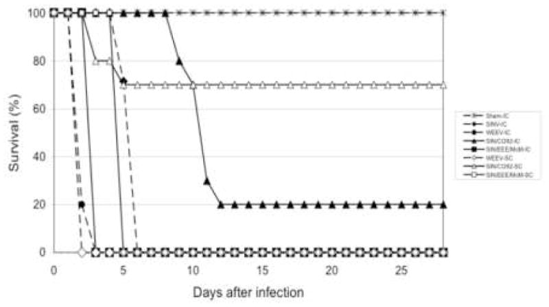
Survival of 6-day-old NIH Swiss mice after intracerebral (IC) inoculation with PBS, WEEV-CO92, SINV-AR339, SIN/CO92 or SIN/EEE/McM (5 log10 PFU) and survival of 4-day-old mice after subcutaneous (SC) inoculation with WEEV-McMillan, SIN/CO92 or SIN/EEE/McM (4.6 log10 PFU). Each cohort was monitored daily for signs of neurologic disease and mortality.
Levels of viral replication in the brains of the 6-day-old mice were quantified by plaque assay on days 1 and 2 after IC infection (Fig. 9A). On day 1, the parental WEEV strains CO92 and McMillan replicated to the highest mean titer of 8.5 and 9.4 log10 PFU/g, respectively. SIN/EEE/McM and SIN/CO92 developed slightly lower mean titers than their respective parental WEEV strains; the mean levels of virus replication of SIN/EEE/McM and SIN/CO92 were 8.1 and 6.2 log10 PFU/g, respectively. Parental SINV had moderate levels of replication in the brains of 6-day-old mice. These results suggest that the vaccine candidates are attenuated when compared to the parental WEEV strains.
Fig. 9.
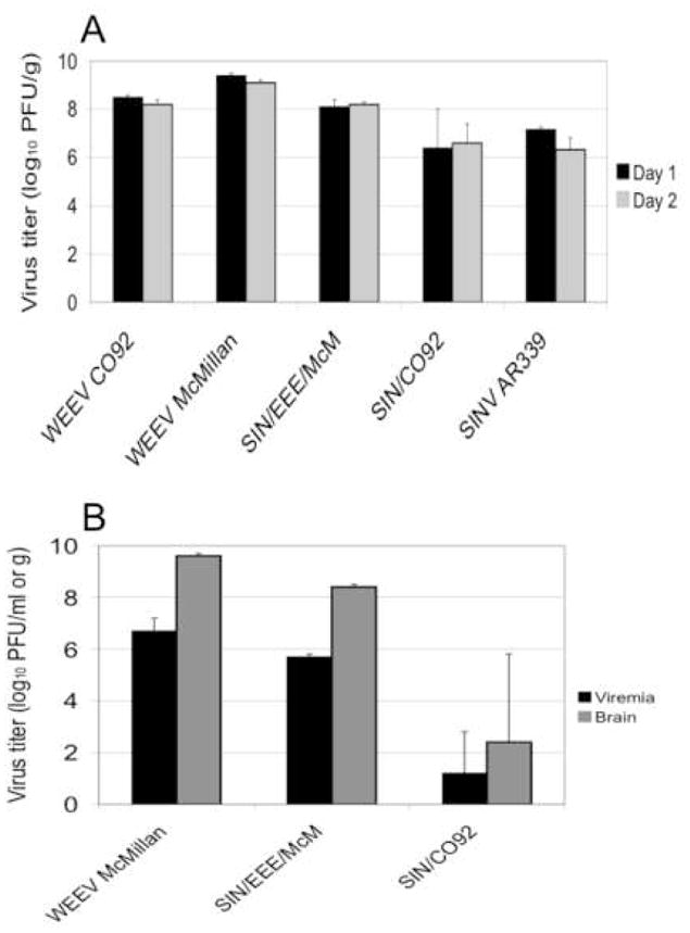
A) Mean virus titer in the brains of 6-day-old mice at days 1 and 2 after intracerebral infection with 5.0 log10 PFU of virus (as indicated on the x-axis). B) Mean viremia and virus titers in the brains of 4-day-old mice after subcutaneous infection with 4.6 log10 PFU of virus (as indicated on the x-axis). Two animals per cohort were sampled at each time point. Bars indicate standard deviations.
Evaluation of 4-day-old mice after SC infection also yielded evidence of attenuation of the WEEV vaccine candidates when compared to parental strain McMillan (Fig. 9B). Chimeras produced lower levels of viremia and virus in the brain than wild-type WEEV. Overall, the results of these experiments demonstrate that, although it is less immunogenic, SIN/CO92 is more attenuated than SIN/EEE/McM.
DISCUSSION
Advantages of the chimeric alphavirus vaccines
WEEV is an important emerging and reemerging veterinary pathogen in the New World, causing severe encephalitis in humans and horses. A formalin-inactivated WEE vaccine is available for veterinary use [28]; however, no human vaccine and antiviral drug is currently available. We used a live-attenuated chimeric alphavirus approach to develop a vaccine that would be: 1) inexpensive to manufacture, 2) effective after a single dose, 3) able to illicit long-lasting immunity without the need for frequent boosters, and 4) stably attenuated by virtue of the chimeric design that cannot easily revert to a wild-type alphavirus. We demonstrated here that chimeric WEE vaccines meet criteria 2 and 4. Our vaccine should also be inexpensive to manufacture because it replicated to high titer in cell culture, producing at least the equivalent of ca. 100–1,000 doses per ml of medium. Additional experiments are underway to assess the duration of immunity and the stability of attenuation after serial passages in mice. The live-attenuated TC-83 VEE vaccine strain produces long-lasting immunity in human vaccinees [29], suggesting that our live vaccine candidate should also. Our chimeric approach has also yielded promising alphavirus vaccine candidates for VEE [14, 15], EEE [16] and chikungunya [27].
Animal model development for vaccine testing
To define a robust animal model for vaccine testing, we compared the virulence of 3 strains of WEEV using 2 routes of infection and 4 mouse strains. Previous studies indicate that WEEV is generally less virulent in adult mice than the two other New World encephalitic alphaviruses (EEEV and VEEV) [30]. WEEV is uniformly only lethal in 3–5-week-old mice infected IP, and IN infection generally results in higher rates of mortality [25]. Depending on the WEEV strain, outbred adult mice infected SC show variable mortality [24]. In our study, we found that the WEEV strain McMillan killed 9–10-week-old female NIH Swiss mice by IP inoculation, and up to 4.5 mo old mice by IN infection (data not shown). Similar to WEEV strain McMillan, WEEV strain TBT235 also killed 9–10-week-old NIH Swiss mice within 4 days after IN infection. Therefore, WEEV strains McMillan and TBT235 were used for our vaccine challenge experiments and grouped into the high virulence phenotype as previously characterized for WEEV strains California and Fleming [25].
Genetic diversity among WEEV strains
By comparing the deduced amino acid sequences among the WEEV strains of this study, four common amino acid differences were identified between the low and high virulence strains. These amino acid differences occurred in both nonstructural and structural protein regions of the viral genome, including positions 334, 365, and 469 in the nsP3 and position 250 in the capsid. A similar study using low and high virulence WEEV strains also suggested that amino acid variation at position 250 in the capsid of WEEV correlates to mouse virulence at the molecular level [25]. By sequencing the complete genomes of the 3 WEEV strains used in our study, we were also able to identify amino acid substitutions in the nonstructural protein region that may be essential for WEEV infectivity. Reverse genetic studies are needed to further assess their relationship with WEEV virulence.
Evaluation of chimeric alphavirus vaccine candidates
Our study demonstrated that the first-generation chimeric SIN/CO92 vaccine, which derives its backbone from SINV and structural protein genes from the relatively benign WEEV strain CO92-1356, provides better cross-protection in mice than an adenovirus-based vaccine [8, 9]. Mice vaccinated with SIN/CO92 were completely protected when challenged IN with two virulent strains of WEEV at doses approximately 1.5-fold higher than those reported in the adenovirus studies [9]. However, because the SIN/CO92 strain required relatively high doses (≥5 log10 PFU/mouse) to achieve robust PRNT titers and complete protection against challenge, we selected the more virulent McMillan WEEV strain for the second-generation vaccine candidates with the expectation that it would be more immunogenic than the SIN/CO92 vaccine strain.
Initial data indicated that the SIN/SIN/McM strain was immunogenic but unacceptably virulent, killing two 6-week-old mice in a cohort of 10. In contrast, SIN/EEE/McM was also highly immunogenic but well attenuated in vaccinated adult mice, with little or no sign of disease. SIN/EEE/McM also provided complete protection against WEEV challenge after IN administration of virulent WEEV strain McMillan.
In conclusion, the SIN/EEE/McM candidate appears to be the efficacious of the 2 chimeric vaccine candidates while SIN/CO92 is less immunogenic but more attenuated. However, the possibility that this limitation in immunogenicity could be overcome with larger SIN/CO92 doses or with boosters should be considered. In addition to its favorable attenuation and immunogenicity profile for adult mice, SIN/EEE/McM also exhibits the important safety characteristic of being unable to be transmitted by the enzootic/epidemic North American mosquito vector, Cx. tarsalis [31]. Further evaluation of these vaccine strains in equids and nonhuman primates is warranted.
Acknowledgments
We thank Olga Petrakova for technical assistance on the first steps of the project. This work was supported by a grant from NIAID through the Western Regional Center of Excellence for Biodefense and Emerging Infectious Diseases Research, NIH grant number U54 AI057156 and R01AI070207 (IF).
Footnotes
Publisher's Disclaimer: This is a PDF file of an unedited manuscript that has been accepted for publication. As a service to our customers we are providing this early version of the manuscript. The manuscript will undergo copyediting, typesetting, and review of the resulting proof before it is published in its final citable form. Please note that during the production process errorsmaybe discovered which could affect the content, and all legal disclaimers that apply to the journal pertain.
References
- 1.Reisen WK. Western equine encephalitis. In: Service MW, editor. The Encyclopedia of Arthropod-transmitted Infections. Wallingford, UK: CAB International; 2001. pp. 558–63. [Google Scholar]
- 2.Schlesinger S, Schlesinger MJ. Togaviridae: The viruses and their replication. In: Knipe DM, Howley PM, editors. Fields' Virology. 4. New York: Lippincott, Williams and Wilkins; 2001. pp. 895–916. [Google Scholar]
- 3.Weaver SC, Kang W, Shirako Y, Rumenapf T, Strauss EG, Strauss JH. Recombinational history and molecular evolution of western equine encephalomyelitis complex alphaviruses. Journal of virology. 1997;71(1):613–23. doi: 10.1128/jvi.71.1.613-623.1997. [DOI] [PMC free article] [PubMed] [Google Scholar]
- 4.Reisen WK, Monath TP. Western equine encephalomyelitis. In: Monath TP, editor. The Arboviruses: Epidemiology and Ecology. V. Boca Raton, Florida: CRC Press; 1988. pp. 89–137. [Google Scholar]
- 5.Reed DS, Larsen T, Sullivan LJ, Lind CM, Lackemeyer MG, Pratt WD, et al. Aerosol exposure to western equine encephalitis virus causes fever and encephalitis in cynomolgus macaques. The Journal of infectious diseases. 2005 Oct 1;192(7):1173–82. doi: 10.1086/444397. [DOI] [PubMed] [Google Scholar]
- 6.Sidwell RW, Smee DF. Viruses of the Bunya- and Togaviridae families: potential as bioterrorism agents and means of control. Antiviral Res. 2003 Jan;57(1–2):101–11. doi: 10.1016/s0166-3542(02)00203-6. [DOI] [PubMed] [Google Scholar]
- 7.Nagata LP, Hu WG, Masri SA, Rayner GA, Schmaltz FL, Das D, et al. Efficacy of DNA vaccination against western equine encephalitis virus infection. Vaccine. 2005 Mar 18;23(17–18):2280–3. doi: 10.1016/j.vaccine.2005.01.032. [DOI] [PubMed] [Google Scholar]
- 8.Wu JQ, Barabe ND, Chau D, Wong C, Rayner GR, Hu WG, et al. Complete protection of mice against a lethal dose challenge of western equine encephalitis virus after immunization with an adenovirus-vectored vaccine. Vaccine. 2007 May 30;25(22):4368–75. doi: 10.1016/j.vaccine.2007.03.042. [DOI] [PubMed] [Google Scholar]
- 9.Barabe ND, Rayner GA, Christopher ME, Nagata LP, Wu JQ. Single-dose, fast-acting vaccine candidate against western equine encephalitis virus completely protects mice from intranasal challenge with different strains of the virus. Vaccine. 2007 Aug 14;25(33):6271–6. doi: 10.1016/j.vaccine.2007.05.054. [DOI] [PubMed] [Google Scholar]
- 10.Kostense S, Koudstaal W, Sprangers M, Weverling GJ, Penders G, Helmus N, et al. Adenovirus types 5 and 35 seroprevalence in AIDS risk groups supports type 35 as a vaccine vector. AIDS. 2004 May 21;18(8):1213–6. doi: 10.1097/00002030-200405210-00019. [DOI] [PubMed] [Google Scholar]
- 11.Vogels R, Zuijdgeest D, van Rijnsoever R, Hartkoorn E, Damen I, de Bethune MP, et al. Replication-deficient human adenovirus type 35 vectors for gene transfer and vaccination: efficient human cell infection and bypass of preexisting adenovirus immunity. Journal of virology. 2003 Aug;77(15):8263–71. doi: 10.1128/JVI.77.15.8263-8271.2003. [DOI] [PMC free article] [PubMed] [Google Scholar]
- 12.Mane VP, Toietta G, McCormack WM, Conde I, Clarke C, Palmer D, et al. Modulation of TNFalpha, a determinant of acute toxicity associated with systemic delivery of first-generation and helper-dependent adenoviral vectors. Gene Ther. 2006 Sep;13(17):1272–80. doi: 10.1038/sj.gt.3302792. [DOI] [PubMed] [Google Scholar]
- 13.Pedersen CE, Jr, Robinson DM, Cole FE., Jr Isolation of the vaccine strain of Venezuelan equine encephalomyelitis virus from mosquitoes in Louisiana. Am J Epidemiol. 1972;95(5):490–6. doi: 10.1093/oxfordjournals.aje.a121416. [DOI] [PubMed] [Google Scholar]
- 14.Paessler S, Fayzulin RZ, Anishchenko M, Greene IP, Weaver SC, Frolov I. Recombinant Sindbis/Venezuelan equine encephalitis virus is highly attenuated and immunogenic. Journal of virology. 2003 Sep;77(17):9278–86. doi: 10.1128/JVI.77.17.9278-9286.2003. [DOI] [PMC free article] [PubMed] [Google Scholar]
- 15.Paessler S, Ni H, Petrakova O, Fayzulin RZ, Yun N, Anishchenko M, et al. Replication and clearance of Venezuelan equine encephalitis virus from the brains of animals vaccinated with chimeric SIN/VEE viruses. Journal of virology. 2006 Mar;80(6):2784–96. doi: 10.1128/JVI.80.6.2784-2796.2006. [DOI] [PMC free article] [PubMed] [Google Scholar]
- 16.Wang E, Petrakova O, Adams AP, Aguilar PV, Kang W, Paessler S, et al. Chimeric Sindbis/eastern equine encephalitis vaccine candidates are highly attenuated and immunogenic in mice. Vaccine. 2007 Oct 23;25(43):7573–81. doi: 10.1016/j.vaccine.2007.07.061. [DOI] [PMC free article] [PubMed] [Google Scholar]
- 17.McKnight KL, Simpson DA, Lin SC, Knott TA, Polo JM, Pence DF, et al. Deduced consensus sequence of Sindbis virus strain AR339: mutations contained in laboratory strains which affect cell culture and in vivo phenotypes. J Virol. 1996;70(3):1981–9. doi: 10.1128/jvi.70.3.1981-1989.1996. [DOI] [PMC free article] [PubMed] [Google Scholar]
- 18.Aguilar PV, Adams AP, Wang E, Kang W, Carrara AS, Anishchenko M, et al. Structural and nonstructural protein genome regions of eastern equine encephalitis virus are determinants of interferon sensitivity and murine virulence. Journal of virology. 2008 May;82(10):4920–30. doi: 10.1128/JVI.02514-07. [DOI] [PMC free article] [PubMed] [Google Scholar]
- 19.Liljeström P, Lusa S, Huylebroeck D, Garoff H. In vitro mutagenesis of a full-length cDNA clone of Semliki Forest virus: the small 6,000-molecular-weight membrane protein modulates virus release. J Virol. 1991;65:4107–13. doi: 10.1128/jvi.65.8.4107-4113.1991. [DOI] [PMC free article] [PubMed] [Google Scholar]
- 20.Lemm JA, Durbin RK, Stollar V, Rice CM. Mutations which alter the level or structure of nsP4 can affect the efficiency of Sindbis virus replication in a host-dependent manner. J Virol. 1990;64:3001–11. doi: 10.1128/jvi.64.6.3001-3011.1990. [DOI] [PMC free article] [PubMed] [Google Scholar]
- 21.Beaty BJ, Calisher CH, Shope RE. Arboviruses. In: Lennete ET, Lennete DA, editors. Diagnostic procedures for viral, rickettsial and chlamydial infections. 7. Washington, D. C: American Public Health Association; 1995. pp. 189–212. [Google Scholar]
- 22.Bianchi TI, Aviles G, Sabattini MS. Biological characteristics of an enzootic subtype of western equine encephalomyelitis virus from Argentina. Acta Virol. 1997;41(1):13–20. [PubMed] [Google Scholar]
- 23.Hardy JL, Presser SB, Chiles RE, Reeves WC. Mouse and baby chicken virulence of enzootic strains of western equine encephalomyelitis virus from California. The American journal of tropical medicine and hygiene. 1997 Aug;57(2):240–4. doi: 10.4269/ajtmh.1997.57.240. [DOI] [PubMed] [Google Scholar]
- 24.Forrester NL, Kenney JL, Deardorff E, Wang E, Weaver SC. Western Equine Encephalitis submergence: lack of evidence for a decline in virus virulence. Virology. 2008 Oct 25;380(2):170–2. doi: 10.1016/j.virol.2008.08.012. [DOI] [PMC free article] [PubMed] [Google Scholar]
- 25.Nagata LP, Hu WG, Parker M, Chau D, Rayner GA, Schmaltz FL, et al. Infectivity variation and genetic diversity among strains of Western equine encephalitis virus. The Journal of general virology. 2006 Aug;87(Pt 8):2353–61. doi: 10.1099/vir.0.81815-0. [DOI] [PubMed] [Google Scholar]
- 26.Netolitzky DJ, Schmaltz FL, Parker MD, Rayner GA, Fisher GR, Trent DW, et al. Complete genomic RNA sequence of western equine encephalitis virus and expression of the structural genes. The Journal of general virology. 2000 Jan;81(Pt 1):151–9. doi: 10.1099/0022-1317-81-1-151. [DOI] [PubMed] [Google Scholar]
- 27.Wang E, Volkova E, Adams AP, Forrester N, Xiao SY, Frolov I, et al. Chimeric alphavirus vaccine candidates for chikungunya. Vaccine. 2008 Sep 15;26(39):5030–9. doi: 10.1016/j.vaccine.2008.07.054. [DOI] [PMC free article] [PubMed] [Google Scholar]
- 28.Barber TL, Walton TE, Lewis KJ. Efficacy of trivalent inactivated encephalomyelitis virus vaccine in horses. Am J Vet Res. 1978 Apr;39(4):621–5. [PubMed] [Google Scholar]
- 29.Pittman PR, Makuch RS, Mangiafico JA, Cannon TL, Gibbs PH, Peters CJ. Long-term duration of detectable neutralizing antibodies after administration of live-attenuated VEE vaccine and following booster vaccination with inactivated VEE vaccine. Vaccine. 1996 Mar;14(4):337–43. doi: 10.1016/0264-410x(95)00168-z. [DOI] [PubMed] [Google Scholar]
- 30.Liu C, Voth DW, Rodina P, Shauf LR, Gonzalez G. A comparative study of the pathogenesis of western equine and eastern equine encephalomyelitis viral infections in mice by intracerebral and subcutaneous inoculations. The Journal of infectious diseases. 1970;122(1):53–63. doi: 10.1093/infdis/122.1-2.53. [DOI] [PubMed] [Google Scholar]
- 31.Kenney JL, Frolov I, Weaver SC. Evaluation of Transmission Potential of Two Chimeric Western Equine Encephalitis Vaccine Candidates in Culex tarsalis. Am J Trop Med Hyg. 2009 doi: 10.4269/ajtmh.2010.09-0092. under review. [DOI] [PMC free article] [PubMed] [Google Scholar]


