Abstract
Clostridium botulinum is a spore-forming bacterium that can produce a very powerful neurotoxin that causes botulism. In this study, we have investigated the Fur transcription regulators in Clostridium botulinum and Fur-regulated genes in Clostridium botulinum A ATCC 3502. We found that gene loss may be the main cause leading to the different numbers of Fur transcription regulators in different Clostridium botulinum strains. Meanwhile, 46 operons were found to be regulated by the Fur transcription regulator in Clostridium botulinum A ATCC 3502, involved in several functional classifications, including iron acquisition, iron utilization, iron transport, and transcription regulator. Under an iron-restricted medium, we experimentally found that a Fur transcription regulator (CBO1372) and two operons (DedA, CBO2610–CBO2614 and ABC transporter, CBO0845–CBO0847) are shown to be differentially expressed in Clostridium botulinum A ATCC 3502. This study has provided-us novel insights into the diversity of Fur transcription regulators in different Clostridium botulinum strains and diversity of Fur-targeted genes, as well as a better understanding of the dynamic changes in iron restriction occurring in response to this stress.
1. Introduction
Iron has been documented to play important roles as an enzymatic cofactor and electron transport component in cellular processes in bacteria [1]. In addition, pathogenic bacteria often use iron as an environmental signal to regulate virulence genes, which encode different virulence determinants, such as toxins, enzymes, and adhesins [2]. It should be noted that the iron in the host usually bounds to host proteins and only a small fraction exists in free form, which is too low to support bacterial growth within the host. Therefore, pathogenic bacteria must compete with its host for the iron.
Iron concentration must be maintained as a delicate balance in the cell. DNA replication will be impeded with too little iron, while too much iron initiates the production of oxygen radicals which are fatal to the cell. In particular, iron is notorious in its ability to catalyze the formation of hydroxyl radicals that can cause cellular death [3]. Therefore, iron uptake must be tightly regulated to avoid the toxic effects of iron over accumulation.
To overcome the iron limitation, bacteria have developed an iron uptake systems to regulate the acquiring of iron from host [2]. The iron uptake systems are often regulated by the ferric uptake regulator (Fur) protein. Fur acts as a transcriptional repressor of iron-regulated genes and is responsible for the regulation of iron acquisition genes of iron uptake system. In Escherichia coli, Fur has been found to be responsible for the regulation of more than 30 genes [4].
The Fur protein has been demonstrated to work in various bacteria by binding intracellular iron as well as a sequence in the promoter region of Fur-Fe2+-regulated genes [5]. These genes are primarily iron acquisition genes that are downregulated when iron levels within the cell are sufficient. Fur protein binds the promoter only when sufficient iron is present within the cell. This binding causes a conformational change that prevents transcription of the regulated gene, thus acting as a repressor. When iron levels fall, Fe2+ dissociates from the Fur protein which then falls off the promoter and restores transcription. Additionally, Fur has been shown to regulate virulence and virulence-associated factors.
Clostridium botulinum is a spore-forming bacterium that produces a very powerful neurotoxin that causes botulism [6], which is among the most toxic of all naturally occurring substances. Botulism is usually associated with consumption of the toxin in food. Although the Fur gene family and Fur-binding sites have been well studied in many bacteria, such as Escherichia coli [7] and Aliivibrio salmonicida [8], less is known about their roles and targeted genes in Clostridium botulinum. In this study, we have firstly investigated the diversity of Fur genes and their evolutionary history in Clostridium botulinum. Then, we have predicted the Fur-binding sites and associated genes in Clostridium botulinum A ATCC 3502. Finally, we have detected the expression level of Fur transcription regulator and associated genes in Clostridium botulinum A ATCC 3502 under the iron-restricted condition.
2. Materials and Methods
2.1. Data Resource
The predicted proteins from eight Clostridium botulinum genomes were obtained from the KEGG database [9]. The eight genomes are Clostridium botulinum A ATCC 19397, Clostridium botulinum A ATCC 3502, Clostridium botulinum A Hall, Clostridium botulinum A3 Loch Maree, Clostridium botulinum B Eklund 17B, Clostridium botulinum B1 Okra, Clostridium botulinum E3, and Clostridium botulinum F Langeland.
2.2. Retrieval of Fur Protein and Phylogenetic Analysis
The identification of Fur transcription regulator was carried out using a local BLASTP program with stringent E-value of E−10. The query sequences were the Fur transcription regulator from a set of well-defined and putative Fur protein sequences (http://www.genome.jp/kegg-bin/get_htext?ko03000+K09825).
We exercised the multiple Fur transcription regulator alignment with ClustalX 1.84 program [10] by defaults. The phylogenetic tree was reconstructed using the neighbor-joining method in the MEGA 4.0 program [11]. Evaluation of the tree was carried out by 1,000 replicates of a bootstrapping test, and its visualization was performed using the MEGA 4.0 program.
2.3. Identification of Fur-Binding Sites and Regulated Genes
The sequences of Fur box binding sites validated in the studies from Pedersen et al. were used to generate a weight matrix [8]. The matrix was then used to be the input for the Patser program (http://gzhertz.home.comcast.net/) to search Fur-binding sites in Clostridium botulinum A ATCC 3502 genome sequences. All sites identified to surpass the threshold of score 5.0 were considered to be the candidate Fur-binding sites. The genes and operon annotation information of Clostridium botulinum A ATCC 3502 were obtained from MicrobesOnline [12] and the genes regulated by the Fur were identified according to the coordinates of the genes and candidate Fur-binding sites. In other words, if one gene was scanned to be at the downstream of a candidate Fur-binding site and both were on the same strand, the gene was thought to be regulated by the Fur-binding site.
The sequences of Fur-binding sites confirmed to regulate other genes were aligned by ClustalX 1.84 program [10], and then used to generate the sequence LOGO using the online WebLogo server [13]. The height of each letter indicates its relative abundance in bits at the according position.
2.4. Bacterial Strains, Growth Conditions and Real-Time Quantitative RT-PCR
Clostridium botulinum A ATCC 3502 strain was routinely grown in deoxygenated TPGY broth at 37°C under anaerobic conditions supplemented with 2.5 ug/mL erythromycin, 250 ug/mL cycloserine, or 15 ug/mL thiamphenicol.
Total RNA was isolated under the experimental conditions using the Trizol reagent (Invitrogen) according to the manufacturer's instructions. The total RNA concentration was evaluated by measurement of the absorption units at the wavelength of 260 nm (A260) using the NanoDrop 1000 Spectrophotometer. For real-time quantitative RT-PCR, two ug of total RNA was used and cDNA was synthesized for 1 h at 41°C with Superscript II Reverse Transcriptase (Invitrogen) and 1 pmol of hexamer oligonucleotide primers (pDN6, Roche). The real-time quantitative PCR reaction volume contains 1 μg cDNAs, 12 μL of the SYBR PCR master mix (Applied Biosystems), and 200 nM of gene-specific primers. Amplification and detection were similar as previous studies [14]. The expression level of each gene was normalized to the quantity of cDNAs of gyrA, which is shown to be stably expressed in our studies (data not shown). The relative expression level was calculated as the ratio of normalized target concentrations (ΔΔct) [15].
3. Results and Discussion
3.1. Distribution of Fur in Clostridium botulinum
Clostridium botulinum, a gram-positive and spore-forming anaerobic bacterium, can cause the severe neuroparalytic illness in humans that named botulism [16]. In this study, through exhaustive sequence similarity searches of the Clostridium botulinum genomes with a set of well-characterized and putative Fur transcription regulator, a total of 35 Fur sequences were retrieved from eight Clostridium botulinum strains (Table 1). Different Clostridium botulinum strain was revealed to have a close phylogenetic relationship with a similar one of genome size [6, 16]. Interestingly, different Clostridium botulinum strain was found to contain different number of Fur transcription regulators, including four in Clostridium botulinum A ATCC 19397, five in Clostridium botulinum A ATCC 3502, four in Clostridium botulinum A Hall, five in Clostridium botulinum A3 Loch Maree, three in Clostridium botulinum B Eklund 17B, six in Clostridium botulinum B1 Okra, three in Clostridium botulinum E3, and five in Clostridium botulinum F Langeland (Table 1). The protein of Fur gene identified from different Clostridium botulinum strain has a length range from 107 to 160 aa, while the genome size ranges from 3.66 Mb in Clostridium botulinum E3 to 3.99 Mb in Clostridium botulinum A3 Loch Maree.
Table 1.
The distribution of Fur in Clostridium botulinum.
| Locus tag | Species | Genome size | Protein (aa) |
|---|---|---|---|
| CLB_1396 | Clostridium botulinum A ATCC 19397 | 3.86 Mb | 157 |
| CLB_3256 | Clostridium botulinum A ATCC 19397 | 3.86 Mb | 142 |
| CLB_2774 | Clostridium botulinum A ATCC 19397 | 3.86 Mb | 155 |
| CLB_2501 | Clostridium botulinum A ATCC 19397 | 3.86 Mb | 150 |
| CBO2830 | Clostridium botulinum A ATCC 3502 | 3.89 Mb | 155 |
| CBO0395 | Clostridium botulinum A ATCC 3502 | 3.89 Mb | 138 |
| CBO3220 | Clostridium botulinum A ATCC 3502 | 3.89 Mb | 142 |
| CBO1372 | Clostridium botulinum A ATCC 3502 | 3.89 Mb | 157 |
| CBO2560 | Clostridium botulinum A ATCC 3502 | 3.89 Mb | 150 |
| CLC_2431 | Clostridium botulinum A Hall | 3.76 Mb | 150 |
| CLB_1407 | Clostridium botulinum A Hall | 3.76 Mb | 157 |
| CLC_3130 | Clostridium botulinum A Hall | 3.76 Mb | 155 |
| CLC_3130 | Clostridium botulinum A Hall | 3.76 Mb | 142 |
| CLK_2617 | Clostridium botulinum A3 Loch Maree | 3.99 Mb | 142 |
| CLK_3586 | Clostridium botulinum A3 Loch Maree | 3.99 Mb | 144 |
| CLK_1945 | Clostridium botulinum A3 Loch Maree | 3.99 Mb | 150 |
| CLK_0814 | Clostridium botulinum A3 Loch Maree | 3.99 Mb | 107 |
| CLK_2231 | Clostridium botulinum A3 Loch Maree | 3.99 Mb | 160 |
| CLL_A0771 | Clostridium botulinum B Eklund 17B | 3.80 Mb | 137 |
| CLL_A1178 | Clostridium botulinum B Eklund 17B | 3.80 Mb | 154 |
| CLL_A2882 | Clostridium botulinum B Eklund 17B | 3.80 Mb | 143 |
| CLD_3166 | Clostridium botulinum B1 Okra | 3.96 Mb | 157 |
| CLD_0353 | Clostridium botulinum B1 Okra | 3.96 Mb | 144 |
| CLD_1728 | Clostridium botulinum B1 Okra | 3.96 Mb | 160 |
| CLD_1312 | Clostridium botulinum B1 Okra | 3.96 Mb | 142 |
| CLD_1744 | Clostridium botulinum B1 Okra | 3.96 Mb | 155 |
| CLD_2005 | Clostridium botulinum B1 Okra | 3.96 Mb | 150 |
| CLH_1129 | Clostridium botulinum E3 | 3.66 Mb | 154 |
| CLH_0737 | Clostridium botulinum E3 | 3.66 Mb | 137 |
| CLH_2610 | Clostridium botulinum E3 | 3.66 Mb | 141 |
| CLI_2623 | Clostridium botulinum F Langeland | 3.99 Mb | 150 |
| CLI_2880 | Clostridium botulinum F Langeland | 3.99 Mb | 155 |
| CLI_3358 | Clostridium botulinum F Langeland | 3.99 Mb | 142 |
| CLI_1468 | Clostridium botulinum F Langeland | 3.99 Mb | 157 |
| CLI_0471 | Clostridium botulinum Langeland | 3.99 Mb | 144 |
3.2. The Evolutional History of Fur in Clostridium botulinum
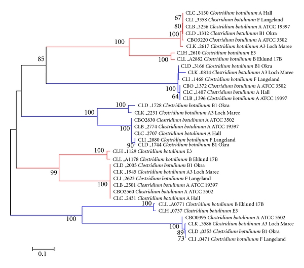
To examine the molecular evolutionary relationship of Fur genes in Clostridium botulinum, phylogenetic tree was constructed based on the multiple sequence alignments of the full-length proteins of identified Fur genes (Figure 1). On the evolutionary tree, we found that the Fur genes are grouped into eight clusters, most of which have 100% bootstrap support, except one of them that has a bootstrap value of 99%. Detailed observation of each cluster shows only two one-on-one monophyletic clusters. In other words, the strains of both the two clusters contain eight Fur genes, which are from eight different Clostridium botulinum strains. The presence of the one-on-one monophyletic clusters in the total 5 clusters uncovered a highly conservative property of the Fur genes of eight strains in these two clusters. The rest of the three clusters missed one or two strains, indicating the evolutionary diversity of Fur genes in Clostridium botulinum, which may be the loss of specific Fur genes after its separation from their common ancestor.
Figure 1.

The phylogenetic tree of Fur transcription regulators in Clostridium botulinum.
One of the most common origins of the divergent number of gene family members in different species is the gene duplication event [17]. The Clostridium botulinum strains shown in five clusters all contain at most one copy of Fur genes, indicating that most of the Fur gene family members are present in their common ancestor and then experience sequence divergence after the divergence of the common ancestor. It is interesting that no gene duplication event was observed in the phylogenetic tree and several Fur genes are located within separate clusters, which indicates that the expansion of Fur genes in Clostridium botulinum is not due to a simple intragenic duplication. Horizontal gene transfer (HGT) event is reported to be an important source to obtain new genes among bacteria species [18]. To test whether genes of these separate clusters were acquired through HGT, we took Fur genes from all the other bacteria into account (as described in Section 2). Then, a phylogenetic tree of all the Fur genes of Clostridium botulinum and other bacteria species were constructed (data not shown). On the tree, all the Clostridium botulinum specific separate clusters are located within group of Clostridium botulinum species. Therefore, neither HGT nor gene duplication event could be regarded as the major reason that caused the number divergence of Fur genes among Clostridium botulinum genera.
3.3. The Fur-Regulated Genes in Clostridium botulinum A ATCC 3502
To shed more lights on the important roles of Fur transcription regulator, we performed the investigation of Fur-regulated genes in Clostridium botulinum. In order to identify the Fur-targeted genes in Clostridium botulinum A ATCC 3502, a genomewide search for conserved Fur-binding sites was carried out using the computational methods [19]. The strategy involves the following. (1) A Fur-binding siteweight matrix was derived from a pool of recognized Fur-binding sites validated in the previous studies. (2) The matrix was then used to search the potential Fur-binding sites on the complete genome of Clostridium botulinum A ATCC 3502 (Figure S1 of the supplementary material available online at doi:10.1155/2011/934756). Subsequently, sites identified to be upstream of the conserved Clostridium botulinum A ATCC 3502 genes which encode Fur-regulated functions were confirmed to be Fur-binding sites. After reducing potential false positives, a set of 46 candidate sites selected for further analysis (Table S1). The genes identified to be regulated by the set of 46 Fur-binding sites consist of 155 genes, which are organized into 46 operons and classified into five mainly function categories (iron acquisition, iron utilization, iron transport, transcription regulator, and unclassified) (Table S1).
Iron acquisition includes an iron-binding protein (CBO0203), a thioredoxin protein (trxC), and two iron-sulfur proteins (CBO261 and CBO0853). In addition, other proteins associated with iron acquisition are also included, such as glycosyl transferase, butyrate kinase, phosphate butyryltransferase, isoaspartyl dipeptidase, fumarate hydratase and 4Fe-4S cluster, and ATPase protein.
Iron utilization contains several genes predicted to encode Fe-S-binding proteins, including 2-ketoisovalerate ferredoxin reductase, ferredoxin-NADP(+) reductase, FMN-dependent dehydrogenase, dihydropyrimidine dehydrogenase, iron-only hydrogenase, and pyruvate formate lyase. Oxidoreductases, such as pyruvate-flavodoxin oxidoreductase, flavodoxin oxidoreductase, and pyridine nucleotide-disulphide oxidoreductase, are also regulated.
ABC transporters were identified to belong to diverse functional families, including two distinct efflux permeases of the ATP-dependent family of transmembrane efflux pumps. One of these systems consists of adjacent genes encoding, respectively. The most abundant proteins of this kind are membrane protein and ATP-binding protein.
In addition, interestingly, several genes involved in flagellar system and cell division were identified to be regulated by Fur transcription regulator. However, further experiment investigation and their possible roles in Fur-regulated activities remains to be addressed.
3.4. The Change of Genes under Iron Restriction in Clostridium botulinum A ATCC 3502
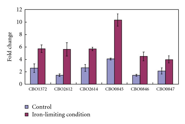
In bacteria, iron has been indicated to play important roles as an enzymatic cofactor and electron transport component in cellular processes. Low availability of iron in the encountered environment represents a major stress of bacterial growth and is often considered a signal leading to significant changes in cell processes [20]. Previous studies suggested that, under iron-limiting condition, bacteria can differentially express about 10–20% of their genes in order to response to the iron starvation stress [21–23]. To extend our knowledge of the dynamic gene expression induced in Clostridium botulinum A ATCC 3502 by iron treatment, the strain was grown in BHI broth with FeCl3 and with iron chelator HBED (ethylenediamine-N,N′-diacetic acid). Under both conditions, we have employed the real-time quantitative RT-PCR method to detect the expression situation of Fur transcription regulators (CBO2830, CBO3220, CBO0395, CBO1372, and CBO2560) in Clostridium botulinum A ATCC 3502 (Figure 2). It is found that, two Fur transcription regulators (CBO2830 and CBO0395) are absent in both conditions examined in the study. The other two Fur transcription regulators (CBO3220 and CBO2560) show no significant difference of the expression level although they are expressed. However, interestingly, we found that a Fur transcription regulator (CBO1372) has upregulated about threefold under iron-limiting condition in Clostridium botulinum A ATCC 3502. Thus, we speculated that the Fur transcription regulator CBO1372 may play important roles in iron starvation stress response.
Figure 2.

The differentially expressed genes under the iron-limitation condition in Clostridium botulinum A ATCC 3502.
To further investigate the regulation role of Fur transcription regulator CBO1372 and its targeted genes, 12 predicted Fur-targeted genes were randomly selected to perform the real-time quantitative RT-PCR analysis. As a result, we found that there are two genes (CBO2612 and CBO0846) that exhibited an expression level change larger than twofold under iron-limiting condition (Figure 2). Subsequently, we further employed the real-time quantitative RT-PCR to detect the expression level of the genes located in the same operon of the two differentially expressed genes. It is found that there are three additional genes (CBO2614, CBO0845, and CBO0847) that show significantly differential expression under the iron-restricted medium. Based on these results, it is considered that the DedA operon consisting of CBO2610–CBO2614 and the ABC transporter consisting of CBO0845–CBO0847 may be the targeted genes of Fur transcription regulator CBO1372 under the iron-limiting condition in Clostridium botulinum A ATCC 3502.
Supplementary Material
Figure S1. The logo of Fur binding sites in Clostridium botulinum A ATCC 3502.
Table S1. The putative association genes of Fur binding sites in Clostridium botulinum A ATCC 3502.
Acknowledgment
Funding for this study was provided by Zhejiang Gongshang University, China.
References
- 1.Wandersman C, Stojiljkovic I. Bacterial heme sources: the role of heme, hemoprotein receptors and hemophores. Current Opinion in Microbiology. 2000;3(2):215–220. doi: 10.1016/s1369-5274(00)00078-3. [DOI] [PubMed] [Google Scholar]
- 2.Braun V. Iron uptake mechanisms and their regulation in pathogenic bacteria. International Journal of Medical Microbiology. 2001;291(2):67–79. doi: 10.1078/1438-4221-00103. [DOI] [PubMed] [Google Scholar]
- 3.Halliwell B, Gutteridge JMC. Oxygen toxicity, oxygen radicals, transition metals and disease. Biochemical Journal. 1984;219(1):1–14. doi: 10.1042/bj2190001. [DOI] [PMC free article] [PubMed] [Google Scholar]
- 4.Vassinova N, Kozyrev D. A method for direct cloning of Fur-regulated genes: Identification of seven new Fur-regulated loci in Escherichia coli. Microbiology. 2000;146(12):3171–3182. doi: 10.1099/00221287-146-12-3171. [DOI] [PubMed] [Google Scholar]
- 5.Pohl E, Haller JC, Mijovilovich A, Meyer-Klaucke W, Garman E, Vasil ML. Architecture of a protein central to iron homeostasis: crystal structure and spectroscopic analysis of the ferric uptake regulator. Molecular Microbiology. 2003;47(4):903–915. doi: 10.1046/j.1365-2958.2003.03337.x. [DOI] [PubMed] [Google Scholar]
- 6.Fang PK, Raphael BH, Maslanka SE, Cai S, Singh BR. Analysis of genomic differences among Clostridium botulinum type A1 strains. BMC Genomics. 2010;11, article 725 doi: 10.1186/1471-2164-11-725. [DOI] [PMC free article] [PubMed] [Google Scholar]
- 7.Chen Z, Lewis KA, Shultzaberger RK, et al. Discovery of Fur binding site clusters in Escherichia coli by information theory models. Nucleic Acids Research. 2007;35(20):6762–6777. doi: 10.1093/nar/gkm631. [DOI] [PMC free article] [PubMed] [Google Scholar]
- 8.Pedersen HL, Ahmad R, Riise EK, et al. Experimental and computational characterization of the ferric uptake regulator from Aliivibrio salmonicida (Vibrio salmonicida) Journal of Microbiology. 2010;48(2):174–183. doi: 10.1007/s12275-010-9199-5. [DOI] [PubMed] [Google Scholar]
- 9.Kanehisa M, Subramaniam The KEGG database. Novartis Foundation Symposium. 2002;247:91–103. [PubMed] [Google Scholar]
- 10.Thompson JD, Gibson TJ, Higgins DG. Multiple sequence alignment using ClustalW and ClustalX. Current Protocols in Bioinformatics. doi: 10.1002/0471250953.bi0203s00. Chapter 2, Unite 2.3, 2002. [DOI] [PubMed] [Google Scholar]
- 11.Tamura K, Dudley J, Nei M, Kumar S. MEGA4: Molecular Evolutionary Genetics Analysis (MEGA) software version 4.0. Molecular Biology and Evolution. 2007;24(8):1596–1599. doi: 10.1093/molbev/msm092. [DOI] [PubMed] [Google Scholar]
- 12.Alm EJ, Huang KH, Price MN, et al. The MicrobesOnline Web site for comparative genomics. Genome Research. 2005;15(7):1015–1022. doi: 10.1101/gr.3844805. [DOI] [PMC free article] [PubMed] [Google Scholar]
- 13.Crooks GE, Hon G, Chandonia JM, Brenner SE. WebLogo: a sequence logo generator. Genome Research. 2004;14(6):1188–1190. doi: 10.1101/gr.849004. [DOI] [PMC free article] [PubMed] [Google Scholar]
- 14.Milohanic E, Glaser P, Coppée J-Y, et al. Transcriptome analysis of Listeria monocytogenes identifies three groups of genes differently regulated by PrfA. Molecular Microbiology. 2003;47(6):1613–1625. doi: 10.1046/j.1365-2958.2003.03413.x. [DOI] [PubMed] [Google Scholar]
- 15.Livak KJ, Schmittgen TD. Analysis of relative gene expression data using real-time quantitative PCR and the 2-ΔΔCT method. Methods. 2001;25(4):402–408. doi: 10.1006/meth.2001.1262. [DOI] [PubMed] [Google Scholar]
- 16.Brüggemann H, Gottschalk G. Comparative genomics of clostridia: link between the ecological niche and cell surface properties. Annals of the New York Academy of Sciences. 2008;1125:73–81. doi: 10.1196/annals.1419.021. [DOI] [PubMed] [Google Scholar]
- 17.Serres MH, Kerr ARW, McCormack TJ, Riley M. Evolution by leaps: gene duplication in bacteria. Biology Direct. 2009;4, article no. 46 doi: 10.1186/1745-6150-4-46. [DOI] [PMC free article] [PubMed] [Google Scholar]
- 18.Dutta C, Pan A. Horizontal gene transfer and bacterial diversity. Journal of Biosciences. 2002;27(1):27–33. doi: 10.1007/BF02703681. [DOI] [PubMed] [Google Scholar]
- 19.Quatrini R, Lefimil C, Veloso FA, Pedroso I, Holmes DS, Jedlicki E. Bioinformatic prediction and experimental verification of Fur-regulated genes in the extreme acidophile Acidithiobacillus ferrooxidans. Nucleic Acids Research. 2007;35(7):2153–2166. doi: 10.1093/nar/gkm068. [DOI] [PMC free article] [PubMed] [Google Scholar]
- 20.Merrell DS, Thompson LJ, Kim CC, et al. Growth phase-dependent response of Helicobacter pylori to iron starvation. Infection and Immunity. 2003;71(11):6510–6525. doi: 10.1128/IAI.71.11.6510-6525.2003. [DOI] [PMC free article] [PubMed] [Google Scholar]
- 21.Deslandes V, Nash JHE, Harel J, Coulton JW, Jacques M. Transcriptional profiling of Actinobacillus pleuropneumoniae under iron-restricted conditions. BMC Genomics. 2007;8, article no. 72 doi: 10.1186/1471-2164-8-72. [DOI] [PMC free article] [PubMed] [Google Scholar]
- 22.Ducey TF, Carson MB, Orvis J, Stintzi AP, Dyer DW. Identification of the iron-responsive genes of Neisseria gonorrhoeae by microarray analysis in defined medium. Journal of Bacteriology. 2005;187(14):4865–4874. doi: 10.1128/JB.187.14.4865-4874.2005. [DOI] [PMC free article] [PubMed] [Google Scholar]
- 23.Palma M, Worgall S, Quadri LEN. Transcriptome analysis of the Pseudomonas aeruginosa response to iron. Archives of Microbiology. 2003;180(5):374–379. doi: 10.1007/s00203-003-0602-z. [DOI] [PubMed] [Google Scholar]
Associated Data
This section collects any data citations, data availability statements, or supplementary materials included in this article.
Supplementary Materials
Figure S1. The logo of Fur binding sites in Clostridium botulinum A ATCC 3502.
Table S1. The putative association genes of Fur binding sites in Clostridium botulinum A ATCC 3502.


