Abstract
Background/Aims
Interferon-γ-inducible protein 10 (IP-10) plays important roles in the pathogenesis of hepatitis C virus (HCV) infection. We investigated the association between serum IP-10 levels and liver pathology in patients with chronic HCV infection.
Methods
The serum IP-10 concentration was assessed in 85 patients with chronic HCV infection using a solid phase sandwich enzyme-linked immunosorbent assay, and a liver biopsy specimen was obtained. The pathology was scored using the Knodell histologic activity index (HAI).
Results
Of the 85 patients, 58 had genotype 1 HCV infection, 21 had genotype non-1, and 6 were undetermined. The serum IP-10 levels did not differ between patients infected with genotype 1 and genotype non-1 (p=0.472). In patients with genotype 1 infection, the total HAI score and the stage of fibrosis were highly correlated with the serum IP-10 level (r=0.555, r=0.578, p<0.001). Furthermore, the serum IP-10 concentrations of patients with severe fibrosis (stages 3, 4) were higher than those of patients with mild fibrosis (stages 0 to 2; 214.4 vs. 72.3 pg/mL, p=0.002) among patients with genotype 1 infection. However, in patients without genotype 1 infection, the histopathology was not associated with the serum IP-10 level. A multivariate analysis showed that serum IP-10 was an independent predictor of fibrosis (stages 3, 4) in patients with genotype 1 infection (odds ratio, 1.034; 95% confidence interval, 1.006 to 1.064; p=0.018).
Conclusions
Serum IP-10 concentration was significantly correlated with the severity of liver histology in genotype 1 HCV infection.
Keywords: Chronic hepatitis C, Fibrosis, Interferon-γ-inducible protein 10
INTRODUCTION
The immunological and pathological mechanisms of liver injury caused by the hepatitis C virus (HCV) remain unclear. Several reports have suggested that HCV-induced hepatic injuries result from the lysis of virus-infected cells by the host immune system, T cells and some cytokines, rather than direct destruction by HCV.1-3 The host immune response to HCV infection can lead to 2 opposite results: spontaneous eradication or chronic HCV infection. A powerful and specific cellular response may clear HCV, but most patients fail to eradicate HCV and become chronically infected with the virus. Persistent HCV infection results in chronic inflammation and fibrosis of the liver, cirrhosis, and hepatocellular carcinoma. Of the patients with chronic HCV infection, 20% progress to cirrhosis and approximately 4% progress to hepatocellular carcinoma.
Chemokines are relatively small cytokines that play various roles under physiological and pathological conditions. They are involved in the immune response, tumor development and progression, and tissue regeneration.4 Chemokines are divided into 4 families according to 2 characteristic N-terminal cysteine residues: CC, CXC, CX3C, and C.5 Chemokine receptors are expressed mainly in leukocytes, and the interaction of chemokines with their receptors induces leukocyte migration and activation.
Many chemokines and chemokine receptors are involved in immune reactions in response to HCV infection. Of these, the non-EXR (Glu-Leu-Arg)-CXC chemokines and CC chemokines may be particularly important in the pathogenesis of HCV infection.5-8 For example, the mRNA of CXCL9, CXCL10 (IP-10), and CXCL11, which are non-EXR-CXC chemokines, and the mRNA of their receptor CXCR3 are highly expressed in HCV infection.9,10 In addition, the mRNA expression of CCL2, CCL3, and CCL5, which are CC chemokines, and their receptors, CCR2 and CCR5, are elevated in HCV infection.9-11 CXCR3 is expressed on activated T and natural killer (NK) cells, and CCR5 is expressed on activated memory T cells.6 Interferon-γ-inducible protein-10 kDa (IP-10/CXCL10) is a non-EXR-CXC chemokine12,13 and is a chemo-attractant for T lymphocytes, monocytes, and NK cells.14,15 Recently, it was found that the expression of IP-10 in liver specimens from patients with chronic hepatitis C was higher than in patients with chronic hepatitis B and normal healthy controls.16 Furthermore, the expression of IP-10 mRNA by hepatocytes strongly correlated with the serum IP-10 levels.17 These studies suggest that IP-10 may be involved in the immune response to chronic hepatitis C. In this study, we evaluated the possible association between serum IP-10 levels and liver histology in terms of inflammation and fibrosis to examine the role of IP-10 in patients with chronic hepatitis C.
MATERIALS AND METHODS
1. Patients
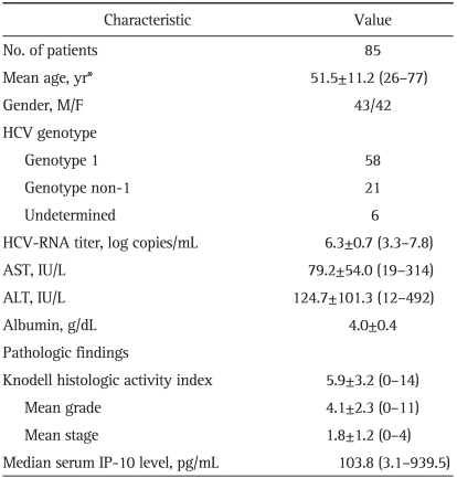
Eighty-five patients with chronic hepatitis C were enrolled in this study. All of the patients were adults (median age, 52 years; range, 26 to 77 years), and consisted of 43 men and 42 women. The diagnosis of chronic hepatitis C was based on seropositivity for anti-HCV antibody using third-generation enzyme immunoassay, the confirmation of HCV-RNA using reverse transcription-polymerase chain reaction (RT-PCR), and the histology of liver biopsy specimens. Patients with a history of alcohol abuse (more than 60 g/day) or taking herbal medicine and those diagnosed with hepatic failure (hepatic encephalopathy within 3 months after onset of jaundice), decompensated cirrhosis, hepatocellular carcinoma, other malignant tumors, or severe systemic diseases were excluded from the study. In addition, patients were excluded if they had taken immunosuppressants within the previous 6 months. The baseline characteristics of the patients are listed in Table 1.
Table 1.
Baseline Characteristics of the Patients
Data are presented as mean±SD (range).
HCV, hepatitis C virus; AST, aspartate transaminase; ALT, alanine transaminase; IP-10, interferon-γ-inducible protein 10.
2. Laboratory tests and HCV-RNA quantification
Within 2 weeks before the liver biopsy, HCV-RNA quantification and other laboratory tests, including liver function tests, were conducted. HCV-RNA titer was determined using RT-PCR hybridization (lower limit 1.62×10³ copies/mL).
3. Liver biopsy
Liver biopsy tissue was obtained from all patients with an ultrasound-guided percutaneous approach. An experienced pathologist evaluated and scored the biopsy specimens according to the Knodell histologic activity index (HAI). The Knodell HAI score (0 to 22 points) consists of grade of inflammation and necrosis (range 0 to 18: piecemeal necrosis, 0 to 10; intralobular degeneration, 0 to 4; and portal inflammation, 0 to 4) and stage of fibrosis (range 0 to 4: no fibrosis, 0; portal fibrosis, 1; periportal fibrosis, 2; septal fibrosis, 3; cirrhosis, 4). The degree of fibrosis was classified into mild (stages 0 to 2) and severe (stages 3, 4) fibrosis.
4. IP-10 quantification
Serum samples for IP-10 quantification were obtained on the day of the liver biopsy and then stored at -70℃ until analysis. The level of secreted IP-10 was measured using a DuoSet ELISA Development kit (R&D Systems, Minneapolis, MN, USA) according to the manufacturer's protocol.
5. Statistical analyses
The Spearman rank test was used to analyze the correlations between serum IP-10 levels and histopathology, HCV-RNA titer, and laboratory results. Logistic regression was used in univariate and multivariate analyses to determine factors related to severe fibrosis. p-values of less than 0.05 were deemed to be significant. All statistical analyses were conducted using SPSS version 14.0 for Windows (SPSS Inc., Chicago, IL, USA).
RESULTS
The median serum IP-10 level for the 85 patients was 103.8 pg/mL (range, 3.1 to 939.5). The serum IP-10 level was not related to age (≤50 years vs. >50 years, 132.1±182.9 vs. 116.4±76.8 pg/mL, p=0.587) or gender (male vs. female, 134.8±163.3 vs. 110.3±81.3 pg/mL, p=0.386).
1. Genotype of HCV and HCV RNA
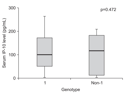
Of the 85 patients, 58 had genotype 1 HCV infection, 21 had non-1 genotypes, and 6 were undetermined. Serum IP-10 levels were similar in HCV patients with genotype 1 and non-1 infections (130.2±148.5 vs. 105.6±76.7 pg/mL, p=0.472, Fig. 1). The mean HCV-RNA titer was 6.3±0.7 log copies/mL (range, 3.3 to 7.8 log copies/mL). The HCV-RNA titer did not correlate with serum IP-10 levels (r=0.063; p=0.579).
Fig. 1.
Serum interferon-γ-inducible protein 10 (IP-10) concentrations did not differ significantly between patients with genotype 1 (n=58) and those without genotype 1 (n=21).
2. Association between histological severity and serum IP-10 level in genotype 1
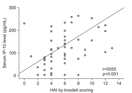
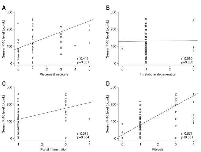
For the 58 patients with HCV genotype 1, a positive correlation between serum IP-10 concentration and the Knodell HAI score was identified (r=0.555, p<0.001, Fig. 2). In terms of the relationship between the grade of necroinflammation and the serum IP-10 concentration in genotype 1 patients, the serum IP-10 concentration correlated with the degree of piecemeal necrosis (r=0.416, p=0.001) and portal inflammation (r=0.381, p=0.004), but not intralobular degeneration (r=0.060, p=0.660, Fig. 3). There was also a significant correlation between the serum IP-10 concentration and the stage of fibrosis (r=0.577, p<0.001, Fig. 3).
Fig. 2.
The serum interferon-γ-inducible protein 10 (IP-10) concentration positively correlated with histopathological severity in patients with genotype 1 hepatitis C virus infection.
HAI, histologic activity index.
Fig. 3.
The correlations between serum interferon-γ-inducible protein 10 (IP-10) concentration and histopathological findings in patients with genotype 1 hepatitis C virus infection: piecemeal necrosis (A), intralobular degeneration (B), portal inflammation (C), and fibrosis (D). In patients with genotype 1 HCV infection, the serum IP-10 concentration was significantly correlated with the scores for piecemeal necrosis, portal inflammation, and fibrosis (A, C, and D).
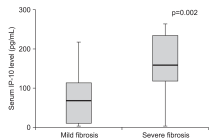
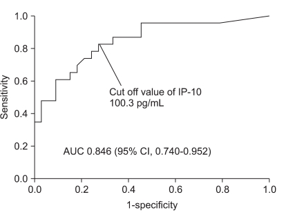
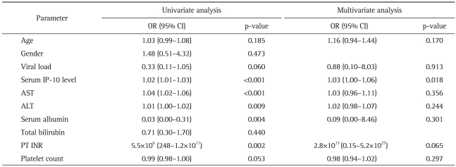
Of the patients infected with HCV genotype 1, the serum IP-10 concentration in the patients with severe fibrosis (stages 3, 4) was significantly higher than in the patients with mild fibrosis (stages 0 to 2, 214.4 vs. 72.3 pg/mL, p=0.002, Fig. 4). The receiver operating characteristic (ROC) curve for severe fibrosis in genotype 1-infected patients had an area under the curve (AUC) of 0.846 (95% confidence interval [CI], 0.740 to 0.952, Fig. 5). The cutoff serum IP-10 level for severe fibrosis in genotype 1-infected patients was 100.3 pg/mL (sensitivity 82.6%, specificity 72.7%). In a multivariate analysis of factors related to severe fibrosis, the serum IP-10 level was an independent predictor of fibrosis in genotype 1-infected patients (odds ratio, 1.034; 95% CI, 1.006 to 1.064; p=0.018, Table 2).
Fig. 4.
The serum interferon-γ-inducible protein 10 (IP-10) concentrations were higher in patients with severe fibrosis (stages 3, 4) than in patients with mild fibrosis (stages 0 to 2, 214.4 vs 72.3 pg/mL, p=0.002).
Fig. 5.
The receiver operating characteristic (ROC) curve of the serum interferon-γ-inducible protein 10 (IP-10) concentrations in patients with genotype 1 infection and severe fibrosis (stages 3, 4). The area under the curve (AUC) was 0.846 (95% confidence interval, 0.740 to 0.952), and the cutoff value of the serum IP-10 concentration was 100.3 pg/mL (sensitivity, 82.6%; specificity, 72.7%).
Table 2.
Factors Related to Severe Fibrosis in Genotype 1 HCV Infection
HCV, hepatitis C virus; OR, odds ratio; CI, confidence interval; IP-10, interferon-γ-inducible protein 10; AST, aspartate transaminase; ALT, alanine transaminase; PT INR, prothrombin time international normalized ratio.
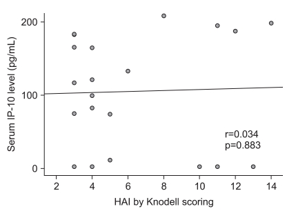
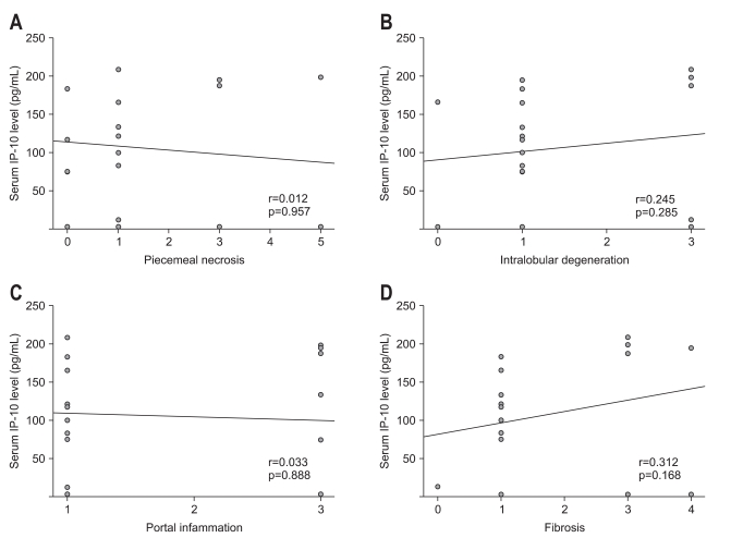
In contrast to patients with genotype-1 HCV infection, no significant correlation was observed between serum IP-10 concentrations and Knodell HAI scores in the 21 patients with genotype non-1 HCV infection (r=0.034, p=0.883, Fig. 6). The serum IP-10 concentration did not correlate with the degree of piecemeal necrosis, intralobular degeneration, portal inflammation, or stage of fibrosis (Fig. 7).
Fig. 6.
The serum interferon-γ-inducible protein 10 (IP-10) concentration did not correlate with the severity of pathology in patients with genotype non-1 hepatitis C virus infection.
HAI, histologic activity index.
Fig. 7.
The correlations between the serum interferon-γ-inducible protein 10 (IP-10) concentration and pathology findings in patients with genotype non-1 hepatitis C virus infection: piecemeal necrosis (A), intralobular degeneration (B), portal inflammation (C), and fibrosis (D). In these patients, the serum IP-10 concentration did not correlate with the scores for piecemeal necrosis, intralobular degeneration, portal inflammation, or fibrosis.
3. Association between liver enzyme and peripheral blood mononuclear cell count and serum IP-10 level
In the patients with genotype-1 HCV infection, as the serum aspartate aminotransferase (AST) and alanine aminotransferase (ALT) levels became elevated, the serum IP-10 concentration also increased, and these correlations were statistically significant (r=0.463, p<0.001; r=0.484, p<0.001, respectively). The serum IP-10 concentration of the patients with AST >100 IU/L was higher than that of the patients with AST ≤100 IU/L (median, 155.4 vs. 83.1 pg/mL, p=0.048). A similar result was obtained for the serum ALT (median, 140.8 vs. 80.5 pg/mL, p=0.014). A slightly positive (statistically nonsignificant) correlation was observed between the peripheral lymphocyte count and serum IP-10 concentration (r=0.250, p=0.058).
In patients with genotype non-1 HCV infection, serum IP-10 concentrations did not correlate with the peripheral lymphocyte count (r=0.117, p=0.615) or with serum AST (r=0.402, p=0.071) or ALT (r=0.233, p=0.308) levels.
DISCUSSION
IP-10 is a CXC chemokine. Unlike other CXC chemokines, IP-10 lacks chemotactic activity for neutrophils, but instead targets T lymphocytes, NK cells, and monocytes.14,15,18 According to recent data, the transcription of IP-10 in hepatocytes and the expression of the IP-10 receptor CXCR3 on lymphocytes infiltrating the liver are increased in patients with chronic HCV infection.11,19 The serum IP-10 levels in patients with HCV infection are higher than in healthy controls, patients with hepatitis B virus infection, and rheumatoid arthritis patients.16,19 These results imply that IP-10 plays an important role in the pathogenesis of HCV infection. In HCV infection, the inflammatory cells that infiltrate the liver are mainly antigen-nonspecific T-helper 1 (Th1) lymphocytes.20,21 These cells secrete cytokines such as interferon-gamma (IFN-γ) and IL-2, which activate monocytes and macrophages. In other words, Th1 lymphocytes initiate a delayed hypersensitivity reaction by producing IFN-γ and IL-2.22-24 This immune reaction results in ongoing tissue damage and progressive liver disease. IP-10 is produced by hepatocytes25,26 and recruits stimulated CD4+ T cells, NK cells, and monocytes to the target site, rather than CD8+ T cells and neutrophils.14,15,18 IP-10 may be involved in the Th1-dominant immune response of chronic hepatitis C.
In our study, the serum IP-10 level correlated with the histological severity of necroinflammation and fibrosis in patients with chronic genotype 1 HCV infection. Interestingly, the increasing of serum IP-10 level was independently related to severe fibrosis in genotype 1-infected patients. Thus, the IP-10 level is closely related to the progression of fibrosis in chronic HCV-mediated disease. A higher concentration of serum IP-10 may recruit more CD4+ T cells to the liver, inducing more vigorous immune reactions. This is supported by the correlation between the serum IP-10 level and the liver enzyme levels. Additionally, our results may indicate a potential association between activation of hepatic stellate cells and chemokines. The activation of hepatic stellate cells is known to be a key pathogenic feature of liver cirrhosis. Many cytokines, platelet-derived growth factors, and chemokines are involved in the activation of hepatic stellate cells.27 However, whether hepatic stellate cells express a specific receptor for IP-10 is unclear. It is a new challenge to discover the exact mechanism as to how IP-10 is involved in hepatic fibrosis.
Several other studies have demonstrated the relationships between the pathological findings and serum IP-10 levels.27,28 Diago et al.27 suggested that the serum IP-10 levels are higher in patients with advanced fibrosis than in those with mild fibrosis with chronic HCV infection, and that this relationship is more significant for genotype 1. Romero et al.28 demonstrated a correlation between IP-10 levels and necroinflammatory activity and fibrosis stage in patients with chronic hepatitis C. Recently, not only serum IP-10, but also intrahepatic IP-10, was reported to correlate with hepatic inflammation and fibrosis in chronic hepatitis C.5 Moreover, elevated intrahepatic mRNA expression of IP-10 in chronic HCV-infected patients was found to be associated with increased necroinflammation and fibrosis.5 This result suggests that serum IP-10 levels correlate with intrahepatic IP-10 level, as suggested by Mihm et al.17 The findings of the three studies mentioned above are consistent with our results. Furthermore, in our study, we evaluated the association between serum IP-10 and the severity of histology in patients with genotype non-1 HCV infection. The result was that serum IP-10 in patients with genotype non-1 HCV infection was not correlated with the severity of necroinflammation or fibrosis, in contrast to patients with genotype-1 HCV infection. The reason for this difference remains unclear, but may be attributable to the different mechanisms of pathogenesis involved under the 2 conditions. Generally, the prognosis of genotype-1 HCV infection is poorer than that of genotype non-1 HCV infection. In genotype-1 HCV infection, more powerful immune reactions may be induced, and IP-10 may be closely related to disease progression. Another issue is that we could not completely evaluate and make comparisons in terms of the association between serum IP-10 levels and the histological severity because of the limited number of patients with genotype non-1 HCV.
Patients with HCV infection show very diverse progressions in terms of hepatic injury. Some patients will only have mild inflammation, without fibrosis, despite the long duration of HCV infection, whereas others may progress to severe fibrosis or decompensated cirrhosis. According to our study, in patients with genotype-1 HCV infection, the serum IP-10 level could be a potential prognostic marker for fibrosis.
In conclusion, we showed that the serum IP-10 concentration was correlated with the severity of fibrosis and necroinflammation and that a high serum IP-10 level was an independent predictor of severe fibrosis in genotype 1 HCV infection. Further studies must determine whether IP-10 has the potential to be an important factor in the clinical management of patients with chronic hepatitis C such as in the development of new therapeutic strategies or the prediction of the prognosis.
Footnotes
No potential conflict of interest relevant to this article was reported.
References
- 1.Ferrari C, Urbani S, Penna A, et al. Immunopathogenesis of hepatitis C virus infection. J Hepatol. 1999;31(Suppl 1):31–38. doi: 10.1016/s0168-8278(99)80371-7. [DOI] [PubMed] [Google Scholar]
- 2.Chazouilleres O, Kim M, Combs C, et al. Quantitation of hepatitis C virus RNA in liver transplant recipients. Gastroenterology. 1994;106:994–999. doi: 10.1016/0016-5085(94)90759-5. [DOI] [PubMed] [Google Scholar]
- 3.Fong TL, Valinluck B, Govindarajan S, Charboneau F, Adkins RH, Redeker AG. Short-term prednisone therapy affects aminotransferase activity and hepatitis C virus RNA levels in chronic hepatitis C. Gastroenterology. 1994;107:196–199. doi: 10.1016/0016-5085(94)90077-9. [DOI] [PubMed] [Google Scholar]
- 4.Wald O, Weiss ID, Galun E, Peled A. Chemokines in hepatitis C virus infection: pathogenesis, prognosis and therapeutics. Cytokine. 2007;39:50–62. doi: 10.1016/j.cyto.2007.05.013. [DOI] [PubMed] [Google Scholar]
- 5.Zeremski M, Petrovic LM, Talal AH. The role of chemokines as inflammatory mediators in chronic hepatitis C virus infection. J Viral Hepat. 2007;14:675–687. doi: 10.1111/j.1365-2893.2006.00838.x. [DOI] [PubMed] [Google Scholar]
- 6.Qin S, Rottman JB, Myers P, et al. The chemokine receptors CXCR3 and CCR5 mark subsets of T cells associated with certain inflammatory reactions. J Clin Invest. 1998;101:746–754. doi: 10.1172/JCI1422. [DOI] [PMC free article] [PubMed] [Google Scholar]
- 7.Bonecchi R, Bianchi G, Bordignon PP, et al. Differential expression of chemokine receptors and chemotactic responsiveness of type 1 T helper cells (Th1s) and Th2s. J Exp Med. 1998;187:129–134. doi: 10.1084/jem.187.1.129. [DOI] [PMC free article] [PubMed] [Google Scholar]
- 8.Loetscher P, Uguccioni M, Bordoli L, et al. CCR5 is characteristic of Th1 lymphocytes. Nature. 1998;391:344–345. doi: 10.1038/34814. [DOI] [PubMed] [Google Scholar]
- 9.Asselah T, Bièche I, Laurendeau I, et al. Liver gene expression signature of mild fibrosis in patients with chronic hepatitis C. Gastroenterology. 2005;129:2064–2075. doi: 10.1053/j.gastro.2005.09.010. [DOI] [PubMed] [Google Scholar]
- 10.Bièche I, Asselah T, Laurendeau I, et al. Molecular profiling of early stage liver fibrosis in patients with chronic hepatitis C virus infection. Virology. 2005;332:130–144. doi: 10.1016/j.virol.2004.11.009. [DOI] [PubMed] [Google Scholar]
- 11.Shields PL, Morland CM, Salmon M, Qin S, Hubscher SG, Adams DH. Chemokine and chemokine receptor interactions provide a mechanism for selective T cell recruitment to specific liver compartments within hepatitis C-infected liver. J Immunol. 1999;163:6236–6243. [PubMed] [Google Scholar]
- 12.Springer TA. Traffic signals for lymphocyte recirculation and leukocyte emigration: the multistep paradigm. Cell. 1994;76:301–314. doi: 10.1016/0092-8674(94)90337-9. [DOI] [PubMed] [Google Scholar]
- 13.Lalor PF, Shields P, Grant A, Adams DH. Recruitment of lymphocytes to the human liver. Immunol Cell Biol. 2002;80:52–64. doi: 10.1046/j.1440-1711.2002.01062.x. [DOI] [PubMed] [Google Scholar]
- 14.Taub DD, Lloyd AR, Conlon K, et al. Recombinant human interferon-inducible protein 10 is a chemoattractant for human monocytes and T lymphocytes and promotes T cell adhesion to endothelial cells. J Exp Med. 1993;177:1809–1814. doi: 10.1084/jem.177.6.1809. [DOI] [PMC free article] [PubMed] [Google Scholar]
- 15.Taub DD, Sayers TJ, Carter CR, Ortaldo JR. Alpha and beta chemokines induce NK cell migration and enhance NK-mediated cytolysis. J Immunol. 1995;155:3877–3888. [PubMed] [Google Scholar]
- 16.Patzwahl R, Meier V, Ramadori G, Mihm S. Enhanced expression of interferon-regulated genes in the liver of patients with chronic hepatitis C virus infection: detection by suppression-subtractive hybridization. J Virol. 2001;75:1332–1338. doi: 10.1128/JVI.75.3.1332-1338.2001. [DOI] [PMC free article] [PubMed] [Google Scholar]
- 17.Mihm S, Schweyer S, Ramadori G. Expression of the chemokine IP-10 correlates with the accumulation of hepatic IFN-gamma and IL-18 mRNA in chronic hepatitis C but not in hepatitis B. J Med Virol. 2003;70:562–570. doi: 10.1002/jmv.10431. [DOI] [PubMed] [Google Scholar]
- 18.Neville LF, Mathiak G, Bagasra O. The immunobiology of interferon-gamma inducible protein 10 kD (IP-10): a novel, pleiotropic member of the C-X-C chemokine superfamily. Cytokine Growth Factor Rev. 1997;8:207–219. doi: 10.1016/s1359-6101(97)00015-4. [DOI] [PubMed] [Google Scholar]
- 19.Narumi S, Tominaga Y, Tamaru M, et al. Expression of IFN-inducible protein-10 in chronic hepatitis. J Immunol. 1997;158:5536–5544. [PubMed] [Google Scholar]
- 20.Bertoletti A, D'Elios MM, Boni C, et al. Different cytokine profiles of intraphepatic T cells in chronic hepatitis B and hepatitis C virus infections. Gastroenterology. 1997;112:193–199. doi: 10.1016/s0016-5085(97)70235-x. [DOI] [PubMed] [Google Scholar]
- 21.Khakoo SI, Soni PN, Savage K, et al. Lymphocyte and macrophage phenotypes in chronic hepatitis C infection. Correlation with disease activity. Am J Pathol. 1997;150:963–970. [PMC free article] [PubMed] [Google Scholar]
- 22.Mihm S, Fayyazi A, Ramadori G. Hepatic expression of inducible nitric oxide synthase transcripts in chronic hepatitis C virus infection: relation to hepatic viral load and liver injury. Hepatology. 1997;26:451–458. doi: 10.1002/hep.510260228. [DOI] [PubMed] [Google Scholar]
- 23.Mihm S, Hutschenreiter A, Fayyazi A, Pingel S, Ramadori G. High inflammatory activity is associated with an increased amount of IFN-gamma transcripts in peripheral blood cells of patients with chronic hepatitis C virus infection. Med Microbiol Immunol. 1996;185:95–102. doi: 10.1007/s004300050020. [DOI] [PubMed] [Google Scholar]
- 24.Mochizuki K, Hayashi N, Katayama K, et al. B7/BB-1 expression and hepatitis activity in liver tissues of patients with chronic hepatitis C. Hepatology. 1997;25:713–718. doi: 10.1002/hep.510250337. [DOI] [PubMed] [Google Scholar]
- 25.Hua LL, Lee SC. Distinct patterns of stimulus-inducible chemokine mRNA accumulation in human fetal astrocytes and microglia. Glia. 2000;30:74–81. doi: 10.1002/(sici)1098-1136(200003)30:1<74::aid-glia8>3.0.co;2-c. [DOI] [PubMed] [Google Scholar]
- 26.Harvey CE, Post JJ, Palladinetti P, et al. Expression of the chemokine IP-10 (CXCL10) by hepatocytes in chronic hepatitis C virus infection correlates with histological severity and lobular inflammation. J Leukoc Biol. 2003;74:360–369. doi: 10.1189/jlb.0303093. [DOI] [PubMed] [Google Scholar]
- 27.Diago M, Castellano G, García-Samaniego J, et al. Association of pretreatment serum interferon gamma inducible protein 10 levels with sustained virological response to peginterferon plus ribavirin therapy in genotype 1 infected patients with chronic hepatitis C. Gut. 2006;55:374–379. doi: 10.1136/gut.2005.074062. [DOI] [PMC free article] [PubMed] [Google Scholar]
- 28.Romero AI, Lagging M, Westin J, et al. Interferon (IFN)-gamma-inducible protein-10: association with histological results, viral kinetics, and outcome during treatment with pegylated IFN-alpha 2a and ribavirin for chronic hepatitis C virus infection. J Infect Dis. 2006;194:895–903. doi: 10.1086/507307. [DOI] [PubMed] [Google Scholar]


