Figure 5.
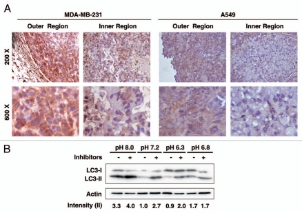
Differential autophagic activity in the outer and inner regions of solid tumors. (A) MDA-MB-231 or A549 cells were injected subcutaneously into the posterior flank of BALB/c athymic nude mice. Two weeks after implantation, tumors were dissected and stained with LC3 antibody. Pictures were taken in both the outer and inner region within the same tumor slide. Two independent tumors were examined for each cancer cell line and representative pictures were shown at magnifications of 200x (upper part) and 600x (lower part). (B) MDA-MB-231 cells were incubated in medium of indicated pH for 6 h in the presence or absence of lysosome inhibitors. Cell lysates were subjected to protein gel blot for LC3. Band densitometry quantification and the calculation of relative LC3-II intensity were performed as described in Materials and Methods.

