Abstract
Background
Sebaceous lymphadenoma is a rare benign salivary gland tumour of uncertain histogenesis. So is synchronous occurrence of two benign or malignant neoplasms.
Case-report
68-year-old female presented with right side parotid swelling associated with pain and gradual increase is size. Fine needle aspiration cytology of parotid swelling was suggestive of pleomorphic adenoma. Total conservative parotidectomy was performed and histopathology of the specimen revealed sebaceous lymphadenoma with squamous cell carcinoma.
Conclusions
Sebaceous lymphadenoma and squamous cell carcinoma are two rare benign and malignant neoplasms arising in parotid gland. Synchronous occurrence of these two entities has not been reported.
Keywords: parotid, tumour, neoplasm, sebaceous, lymphadenoma, squamous cell carcinoma
Background
Synchronous occurrence of two or more pathologically distinct benign and malignant neoplasm in a single organ is rare and accounts for < 0.1% of all salivary gland neoplasms. We report here a rare case of synchronous occurrence of sebaceous lymphadenoma, a rare benign salivary gland neoplasm predominantly seen in elderly females [1] and primary squamous cell carcinoma too is of rare occurrence in parotid glad. The limited number of reported cases of synchronous carcinoma in the salivary glands makes clinical management of these lesions difficult.
Case Report
A 68-year-old woman presented with swelling in the right parotid region of 8 years duration with history of gradual increase in size over last 1 year with pain and tinnitus for past 2 months. There was no difficulty in opening the mouth and no history suggestive of facial nerve palsy. Medical or family history was not contributory.
Examination showed a non tender swelling with variegated consistency in parotid region raising the ear lobule. Overlying skin showed dilated veins. The swelling was partly fixed to underlying structures and clinically there was no evidence of facial nerve involvement. There were no palpable cervical lymph nodes and haematological and biochemical investigation were within normal limits. A fine needle aspiration cytology (FNAC) was carried out which was suggestive of pleomorphic adenoma. With a diagnosis of pleomorphic adenoma patient underwent parotidectomy.
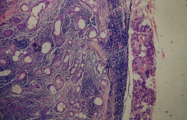
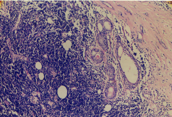
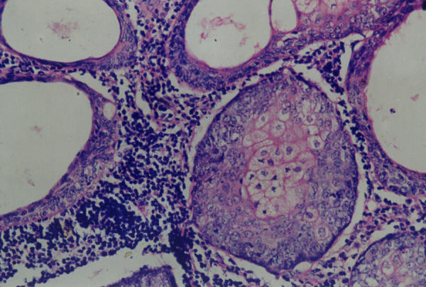
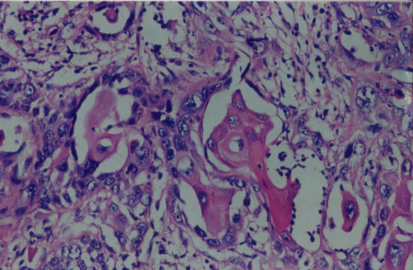
On gross examination the parotidectomy specimen measured 7 × 5 × 2 cm with irregular and nodular surface. Cut section showed grey white appearance with cystic spaces and foci of haemorrhage and calcification. Microscopic examination of the sections from salivary gland revealed partially circumscribed neoplasm (Figure 1) composed of numerous duct like structures and cystic spaces lined by flat to cuboidal epithelium and filled with keratin flakes intricately mixed with lymphoid stroma showing follicular formation in areas (Figure 2). Duct like space were lined by outer basaloid cells and inner mature sebaceous cells were present (Figure 3) focal squamous and mucinous metaplasia of lining epithelium was also seen along with areas of fibrosis and calcification. In one area neoplasm was seen to arise from lining epithelium of duct like spaces composed of polygonal cells with moderate cytoplasm and hyperchromatic pleomorphic nuclei, arranged in sheets, nests and group foci of keratinsation seen in between cell groups (Figure 4). A diagnosis of sebaceous lymphadenoma with moderately differentiated squamous carcinoma was made.
Figure 1.

Photomicrograph showing partially circumscribed neoplasm composed of numerous duct like structures, cystic spaces mixed with lymphoid stroma (Haematoxylin & Eosin × 40)
Figure 2.

Photomicrograph showing neoplasm with intense lymphoid infiltrate (Haematoxylin & Eosin x 40).
Figure 3.

Photomicrograph showing duct like spaces lined by outer basaloid and inner mature sebaceous cells (Haematoxylin & Eosin × 100)
Figure 4.

Photomicrograph of squamous cell carcinoma showing foci of keratinisation (Haematoxylin & Eosin × 100)
Discussion
Sebaceous lymphadenoma is rare benign salivary gland neoplasm [1]. There have been several care reports describing the tumour and its association with other salivary gland neoplasm. Sebaceous lymphadenoma is predominantly seen in elderly females and age at presentation ranges between 25 to 89 years.
Microscopy in sebaceous lymphadenoma show variable sized sebaceous glands admixed with salivary ducts surrounded by dense lymphoid stroma. Lymphoid background has well developed germinal centres. Histiocytes, foreign body giant cell and inflammatory reaction are seen. Focal necrosis can be observed occasionally [2,3]
Histogenesis of Sebaceous lymphadenoma is unclear. Possible theories are that it develops within the ectopic salivary gland in intraparotid lymph node [1] or may arise due to sebaceous differentiation in other tumours [1,4]. Other tumours to consider with similar histological characteristic include tumours with focal sebaceous differentiation, Warthin's tumour, mucoepidermoid carcinoma, pleomorphic adenoma (malignant), adenoid cystic carcinoma, benign oncocytoma and basal cell adenoma. [5,6]
Malignant transformation of the sebaceous lymphadenoma, although rare, should be considered along with possibility of a synchronous second primary malignant neoplasm in enlarging, locally invasive parotid lesions, considering that clinical behaviour and prognosis will be determined by the nature of the malignant component [7-9].
Parotidectomy is the treatment of choice in sebaceous lymphadenoma, however in presence of synchronous squamous cell carcinoma neck dissection should also be carried out if cervical lymph nodes are involved and we feel that postoperative adjuvant radiotherapy should improve the survival.
Conflict of interest
None declared.
Contributor Information
Mridula Shukla, Email: mridushukla@hotmail.com.
Sathibai Panicker, Email: mridushukla@hotmail.com.
References
- Chen K, Chan JKC. Salivary gland tumours. In: Fletcher CDM, editor. In Diagnostic Histopathology of Tumours. 2. Vol. 1. Hong Kong Churchill Livingstone; 2000. p. 259. [Google Scholar]
- Auclair PL, Ellis GL, Gnepp DR. Other benign epithelial neoplasms. In: Ellis GL, Auclair PL, Gnepp DR, editor. Surgical Pathology of Salivary Gland. Philadelphia, WB Saunders Company; 1991. pp. 252–268. [Google Scholar]
- Firat P, Hosal S, Tutar E, Ruacan S. Sebaceous lymphadenoma of the parotid gland. J Otolaryngol. 2000;29:114–116. [PubMed] [Google Scholar]
- Gnepp DR, Brannon R. Sebaceous neoplasms of salivary gland origin. Report of 21 cases. Cancer. 1984;53:2155–2170. doi: 10.1002/1097-0142(19840515)53:10<2155::aid-cncr2820531026>3.0.co;2-f. [DOI] [PubMed] [Google Scholar]
- Kwon GY, Kim EJ, Go JH. Lymphadenoma arising in the parotid gland: a case report. Yonsei Med J. 2002;43:536–538. doi: 10.3349/ymj.2002.43.4.536. [DOI] [PubMed] [Google Scholar]
- Gnepp DR. Sebaceous neoplasms of salivary gland origin: a review. Pathol Annu. 1983;18:71–102. [PubMed] [Google Scholar]
- Croitoru CM, Mooney JE, Luna MA. Sebaceous lymphadenocarcinoma of salivary glands. Ann Diagn Pathol. 2003;7:236–239. doi: 10.1016/S1092-9134(03)00052-2. [DOI] [PubMed] [Google Scholar]
- Mayorga M, Fernandez N, Val-Bernal JF. Synchronous ipsilateral sebaceous lymphadenoma and acinic cell adenocarcinoma of the parotid gland. Oral Surg Oral Med Oral Pathol Oral Radiol Endod. 1999;88:593–596. doi: 10.1016/s1079-2104(99)70091-0. [DOI] [PubMed] [Google Scholar]
- Dreyer T, Battmann A, Silberzahn J, Glanz H, Schulz A. Unusual differentiation of a combination tumour of the parotid gland. A case report. Pathol Res Pract. 1993;189:577–581. doi: 10.1016/S0344-0338(11)80369-9. discussion 581–585. [DOI] [PubMed] [Google Scholar]


