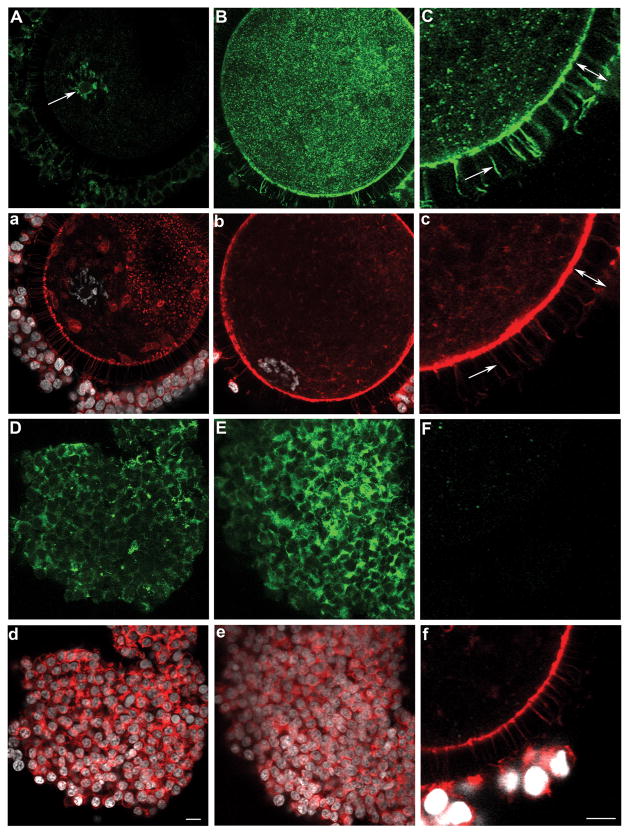
Figure 5.
Expression and localization of SOD3 in oocytes (A–B), trans-zonal projections (C), and cumulus cells (D–E) upon isolation from sized antral follicles. Corresponding microfilament (red) and DNA (white) stains are shown (a–f), and all processing and imaging conditions were kept identical across panels. SOD3 was organized as small aggregates (reminiscent of a vesicular labelling) in the oocyte cortex of oocytes from small (A), medium (C), and large (B) antral follicles. SOD3 was also abundant in cumulus cells and trans-zonal projections (C, c; arrow) with the width of the zona pellucida delineated by a double-ended arrow (C, c). All oocytes had nuclear SOD3, but ones from small (A, a) when compared to large (B, b) follicles showed more SOD3 in the nucleoplasm than ooplasm; SOD3 also co-localized (A, arrow) with chromatin (a). The use of control rabbit IgG antibody (F, f) confirmed the specificity of SOD labelling. (d) Scale bar for all panels except C, c, F, and f for which a scale bar is provided in f: 10μm.

