Abstract
Background
The expression of the Prospero homeodomain transcription factor (Prox1) in a subset of cardinal venous cells specifies the lymphatic lineage in mice. Prox1 is also indispensible for the maintenance of lymphatic cell fate, and is therefore considered a master control gene for lymphangiogenesis in mammals. In zebrafish, there are two prox1 paralogues, the previously described prox1 (also known as prox1a) and the newly identified prox1b.
Principal Findings
To investigate the role of the prox1b gene in zebrafish lymphangiogenesis, we knocked-down prox1b and found that depletion of prox1b mRNA did not cause lymphatic defects. We also generated two different prox1b mutant alleles, and maternal-zygotic homozygous mutant embryos were viable and did not show any lymphatic defects. Furthermore, the expression of prox1b was not restricted to lymphatic vessels during zebrafish development.
Conclusion
We conclude that Prox1b activity is not essential for embryonic lymphatic development in zebrafish.
Introduction
In vertebrates, in addition to the blood vasculature, the lymphatic vasculature plays important roles in maintaining an effective circulation. It is responsible for returning the protein-rich interstitial lymph fluid to the blood stream. Lymphatic vessels also contribute to the immune system that protects the body against infectious agents, and absorb lipids from the intestinal tract. Furthermore, there are a large number of inherited or acquired diseases that are associated with lymphatic vessel malfunction and lymphatic vessels may provide a primary route for the metastatic spread of certain tumors [1], [2], [3].
The lymphatic vessels were first described in 1627 by Aselli [4]. In 1902, Sabin proposed the origin of the lymphatic system from the veins [5]. The recent identification of several transcription factors, Prox1, Sox18, and CoupTF-II [6], [7], [8], which are essential for the specification of lymphatic cell fate, now provides a better understanding of the molecular mechanism of lymphatic development in metazoans.
In mice, the lymphatic system arises from a subset of venous endothelial cells (ECs) which start to express the homeodomain gene Prox1 from E9.75. By subsequent polarized budding and sprouting, Prox1-expressing cells migrate away from the cardinal vein and give rise to the entire lymphatic system [6], [9]. In Prox1 null mice, lymphatic development is arrested, while the development of the blood vascular system is largely unaffected [6].
It has been reported that Prox1 plays similar roles in other vertebrate organisms, such as Xenopus and zebrafish [10], [11]. In zebrafish, the initial process of vasculogenesis leads to the formation of a primitive circulatory loop by 26 hpf (hours post fertilization), which consists of the posterior cardinal vein (PCV) and dorsal aorta (DA). A primary wave of angiogenesis, emanating from the DA, produces intersegmental arteries (ISAs), while a secondary wave of venous angiogenesis gives rise to sprouts that either remodel half of the existing ISAs into ISVs (intersegmental veins) by 2.5 dpf, or become parachordal lymphangioblasts (PLs), constituting a precursor pool of lymphangioblasts formed around 50 hpf at the horizontal myoseptum [12], [13]. At 60 hpf, the PLs start to migrate along ISAs, and move dorsally or ventrally to form the dorsal longitudinal lymphatic vessel (DLLV), the thoracic duct (TD) and the inter-segmental lymphatic vessels (ISLVs) that connect the DLLV and TD [14]. The presence of the TD is a commonly used indicator for normal lymphatic development. The prox1b gene was recently identified in zebrafish and its activity was claimed to be essential for zebrafish lymphangiogenesis and TD formation, using a morpholino knock-down approach [15]. Furthermore, prox1b was suggested to provide a molecular marker for lymphatic endothelial cells. We had performed similar experiments in the past, with different results. Here, we show that two mutant alleles of prox1b do not alter lymphatic development, and that prox1b is not a useful marker for lymphatic endothelial cells in zebrafish.
Results
Prox1b is expressed in endothelial cells and the cranial central nervous system
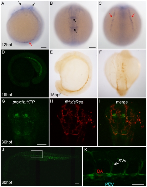
In mice, Prox1 is expressed in a variety of tissues other than lymphatic endothelial cells, including the lens, the heart, liver, pancreas, and central nervous system (CNS) [16]. By whole mount in situ hybridization, we were able to detect prox1b expression in the lateral plate mesoderm (LPM) and in the CNS of the head at 12 hpf stage (Figure 1 A–C). Since zebrafish endothelial cells (which provide the inner lining of blood and lymphatic vessels) originate from the LPM, we intended to follow prox1b over time. To that end, and in order to conveniently visualize the dynamic expression of prox1b gene in live embryo, we generated a Tol2-mediated bacterial artificial chromosome (BAC) transgenic line in zebrafish [17]. The overall morphology of the transgenic embryos appeared normal and was indistinguishable from wild type embryos (Figure 1D and J, and Figure 2D and E).
Figure 1. Prox1b is expressed in the endothelial cells and the central nervous system of the head.
(A–C) shows prox1b transcript expression by whole mount in situ hybridization in wild-type embryos, at 12 hpf. Black arrows point to prox1b expression in the head; red arrows indicate prox1b expression in lateral plate mesoderm. Confocal image (D) shows YFP expression in a prox1b BAC:YFP embryo at 19 hpf stage. (E) and (F) show YFP expression, enhanced by DAB immunostaining, is detected in prox1b BAC:YFP embryos in migrating angioblasts at 15 hpf. (G–I) shows prox1b:YFP expression in the head region of a prox1b BAC:YFP, fli1:DsRed embryo. Note overlapping (endothelial cells) and non-overlapping expression domains. (J) shows prox1b:YFP expression in the trunk vasculature. (K) shows enlarged view of the boxed area in (J). Scale bars represent 50 µm in (K), and 100 µm in other figures.
Figure 2. Prox1b does not specifically mark lymphatic aspects of the vasculature.
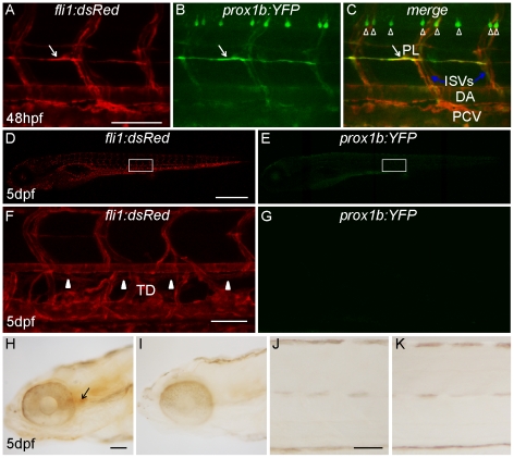
(A–C) shows prox1b:YFP expression in motor neurons and all endothelial cells of a prox1b BAC:YFP, fli1:DsRed embryo at 48 hpf. White arrows point to parachordal lymphangioblasts. The white open arrowheads label motor neurons. Note that while there is expression of prox1:YFP in PLs, prox1b is also expressed in other (non-lymphatic) aspects of the vasculature. (D) and (E) show the fluorescence images of the same 5-day prox1b BAC:YFP, fli1:DsRed embryo. There is no detectable prox1b:YFP expression in the trunk region of the transgenic embryos (E). (F) and (G) show the enlarged views of the boxed area in (D) and (E). (F) White arrowheads indicate the TD, which resides between DA and PCV. (G) prox1b:YFP expression cannot be detected in TD. (H) and (J) show weak DAB immunostaining against YFP expression in the head (H, indicated by the black arrow), but not in the trunk of transgenic embryos (J). (I) and (K) are DAB staining controls without primary antibody. Scale bars represent 250 µm in (D), and 50 µm in other figures.
The prox1b BAC:YFP construct was created by inserting the yellow fluorescence protein (YFP) coding sequences at the position of the first translated ATG in a prox1b-harboring BAC (CH73-247L15) through homologous recombination [17]. This BAC contains approximately a 116-kb long zebrafish genomic contig, spanning from ∼97.7-kb upstream of the prox1b start codon to ∼18.6-kb downstream of the start codon, and harbors the entire prox1b coding sequence. We verified the correct knock-in of YFP in the prox1b gene based on diagnostic PCR (data not shown) and performed extensive analyses of the YFP expression in the prox1b:YFP embryos.
YFP expression in live embryo was not detected via confocal microscopy until late somitogenesis stages, when it was evident in the intermediate cell mass (derived from the earlier LPM and containing the trunk vascular precursors), but not in the head region (Figure 1D). To amplify the signal, we applied anti-GFP immunostaining with diaminobenzidin (DAB) solution in fixed embryos and were able to detect YFP expression in LPM as early as 15hpf (Figure 1E and F). Using standard in situ hybridization (ISH), we could confirm that prox1b is expressed, at 24 hpf, in the caudal vein (Figure S1A–C). As the development of the embryos proceeded, the expression level of YFP increased and signals also became apparent in more anterior aspects of the embryos (Figure 1G–I). At 30 hpf, in the head of prox1b BAC:YFP; fli1:DsRed double transgenic embryos [13], YFP expression partially overlapped with the red signals which marked all endothelial cells (Figure 1G–I). This indicates that the prox1b gene is expressed in both a subset of endothelial cells and some non-endothelial aspects of the central nervous system of the head. At the same stage, we detected bright YFP expression in all trunk endothelial cells: posterior cardinal vein (PCV), dorsal aorta (DA), and inter-segmental vessels (ISVs) (Figure 1J). In transgenic embryos we noted that the YFP signal level in the DA often appeared brighter than in the PCV (Figure 1K), however, using ISHs, we routinely observed that mRNA levels were equally high in the PCV (Figure S1D–F), sometimes even higher in the PCV than in the DA. Both YFP expression in transgenic embryos (Figure 2A–C) and ISHs confirm a rather broad expression in the caudal vein, the PCV and the DA. This dynamic prox1b:YFP expression in zebrafish ECs is hence different from Prox1 expression in mice [6], which is, within endothelial cells, restricted to lymphatics, venous lymphatic precursors and venous valves [18].
Prox1b expression is not restricted to lymphatic endothelial cells
In mice, Prox1 is required for both the specification of LEC fate and the maintenance of LEC cell identity. Prox1 expression is not detected in the blood endothelial cells (BECs) after E11,5, but persists in the lymphatic vasculature until and throughout adulthood [18], [19]. We therefore examined the distribution of zebrafish prox1b:YFP expression and prox1b mRNA expression in late stage LECs. At 5 dpf, when the thoracic duct and intersegmental lymphatic vessels have formed, we were not able to detect any YFP expression in the trunk of prox1b:YFP embryos (Figure 2D–G and J, K), but observed weak signals in the head (Figure 2H and I).
prox1b ISH signals in the trunk of embryos at 48 hpf and 72 hpf had previously been reported [15]. Using a prox1b probe, we detected only diffuse expression in the trunk at these stages (Figure S1G–L), which we would normally consider non-specific. To test the specificity of this staining, we performed ISH for prox1b in cloche mutant embryos, which are devoid of all ECs (Figure S2A and B) [20], [21]. Since the staining pattern was unaltered in mutants, compared to wild-type embryos at 48 hpf and 72 hpf (Figure S2C, D and S1G, H, J, and K), we conclude that prox1b expression in ECs is no longer detectable by ISH from 48 hpf and after 72 hpf (Figure S1G–O). Therefore, different from Prox1 expression in mice, prox1b is not a specific and persistent lineage marker for LECs in zebrafish.
Knock-down of prox1b does not lead to impaired lymphatic development
As prox1b expression in the vasculature is not restricted to LECs, we wondered whether Prox1b is required for lymphatic development of zebrafish.
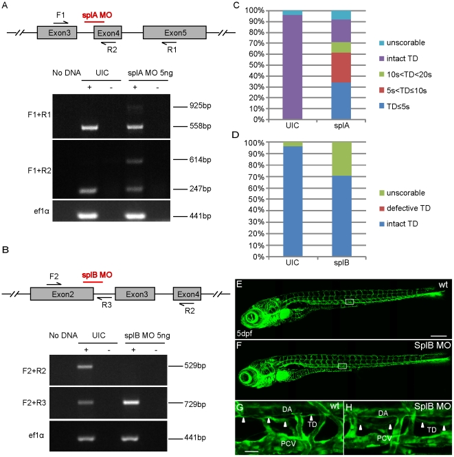
Initially, we used two different antisense morpholinos (splA MO and splB MO) to knock down prox1b gene activity. Both morpholinos were designed to target the splice sites of the prox1b gene (Figure 3A and B). The results of reverse transcription-PCR (RT-PCR) analysis showed that splA MO inefficiently blocked the excision of intron 3–4 (367 bp), leaving a substantial amount of correctly processed transcripts in morpholino injected embryos (Figure 3A), while injection of splB MO caused a loss of detectable wild-type prox1b transcripts, leading to the accumulation of non-spliced prox1b transcripts (retaining of intron2-3 leads to frame shift after aa494) (Figure 3B). However, ∼70% of splA MO injected embryos developed lymphatic defects: a complete or partial loss of TD at 5 dpf (Figure 3C). In contrast, we could not detect a similar phenotype in splB MO injected embryos (Figure 3D–H).
Figure 3. Morpholino-mediated knock-down of prox1b does not cause lymphatic phenotypes.
(A) and (B) show schematics of the prox1b genomic locus with the red bars indicating target sites of the respective splice morpholinos. The numbered black arrows show the position of the primers used in RT-PCRs for examining the splicing of prox1b transcripts. (A) RT-PCR to detect splicing of prox1b in un-injected (UIC) and splA MO injected embryos. The expression of elongation factor 1-alpha (ef1a) gene represents the loading control. Primer pair F1 and R1 amplifies wild-type transcript band (558 bp) and incorrectly spliced transcripts (925 bp), which fail to excise the intron3-4 (367 bp). Primer pair F1 and R2 amplifies wild-type transcript (247 bp) and non-spliced transcripts (614 bp), which retain intron3-4. (B) RT-PCR to detect splicing of prox1b in un-injected and embryos injected with splB MO. Primer pair F2 and R2 amplifies wild-type transcripts (529 bp), which are missing in splB MO injected embryos. Primer pair F2 and R3 amplifies non-spliced transcripts (729 bp), which preferentially accumulated in morphant embryos. (C) Histograms showing the percentage of fli1:GFP embryos with different lengths of TD (10 s<TD<20 s means the partial TD covers the length of 10 to 20 somites in the trunk). Up to 70% of splA MO injected embryos displayed complete or partial loss of TD, even though splA MO seems not to affect prox1b splicing efficiently. Embryos were scored at 5 dpf. (D) Histograms showing the percentage of fli1:GFP embryos with intact or defective TD, and all the scorable embryos (their overall morphology was all right and they had normal blood circulation and did not develop edema at 5 dpf) developed complete TD after injection with splB MO. (E) and (F) show the full images of 5-day UIC (E) and splB MO injected embryos (F). (G) and (H) show enlarged views of the boxed areas in (E) and (F). The white arrowheads indicate the presence of TD in both control embryos (G) and morphants (H). Scale bars represent 250 µm in (E), and 25 µm in (G).
The presence of a TD in splB MO injected embryos indicates that loss of prox1b still allows for normal lymphatic development. The phenotypes observed in splA MO injected embryos are likely to be side-effects due to toxicity, and indeed we have in the past seen many examples where MO-injection affects TD-formation (unpublished observation). Since our data are in conflict with recently published information [15], we decided to generate stable mutant loss-of-function models, alleviating the need to have to rely on morpholinos.
Prox1b mutants develop normally
We screened for prox1b mutants from an in-house TILLING (targeting induced local lesion in genome) library [22] and identified the prox1bhu3510 allele (Figure S3A), with a stop mutation in the homeodomain of the gene. The mutant line was maintained in the fli1:GFP background, where all endothelial cells are marked by GFP expression [23], [24]. We observed that, at 48 hpf, the secondary venous sprouts occurred normally and the formation of PLs was unaffected in homozygous mutant embryos (Figure S3B and C). Indistinguishable from wild-type embryos, the homozygous mutants developed a completely normal TD at 5 dpf (Figure S3D–G). Homozygous prox1bhu3510 mutant embryos were viable, although the mutation changed a highly conserved amino acid (W627) into a stop codon, which would be predicted to alter the function of the Prox1b Homeo-Prospero (HPD) domain by removing residues that bind the DNA phosphate backbone and by unmasking the C-terminal nuclear export sequence [25].
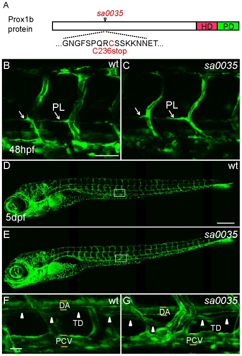
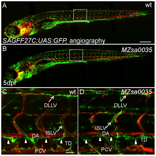
In an effort to obtain an additional allele, we received the prox1bsa0035 line from the Sanger Centre. This allele harbors a mutation at amino acid 236. This mutation would predict a truncated Prox1 protein with the loss of the entire Homeo-Prospero domain, which is indispensable for Prox1 homologues to function as transcription factors (Figure 4A). We maintained the prox1bsa0035 allele in fli1:GFP and SAGFF27C;UAS:GFP (heterozygous SAGFF27C;UAS:GFP line displays high reporter expression in lymphatic vasculature) transgenic background [12]. The prox1bsa0035 homozygous mutant embryos were also viable and developed normally (Figure 4B–G). Both the specification and migration of the lymphatic endothelial cells were unaffected in the mutants, as the formation of PLs (Figure 4B and C) and TD was indistinguishable from wild type embryos (Figure 4F and G). The prox1bsa0035 homozygous fish are fertile. To exclude the possibility that maternal prox1b mRNA might rescue a possible homozygous zygotic phenotype, we crossed prox1bsa0035−/−, SAGFF27C;UAS:GFP fish, and generated maternal-zygotic (MZ) prox1b mutant progeny. These MZ mutant embryos have no visible defects (Figure 5A and B) and we observed a properly formed embryonic lymphatic network, including DLLV, ISLVs and TD, at 5 dpf (Figure 5C and D).
Figure 4. The lymphatic development of homozygous prox1bsa0035 mutants appears normal.
(A) Schematic representation of the Prox1b protein, with the position of the prox1bsa0035 allele indicated. The homeodomain region (HD) is shown in red, the Prospero domain (PD) in green. The predicted stop mutation occurs at C236 in prox1bsa0035. (B) and (C) show vascular structures in the trunk region of wild-type (wt, B) and homozygous prox1bsa0035 mutant embryos (C) in fli1:GFP background. The white arrows indicate PLs. (D) and (E) show whole embryo lateral view images of 5-day wt (D) and homozygous prox1bsa0035 mutant embryos (E). (F) and (G) show enlarged views of the boxed areas in (D) and (E). The white arrowheads indicate the presence of TD in both control (F) and homozygous prox1bsa0035 embryos (G). Scale bars represent 50 µm in (B), 250 µm in (D) and 25 µm in (F).
Figure 5. The lymphatic development of maternal-zygotic prox1bsa0035 mutant is unaltered.
(A) and (B) show whole embryo lateral view images of 5-day wt (A) and MZ prox1bsa0035 mutant (B) in a SAGFF27C;UAS:GFP background. Perfused blood vessels were labeled by angiography (in red). (C) and (D) show enlarged views of the boxed areas in (A) and (B). The entire lymphatic network in the trunk of zebrafish, which is composed of the GFP-expressing lymphatic vessels-DLLV, ISLV and TD (marked by white arrowheads), is properly formed in wt (C) and MZ prox1bsa0035 mutant embryos (D). Scale bars represent 250 µm in (A), and 25 µm in (C).
The analysis of two prox1b mutant alleles demonstrates that prox1b gene activity is not necessary for any known indispensible functional process during the development of zebrafish. In combination with the prox1b expression and knock-down analysis, we conclude that Prox1b activity is not essential for zebrafish embryonic lymphatic development.
Discussion
It has been recently reported that zebrafish prox1b expression marks lymphatic endothelial cells, and that knock-down of prox1b via morpholinos completely abolishes lymphangiogenesis [15]. We here report that two mutant alleles of prox1b undergo normal lymphangiogenesis, and that, at least at later stages of development, prox1b expression does not mark lymphatic endothelial cells.
In mammals, the activities of the transcription factors Coup-TFII [8], Sox 18 [7] and Prox1 [6] lead to the specification of a subpopulation of anterior cardinal vein cells into future lymphatic endothelial cells (LECs) [6], [7], [8]. Subsequently, Ccbe1, VEGF-C and its receptor, VEGFR3, are required for outgrowth of these prospective LECs. For the latter process, there is excellent genetic evidence for strong evolutionary conservation between mice and zebrafish: mutations in Ccbe1 [14], [26], VegfC and Vegfr3 [11], [27], [28], [29] are deficient in most aspects of venous sprouting and consequently lack a thoracic duct. The phenotypic effects of lacking Coup-TFII, Sox 18 and Prox1 are less well supported in zebrafish (no mutants for either of these genes have been reported) and have started to be elucidated only recently. Knock-down of Coup-TFII results in lymphatic defects [30], but lack of Sox18 has been independently reported to have no phenotype by three groups [31], [32], [33]. Since the effect of Sox18 in mice is context-dependent, this might not be completely surprising [7]. Finally, in the case of Prox1, a recent study reported a requirement for prox1b in embryonic lymphatic formation [15].
Our conclusion that Prox1b activity is not essential for lymphatic development, is contradictory to a previous report [15]. In previous work, the function of Prox1b was studied by using a morpholino knock-down approach. Only a single morpholino covering the first translated ATG was employed in the study, however, the authors demonstrated rescue of the morphant phenotype by injection of mRNA. Here, we show analysis with a splice (splB) morpholino, which we demonstrate to deplete nearly all wild-type prox1b transcripts, while not causing any lymphatic defects. The presence of a TD in splB MO injected embryos indicates that loss of prox1b still allows normal lymphatic development. Although we cannot explain the published mRNA rescue experiments, all our results indicate that the lymphatic defects observed in ATG and splA MO injected embryos are most likely not due to loss of Prox1b function. Of note, the prox1b morpholino used in the previous study causes severe edema at 4 dpf. We take this as evidence of considerable toxicity of the morpholino, as from previous studies [14], [29], it is clear that lack of lymphatic tissue does not cause edema at this early stage. The toxicity might be the cause for the reduction of lyve-1 expression in morphants.
Furthermore, we analyzed two mutant alleles that provide an additional method to study prox1b function. Homozygous mutants for both alleles, and even maternal-zygotic mutants, develop normally without visible defects. Prox1b shares high homology with Drosophila Prospero (Pros) in the homeodomain and prospero domain (PD). Both proteins contain a nuclear export signal (NES) in the homeodomain. Based on a previous study [25], a physiological signal induces Pros to adopt a closed conformation so that the PD shields the NES and Pros enters the nucleus and is prepared for DNA binding. If the NES masking is turned off or blocked, Pros adopts an open structure that exposes NES and exits the nucleus [25]. The prox1bhu3510 allele harbors a non-sense mutation, which deletes the C-terminal part of the PD, which contains a highly conserved DNA binding residue Lys629, and is also essential to physically cover the NES in the homeodomain, so the predicted truncated protein is unlikely to adopt the closed conformation and be kept in the nucleus for proper DNA binding.
The other allele, prox1bsa0035, is predicted to be truncated after the coil-coil domain, and does not contain the homeodomain and prospero domain. Therefore, we believe that both mutants almost certainly lack wild-type activity of Prox1b, and that at least the prox1bsa0035 allele represents a loss-of-function situation. Although we cannot exclude the possibility of a compensatory transcriptional mechanism involving other prox homologues, or of a complex transcript processing event to remove these coding mutations, we have not been able to detect any evidence to support either of these hypotheses. Injections of prox1a morpholinos into prox1b mutants at lower dose did not affect TD formation (data not shown; low doses of morpholinos had to be used since prox1a morpholinos are toxic). Based on two mutant alleles and the splB morpholino, we conclude that the activity of Prox1b is dispensable during zebrafish development.
In zebrafish, LECs arise from the PCV sprouts that do not connect to ISAs, and approximately half of the venous spouts fall into this class. These sprouts migrate to the horizontal midline and become lymphatic precursors, dubbed PLs [14]. It is unclear at present whether the lymphatic fate of these venous sprouts is determined before they emanate from the PCV, or whether they get specified during their migration from PCV to horizontal myoseptum. If prox1b was a specific LEC lineage marker comparable to mammalian Prox1, its early expression around 30 hpf, would be expected to be found in a polarized manner, present in a salt-and-pepper pattern in the dorsal side of the PCV or restricted to half of the venous spouts that constitute future PLs. At later stages, prox1b expression should only be observed in lymphatic endothelial cells (PLs and TD, DLLV, and ISLVs) but not blood endothelial cells. This is not what we observe: rather, prox1b expression is detected in all endothelial cells and is not enriched at the dorsal side of the PCV around 30 hpf. After that point in time, prox1b expression in endothelial cells becomes increasingly weaker. Although a few PLs seem to have brighter expression at 48 hpf, prox1b expression is never exclusively observed in LECs, instead it is also present in BECs (Figure 2A–C). Furthermore, prox1b expression is not maintained in LECs, and eventually it completely disappears from the zebrafish trunk vasculature, and at 5 days there was no expression detectable in the TD or any other trunk endothelial tissue (Figure 2G, J and S1M–O). Based on our results, prox1b is not a LEC-specific marker in zebrafish.
In mice, the expression of Coup-TFII in the blood vasculature is restricted to veins and it inhibits Notch activity and blocks an arterial signaling cascade in venous endothelial cells (VECs) [34]. At embryonic day E9, Sox18 expression becomes apparent in a subset of cells in the anterior cardinal vein and induces Prox1 expression in these cells around E9.75 [7], [35], in synergy with Coup-TFII [8]. The direct interaction between Coup-TFII and Prox1 is also necessary for the maintenance of Prox1 expression during early stages of LEC specification and differentiation [8], [36], [37]. In zebrafish, the lymphatic cells also arise from VECs, the secondary sprouts of the PCV [12], [13]. However, the molecular mechanisms that initiate lymphatic specification remain unclear and might be different from mammals. Recent work from Aranguren et al., indicates a conserved role of Coup-TFII for venous and lymphatic development in zebrafish [30]. Knockdown of Sox18 alone failed to reveal any phenotype [32], [33], and the simultaneous knockdown of sox18 and sox7 severely affected the arteriovenous identity and led to dramatic fusion between the major axial vessels [31], [32]. So far, there is no clear evidence to show that SoxF proteins have a role in zebrafish lymphatic development; however, in mice the function of Sox18 in early aspects of lymphangiogenesis is also context dependent [7]. It remains possible that a homeodomain transcription factor, perhaps a Prox homologue, functions directly downstream of Coup-TFII to switch on the lymphatic lineage in zebrafish. Since we demonstrate here that Prox1b is not required for normal lymphatic development, Prox1a becomes the most likely candidate. It was previously reported that the morpholinos targeting zebrafish prox1a gene usually led to severely delayed or impaired growth at early stages and eventually caused massive malformations before a lymphatic phenotype could reasonably be scored [27]. Therefore, a yet to be identified prox1a mutant would be required to shed further light onto the molecular pathway that initiates the lymphatic fate in zebrafish.
Methods
Ethics statement
All zebrafish strains were maintained under standard husbandry conditions at the Hubrecht Institute, and animal experiments were approved by the Animal Experimentation Committee (DEC) of the Royal Netherlands Academy of Arts and Sciences. Permit number for this study is: 80101.
Morpholinos
All morpholinos were obtained from GeneTools. Prox1b splA MO (5′-CACAGCGATTGAACTGTGTAGCGAG-3′) was designed to target the intron3-4/exon4 boundary, while prox1b splB MO (5′-GATAAAAGGATATTGAACCTGCAGC-3′) was designed to target the exon2/intron2-3 boundary. Both of the morpholinos were injected at a concentration of 5 ng/embryo. All morpholinos and primers were designed according to the prox1b sequence in GenBank/EMBL database under the accession number FJ544314.
RT-PCR
Total RNA (1 µg) from wildtype and morpholino injected embryos was reverse transcribed by using M-MLV Reverse Transcriptase and random hexamers (Promega). PCR followed by sequencing was performed with the following primers:
F1: 5′-GCCACTTGAAGAAAGCCAAG-3′
F2: 5′-GCCCCTTCTTCACTACACCA-3′
R1: 5′-CCTCCAGAACCAGCAATAAG-3′
R2:5′-CGGTAAAGCTCGGTGTCTCT-3′
R3: 5′-GTGTGGTCCCTGTTGATCCT-3′.
In situ hybridization
In situ hybridization was performed as previously described [38]. Prox1b probe was synthesized by in vitro transcription from the HindIII-digested full length cDNA in pBSK, using T7 RNA polymerase (Promega).
Embryos were subjected to ISH and paraffin sections (6 µm) were counter-stained with neutral red.
Microangiography
We performed microangiography as described [27]. Embryos were anesthetized in 0.015% MS-222 at 5 dpf and were embedded in 0.5% low melting point agarose. A small bulb of tetramethylrhodamine (TAMRA; Molecular Probes, 2,000,000 MW, 10 mg/ml) was administered by cardiac injection using a conventional microinjection setup. Only embryos exhibiting TAMRA throughout the cardiovascular system immediately after the injection were further analyzed [27].
Microscopy
Embryos were mounted in 0.5% low melting point agarose in a culture dish with a cover slip replacing the bottom. Imaging was performed with a Leica SPE confocal microscope using a 10× objective with digital zoom.
Diaminobenzidine (DAB) histochemistry
DAB staining was performed using VECTASTAIN ABC kit (VECTOR) and DAB tablet (SIGMA). The expression of YFP was detected by using rabbit polyclonal anti-GFP (1∶500, Torrey Pines Biolabs, Acris TP401) as primary antibody and polyclonal swine anti-rabbit Ig/Biotinylated (1∶500, DAKO, E0353) as secondary antibody.
Mutants
prox1bhu3510 allele was identified from an ENU mutagenesis library in Hubrecht Institute by Tilling. The prox1bsa0035 allele was ordered from Zebrafish Mutation Resource of Sanger Institute. Mutants were genotyped by using the following KASPAR primers:
prox1bhu3510 ID1_ALG 5′-GAAGGTGACCAAGTTCATGCTCAGATGATCTTATAGATGGACTTCTTC-3′
prox1bhu3510 ID2_ALA 5′-GAAGGTCGGAGTCAACGGATTACAGATGATCTTATAGATGGACTTCTTT-3′
prox1bhu3510 ID-C1 5′-GCCATCCAGAGCGGTCGGGAT-3′
prox1bsa0035 ID_ALT 5′-GAAGGTGACCAAGTTCATGCTGCAATGGATTTTCTCCTCAACGTTGT-3′
prox1bsa0035 ID_ALA 5′-GAAGGTCGGAGTCAACGGATTGCAATGGATTTTCTCCTCAACGTTGA-3′
prox1bsa0035 ID_C1 5′-GGAAAAATTCCAGCATTGCCATTTCCATT-3′
The clochet22499 allele has been previously described [32].
Generation of transgenic line
Generation of transgenic lines with bacterial artificial chromosome (BAC) was described previously [17]. For the prox1b BAC:YFP transgenic line, a citrine-neomycin cassette was recombined by using Red/ET Recombination Technology (Gene Bridges), into the bacterial artificial chromosome (BAC) clones CH73-247L15 using homology arm tagged PCR primers. DNA was injected into one cell-stage embryos at a concentration of 500 pg/embryo. A transgenic carrier adult was selected by screening for fluorescent progeny.
Supporting Information
Prox1b transcript expression in zebrafish. (A–O) shows prox1b expression analyzed by in situ hybridization in whole mount embryos (A, D, E, G, H, J, K, M and N) and transverse sections (B, C, F, I, L, and O), at different stages: 24 hpf (A–C), 32 hpf (D–F), 48 hpf (G–I), 72 hpf (J–L) and 5 dpf (M–O). (E), (H), (K) and (N) individually show the enlarged views of the boxed area in (D), (G), (J) and (M). The blue and red bars in (A) represent the positions of the sections in (B) and (C). prox1b expression is prominent in the caudal vein of embryos at 24 hpf (C) and in both the DA and PCV at 32 hpf stage, shown by (E) and (F). (G–O) However, there is no signal in the blood and lymphatic endothelial cells of older embryos at 48 hpf, 72 hpf and 5 dpf. The black arrows point to the dorsal aorta; the blue arrows point to the posterior cardinal vein or caudal vein; and the red arrows point to thoracic duct. The black and blue arrow heads point to the prox1b expression in the DA and PCV separately. NT: neural tube; NC: notochord. Scale bars represent 200 µm in (A), (D), (G), (J), (M); 50 µm in (E), (H), (K) and (N); and 20 µm in (B), (C), (F), (I), (L) and (O).
(TIF)
Prox1b expression in cloche mutant. (A–D) shows prox1b expression in cloche mutants and siblings. The red and blue arrows point to prox1b expression in the DA and PCV of a sibling embryo separately (A), while this prox1b expression is absent in cloche mutant (B). The black arrows point to signals in the ventral region of the trunk (C) and along the somite boundaries of homozygous cloche mutants (D). (E) The inset shows an enlarged view of the boxed area in (D). The staining indicated by black arrows in C, D and E is either non-endothelial or non-specific because cloche mutant embryos lack endothelial cells. Given the diffuse nature of the staining, the black arrow indicated signal is probably a non-specific artifact of over-staining. Scale bars represent 100 µm in (A), and 50 µm in (E).
(TIF)
The lymphatic development of homozygous prox1bhu3510 mutant is normal. (A) Schematic representation of the Prox1b protein, with the position of the prox1bhu3510 allele indicated. The homeodomain region is shown in red, the Prospero domain in green. The predicted stop mutation occurs at W627 in prox1bhu3510 and multiple sequence alignment shows the conservation of zebrafish W627 in the Prospero domain of Prox proteins. (B) and (C) show the vascular structures in the trunk region of wt (B) and homozygous prox1bhu3510 mutant embryos (C) in fli1:GFP background. The white arrows indicate PLs. (D) and (E) show the full images of 5-day wt (D) and homozygous prox1bhu3510 mutant embryos (E). (F) and (G) show enlarged views of the boxed areas in (D) and (E). The white arrowheads indicate the presence of TD in both control (F) and homozygous prox1bhu3510 embryos (G). Scale bars represent 50 µm in (B), 250 µm in (D) and 25 µm in (F).
(TIF)
Acknowledgments
J. Peterson-Maduro provided the prox1b BAC: YFP construct. B. M. H. thanks Graham Lieschke for his support during the preliminary investigations in his lab. The fli:DsRed [13] line was obtained from the Dewerchin lab in Leuven, Belgium. The Hubrecht Imaging Center supported all imaging efforts.
For providing the zebrafish knockout alleles, hu3510 and sa0035, we thank Edwin Cuppen, Henk Roeckel and Ewart de Bruin (Hubrecht Instuitute) and the Sanger Institute Zebrafish Mutation Resource.
Footnotes
Competing Interests: The authors have declared that no competing interests exist.
Funding: The study was financed by the Koninklijke Nederlandse Akademie van Wetenschappen (http://www.knaw.nl/). The funders had no role in study design, data collection and analysis, decision to publish, or preparation of the manuscript.
References
- 1.Oliver G, Alitalo K. The lymphatic vasculature: recent progress and paradigms. Annu Rev Cell Dev Biol. 2005;21:457–483. doi: 10.1146/annurev.cellbio.21.012704.132338. [DOI] [PubMed] [Google Scholar]
- 2.Schulte-Merker S, Sabine A, Petrova TV. Lymphatic vascular morphogenesis in development, physiology, and disease. J Cell Biol. 2011;193:607–618. doi: 10.1083/jcb.201012094. [DOI] [PMC free article] [PubMed] [Google Scholar]
- 3.Alitalo K, Tammela T, Petrova TV. Lymphangiogenesis in development and human disease. Nature. 2005;438:946–953. doi: 10.1038/nature04480. [DOI] [PubMed] [Google Scholar]
- 4.Aselli G. De Lacteibus sive Lacteis Venis, Quarto Vasorum Mesarai corum Genere novo invento. Milan: Mediolani; 1627. [Google Scholar]
- 5.Sabin FR. On the origin of the lymphatic system from the veins, and the development of the lymph hearts and thoracic duct in the pig. Am J Anat. 1902;1:367–389. [Google Scholar]
- 6.Wigle JT, Oliver G. Prox1 function is required for the development of the murine lymphatic system. Cell. 1999;98:769–778. doi: 10.1016/s0092-8674(00)81511-1. [DOI] [PubMed] [Google Scholar]
- 7.Francois M, Caprini A, Hosking B, Orsenigo F, Wilhelm D, et al. Sox18 induces development of the lymphatic vasculature in mice. Nature. 2008;456:643–647. doi: 10.1038/nature07391. [DOI] [PubMed] [Google Scholar]
- 8.Srinivasan RS, Geng X, Yang Y, Wang Y, Mukatira S, et al. The nuclear hormone receptor Coup-TFII is required for the initiation and early maintenance of Prox1 expression in lymphatic endothelial cells. Genes Dev. 2010;24:696–707. doi: 10.1101/gad.1859310. [DOI] [PMC free article] [PubMed] [Google Scholar]
- 9.Oliver G, Srinivasan RS. Endothelial cell plasticity: how to become and remain a lymphatic endothelial cell. Development. 2010;137:363–372. doi: 10.1242/dev.035360. [DOI] [PMC free article] [PubMed] [Google Scholar]
- 10.Ny A, Koch M, Schneider M, Neven E, Tong RT, et al. A genetic Xenopus laevis tadpole model to study lymphangiogenesis. Nat Med. 2005;11:998–1004. doi: 10.1038/nm1285. [DOI] [PubMed] [Google Scholar]
- 11.Yaniv K, Isogai S, Castranova D, Dye L, Hitomi J, et al. Live imaging of lymphatic development in the zebrafish. Nat Med. 2006;12:711–716. doi: 10.1038/nm1427. [DOI] [PubMed] [Google Scholar]
- 12.Bussmann J, Bos FL, Urasaki A, Kawakami K, Duckers HJ, et al. Arteries provide essential guidance cues for lymphatic endothelial cells in the zebrafish trunk. Development. 2010;137:2653–2657. doi: 10.1242/dev.048207. [DOI] [PubMed] [Google Scholar]
- 13.Geudens I, Herpers R, Hermans K, Segura I, Ruiz de Almodovar C, et al. Role of delta-like-4/Notch in the formation and wiring of the lymphatic network in zebrafish. Arterioscler Thromb Vasc Biol. 2010;30:1695–1702. doi: 10.1161/ATVBAHA.110.203034. [DOI] [PMC free article] [PubMed] [Google Scholar]
- 14.Hogan BM, Bos FL, Bussmann J, Witte M, Chi NC, et al. Ccbe1 is required for embryonic lymphangiogenesis and venous sprouting. Nat Genet. 2009;41:396–398. doi: 10.1038/ng.321. [DOI] [PubMed] [Google Scholar]
- 15.Del Giacco L, Pistocchi A, Ghilardi A. prox1b Activity is essential in zebrafish lymphangiogenesis. PLoS One. 2010;5:e13170. doi: 10.1371/journal.pone.0013170. [DOI] [PMC free article] [PubMed] [Google Scholar]
- 16.Oliver G, Sosa-Pineda B, Geisendorf S, Spana EP, Doe CQ, et al. Prox 1, a prospero-related homeobox gene expressed during mouse development. Mech Dev. 1993;44:3–16. doi: 10.1016/0925-4773(93)90012-m. [DOI] [PubMed] [Google Scholar]
- 17.Bussmann J, Schulte-Merker S. Rapid BAC selection for tol2-mediated transgenesis in zebrafish. Development. 2011;138:4327–4332. doi: 10.1242/dev.068080. [DOI] [PubMed] [Google Scholar]
- 18.Choi I, Chung HK, Ramu S, Lee HN, Kim KE, et al. Visualization of lymphatic vessels by Prox1-promoter directed GFP reporter in a bacterial artificial chromosome-based transgenic mouse. Blood. 2011;117:362–365. doi: 10.1182/blood-2010-07-298562. [DOI] [PMC free article] [PubMed] [Google Scholar]
- 19.Johnson NC, Dillard ME, Baluk P, McDonald DM, Harvey NL, et al. Lymphatic endothelial cell identity is reversible and its maintenance requires Prox1 activity. Genes Dev. 2008;22:3282–3291. doi: 10.1101/gad.1727208. [DOI] [PMC free article] [PubMed] [Google Scholar]
- 20.Stainier DY, Weinstein BM, Detrich HW, 3rd, Zon LI, Fishman MC. Cloche, an early acting zebrafish gene, is required by both the endothelial and hematopoietic lineages. Development. 1995;121:3141–3150. doi: 10.1242/dev.121.10.3141. [DOI] [PubMed] [Google Scholar]
- 21.Liao W, Bisgrove BW, Sawyer H, Hug B, Bell B, et al. The zebrafish gene cloche acts upstream of a flk-1 homologue to regulate endothelial cell differentiation. Development. 1997;124:381–389. doi: 10.1242/dev.124.2.381. [DOI] [PubMed] [Google Scholar]
- 22.Wienholds E, Schulte-Merker S, Walderich B, Plasterk RH. Target-selected inactivation of the zebrafish rag1 gene. Science. 2002;297:99–102. doi: 10.1126/science.1071762. [DOI] [PubMed] [Google Scholar]
- 23.Isogai S, Lawson ND, Torrealday S, Horiguchi M, Weinstein BM. Angiogenic network formation in the developing vertebrate trunk. Development. 2003;130:5281–5290. doi: 10.1242/dev.00733. [DOI] [PubMed] [Google Scholar]
- 24.Isogai S, Hitomi J, Yaniv K, Weinstein BM. Zebrafish as a new animal model to study lymphangiogenesis. Anat Sci Int. 2009;84:102–111. doi: 10.1007/s12565-009-0024-3. [DOI] [PubMed] [Google Scholar]
- 25.Yousef MS, Matthews BW. Structural basis of Prospero-DNA interaction: implications for transcription regulation in developing cells. Structure. 2005;13:601–607. doi: 10.1016/j.str.2005.01.023. [DOI] [PubMed] [Google Scholar]
- 26.Bos FL, Caunt M, Peterson-Maduro J, Planas-Paz L, Kowalski J, et al. CCBE1 Is Essential for Mammalian Lymphatic Vascular Development and Enhances the Lymphangiogenic Effect of Vascular Endothelial Growth Factor-C In Vivo. Circ Res. 2011 doi: 10.1161/CIRCRESAHA.111.250738. [DOI] [PubMed] [Google Scholar]
- 27.Kuchler AM, Gjini E, Peterson-Maduro J, Cancilla B, Wolburg H, et al. Development of the zebrafish lymphatic system requires VEGFC signaling. Curr Biol. 2006;16:1244–1248. doi: 10.1016/j.cub.2006.05.026. [DOI] [PubMed] [Google Scholar]
- 28.Karkkainen MJ, Haiko P, Sainio K, Partanen J, Taipale J, et al. Vascular endothelial growth factor C is required for sprouting of the first lymphatic vessels from embryonic veins. Nat Immunol. 2004;5:74–80. doi: 10.1038/ni1013. [DOI] [PubMed] [Google Scholar]
- 29.Hogan BM, Herpers R, Witte M, Helotera H, Alitalo K, et al. Vegfc/Flt4 signalling is suppressed by Dll4 in developing zebrafish intersegmental arteries. Development. 2009;136:4001–4009. doi: 10.1242/dev.039990. [DOI] [PubMed] [Google Scholar]
- 30.Aranguren XL, Beerens M, Vandevelde W, Dewerchin M, Carmeliet P, et al. Transcription factor COUP-TFII is indispensable for venous and lymphatic development in zebrafish and Xenopus laevis. Biochem Biophys Res Commun. 2011;410:121–126. doi: 10.1016/j.bbrc.2011.05.117. [DOI] [PubMed] [Google Scholar]
- 31.Cermenati S, Moleri S, Cimbro S, Corti P, Del Giacco L, et al. Sox18 and Sox7 play redundant roles in vascular development. Blood. 2008;111:2657–2666. doi: 10.1182/blood-2007-07-100412. [DOI] [PubMed] [Google Scholar]
- 32.Herpers R, van de Kamp E, Duckers HJ, Schulte-Merker S. Redundant roles for sox7 and sox18 in arteriovenous specification in zebrafish. Circ Res. 2008;102:12–15. doi: 10.1161/CIRCRESAHA.107.166066. [DOI] [PubMed] [Google Scholar]
- 33.Pendeville H, Winandy M, Manfroid I, Nivelles O, Motte P, et al. Zebrafish Sox7 and Sox18 function together to control arterial-venous identity. Dev Biol. 2008;317:405–416. doi: 10.1016/j.ydbio.2008.01.028. [DOI] [PubMed] [Google Scholar]
- 34.You LR, Lin FJ, Lee CT, DeMayo FJ, Tsai MJ, et al. Suppression of Notch signalling by the COUP-TFII transcription factor regulates vein identity. Nature. 2005;435:98–104. doi: 10.1038/nature03511. [DOI] [PubMed] [Google Scholar]
- 35.Srinivasan RS, Dillard ME, Lagutin OV, Lin FJ, Tsai S, et al. Lineage tracing demonstrates the venous origin of the mammalian lymphatic vasculature. Genes Dev. 2007;21:2422–2432. doi: 10.1101/gad.1588407. [DOI] [PMC free article] [PubMed] [Google Scholar]
- 36.Lee S, Kang J, Yoo J, Ganesan SK, Cook SC, et al. Prox1 physically and functionally interacts with COUP-TFII to specify lymphatic endothelial cell fate. Blood. 2009;113:1856–1859. doi: 10.1182/blood-2008-03-145789. [DOI] [PMC free article] [PubMed] [Google Scholar]
- 37.Yamazaki T, Yoshimatsu Y, Morishita Y, Miyazono K, Watabe T. COUP-TFII regulates the functions of Prox1 in lymphatic endothelial cells through direct interaction. Genes Cells. 2009;14:425–434. doi: 10.1111/j.1365-2443.2008.01279.x. [DOI] [PubMed] [Google Scholar]
- 38.Schulte-Merker S. Looking at embryos. In: Nüsslein-Volhard C, Dahm R, editors. Zebrafish: Oxford University Press; 2002. pp. 39–43. [Google Scholar]
Associated Data
This section collects any data citations, data availability statements, or supplementary materials included in this article.
Supplementary Materials
Prox1b transcript expression in zebrafish. (A–O) shows prox1b expression analyzed by in situ hybridization in whole mount embryos (A, D, E, G, H, J, K, M and N) and transverse sections (B, C, F, I, L, and O), at different stages: 24 hpf (A–C), 32 hpf (D–F), 48 hpf (G–I), 72 hpf (J–L) and 5 dpf (M–O). (E), (H), (K) and (N) individually show the enlarged views of the boxed area in (D), (G), (J) and (M). The blue and red bars in (A) represent the positions of the sections in (B) and (C). prox1b expression is prominent in the caudal vein of embryos at 24 hpf (C) and in both the DA and PCV at 32 hpf stage, shown by (E) and (F). (G–O) However, there is no signal in the blood and lymphatic endothelial cells of older embryos at 48 hpf, 72 hpf and 5 dpf. The black arrows point to the dorsal aorta; the blue arrows point to the posterior cardinal vein or caudal vein; and the red arrows point to thoracic duct. The black and blue arrow heads point to the prox1b expression in the DA and PCV separately. NT: neural tube; NC: notochord. Scale bars represent 200 µm in (A), (D), (G), (J), (M); 50 µm in (E), (H), (K) and (N); and 20 µm in (B), (C), (F), (I), (L) and (O).
(TIF)
Prox1b expression in cloche mutant. (A–D) shows prox1b expression in cloche mutants and siblings. The red and blue arrows point to prox1b expression in the DA and PCV of a sibling embryo separately (A), while this prox1b expression is absent in cloche mutant (B). The black arrows point to signals in the ventral region of the trunk (C) and along the somite boundaries of homozygous cloche mutants (D). (E) The inset shows an enlarged view of the boxed area in (D). The staining indicated by black arrows in C, D and E is either non-endothelial or non-specific because cloche mutant embryos lack endothelial cells. Given the diffuse nature of the staining, the black arrow indicated signal is probably a non-specific artifact of over-staining. Scale bars represent 100 µm in (A), and 50 µm in (E).
(TIF)
The lymphatic development of homozygous prox1bhu3510 mutant is normal. (A) Schematic representation of the Prox1b protein, with the position of the prox1bhu3510 allele indicated. The homeodomain region is shown in red, the Prospero domain in green. The predicted stop mutation occurs at W627 in prox1bhu3510 and multiple sequence alignment shows the conservation of zebrafish W627 in the Prospero domain of Prox proteins. (B) and (C) show the vascular structures in the trunk region of wt (B) and homozygous prox1bhu3510 mutant embryos (C) in fli1:GFP background. The white arrows indicate PLs. (D) and (E) show the full images of 5-day wt (D) and homozygous prox1bhu3510 mutant embryos (E). (F) and (G) show enlarged views of the boxed areas in (D) and (E). The white arrowheads indicate the presence of TD in both control (F) and homozygous prox1bhu3510 embryos (G). Scale bars represent 50 µm in (B), 250 µm in (D) and 25 µm in (F).
(TIF)


