Abstract
The thoracic duct cyst is an extremely rare cystic lesion in the mediastinum. Surgical treatment of the cyst is necessary to confirm histologic diagnosis and prevent potential complications such as spontaneous or traumatic rupture of the cyst and chylothorax.
Keywords: Thoracic duct, Mediastinum, Surgical operation
CASE REPORT
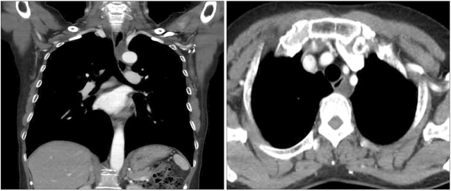
A 53-year-old woman was transferred to our hospital with abnormal findings on chest computed tomography. She had been taking medications for reflux esophagitis for 3 years but her symptoms persisted. She had no unusual past histories and had undergone gastrofibroscopy several times in the past with no abnormalities. Chest CT scan showed a cystic mass at the posterior mediastinum (Fig. 1).
Fig. 1.
Chest CT shows a posterior mediastinal cyst next to the esophagus.
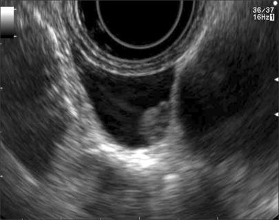
Laboratory findings and a chest X-ray showed no abnormalities. Gastrofibroscopy showed reflux esophagitis and erythematous change with a suspicious submucosal mass in the upper thoracic esophagus. The gastric body and fundus also showed erythematous change. Endoscopic ultrasonography showed a normal esophageal wall and a cystic mass next to the aortic arch. A small polypoid mass was found in the cyst (Fig. 2).
Fig. 2.
Cystic lesion in endoscopic ultrasonography.
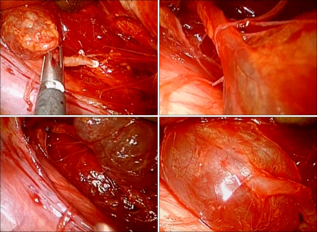
With general anesthesia, thoracoscopic resection of the mass was done. A vascular structure that seemed to be the thoracic duct was found in connection with the cystic mass superiorly and inferiorly (Fig. 3). Careful dissection of the cystic mass and the thoracic duct was done with care not to rupture the cyst or the duct. The thoracic duct was ligated above and below the mass and the cystic mass was successfully removed. The content of the mass was clear fluid.
Fig. 3.
Intraoperative findings.
The patient was sent to the general ward without any problems or hemodynamic instability. Postoperatively, the patient was well without any complications such as hemothorax or chylothorax. The chest tube was removed on the second postoperative day and she was discharged the next day.
DISCUSSION
Cysts of the thoracic duct can occur either above or below the diaphragm. Supradiaphragmatic thoracic duct cysts are typically found in the neck. Mediastinal thoracic duct cysts are quite uncommon [1-3]. The cysts are symptomatic because of the pressure on adjacent structures. Symptoms such as coughing, dyspnea, and chest discomfort may be experienced. Symptoms of dysphagia are often associated with ingestion of fatty foods. Furthermore, acute respiratory insufficiency after ingestion of a fatty meal can be seen in some patients [3-5].
Radiographically, these cysts appear as a round or oval sharply circumscribed mass in the visceral compartment. Although computed tomography shows the cystic nature of the lesion, it does not differentiate it from any other mediastinal cystic lesion [2,4]. Magnetic resonance imaging, especially T2-weighted images, better delineates the anatomic boundaries. The high signal intensity has been attributed to the high concentration of lipids and proteinaceous material in the cyst [3-5]. Fine-needle aspiration is a valuable diagnostic tool, yet its efficiency is debatable because of the hypocellularity of the aspirate. Differential diagnosis includes pericardial or pleural mesothelial cysts, teratomatous cysts, bronchial or esophageal cysts, and neurenteric and lymphangiomatous cysts [2,3].
Definitive diagnosis of mediastinal thoracic duct cysts is based on surgical findings and histopathologic analysis--finding a thick wall, including the endothelium and connective tissue, without elastic fiber [2,3,5].
Surgical resection of the cyst is usually recommended. The surgical treatment should be performed to confirm the diagnosis by histologic analysis and to prevent potential complications such as the spontaneous or traumatic rupture of the cyst. Surgical treatment consists of removal of the cyst and ligation of all lymphatics connected to it [3,5,6].
When the localization of the cyst or invasion to vital structures prevent total excision, partial cystectomy is the treatment of choice [2,4].
The most common complication seen after surgery is chylothorax requiring reoperation. To prevent this complication, the entire connection of the thoracic duct should be ligated. Video-assisted thoracic surgery may be an acceptable surgical procedure. Although surgery is the treatment of choice, ethanol sclerotherapy and an expectative approach in asymptomatic patients with a confirmed diagnosis may represent an alternative therapy [5,6].
In our case, an incidentally found mediastinal cyst turned out to be a thoracic duct cyst. We report the case with a literature review, as we have successfully treated the patient by removing the cyst and ligating the thoracic duct superiorly and inferiorly through thoracoscopic surgery.
References
- 1.Mattila PS, Tarkkanen J, Mattila S. Thoracic duct cyst: a case report and review of 29 cases. Ann Otol Rhinol Laryngol. 1999;108:505–508. doi: 10.1177/000348949910800516. [DOI] [PubMed] [Google Scholar]
- 2.Okabe K, Miura K, Konishi H, et al. Thoracic duct cyst of the mediastinum. Scand J Thorac Cardiovasc Surg. 1993;27:175–177. doi: 10.3109/14017439309099107. [DOI] [PubMed] [Google Scholar]
- 3.Karajiannis A, Kruegetb T, Stauffer E, et al. Large thoracic duct cyst. Eur J Cardiothorac Surg. 2000;17:754–756. doi: 10.1016/s1010-7940(00)00447-4. [DOI] [PubMed] [Google Scholar]
- 4.Morettin LB, Allen TE. Thoracic duct cyst: diagnosis with needle aspiration. Radiology. 1986;161:437–438. doi: 10.1148/radiology.161.2.3763916. [DOI] [PubMed] [Google Scholar]
- 5.Gottwald F, Iro H, Zenk J, et al. Thoracic duct cysts: a rare differential diagnosis. Otolaryngol Head Neck Surg. 2005;132:330–333. doi: 10.1016/j.otohns.2004.09.002. [DOI] [PubMed] [Google Scholar]
- 6.Gomez E, Tejerina E, Martorell V, et al. Thoracic duct cyst presenting as an hour-glass shaped mass in the left supraclavicular area: case report. Acta Otorhinolaryngol Belg. 2001;55:223–225. [PubMed] [Google Scholar]


