Abstract
Purpose
This study investigated the role of death receptor 3 (DR3) in experimental autoimmune uveitis (EAU).
Methods
EAU was induced in B10.RIII mice by subcutaneous injection of interphotoreceptor retinoid-binding protein (IRBP) 161–180 emulsified with complete Freund’s adjuvant and evaluated with clinical and histopathologic observation. Total protein of draining lymph nodes (DLNs) was extracted from the control, EAU, or recovery phase mice. CD4+ T cells were separated from lymphocytes with magnetic-assisted cell sorting. At the same time, some of the CD4+ T cells were cultured with or without recombinant TL1A (rTL1A, the DR3 ligand) for three days, and the supernatants were collected for the interleukin-17 (IL-17) test. DR3 mRNA and protein levels in CD4+ T cells and the endogenous concentration of TL1A in mice DLNs were assessed with real-time PCR or western blotting. Levels of IL-17 in the supernatants were determined with enzyme-linked immunosorbent assay.
Results
Histopathological and clinical data revealed severe intraocular inflammation in the immunized mice. The inflammation reached its peak on day 14 in EAU and had resolved in the recovery phase (weeks 4–5 or more after IRBP immunization). CD4+ T cells obtained from EAU (day 7 or 14) had higher levels of DR3 mRNA and protein expression compared with the control group treated with complete Freund’s adjuvant alone and the recovery group. However, the DR3 mRNA and protein levels on day 21 in EAU were similar to those observed in the control and recovery groups. The endogenous levels of TL1A were upregulated in EAU, and decreased in the recovery phase mice. Adding rTL1A increased the production of IL-17 by CD4+ T cells isolated from mice DLNs. Moreover, the increased IL-17 levels in the culture supernatant of CD4+ T cells from EAU were much higher than those from the control and recovery phase mice. However, the effects on promoting IL-17 production in TL1A-stimulated CD4+ T cells were similar between the controland recovery groups.
Conclusions
Our data suggest that DR3 expression is induced during EAU and may be involved in the development of this disease, possibly by promoting IL-17 secretion.
Introduction
Experimental autoimmune uveitis (EAU) is a T cell–mediated autoimmune disease that serves as a model for several human posterior uveitis [1], such as Behcet’s disease, Vogt-Koyanagi-Harada syndrome (VKH), birdshot retinochoroidopathy, and sympathetic ophthalmia [2,3]. EAU is induced in animals by the adoptive transfer of retinal antigen-specific T lymphocytes [4,5] between syngeneic rodents [6,7], or by immunization with retinal antigens, such as the soluble retinal antigen (S-antigen) and interphotoreceptor retinoid-binding protein (IRBP) [8-14]. In addition, patients with uveitis have serum auto-antibodies to retinal antigens, including S-Ag and IRBP [15,16], and EAU can be induced by immunizing animals with the retinal antigens known to elicit responses in lymphocytes isolated from patients with uveitis [17].
During EAU, the integrity of the blood-retinal barrier is compromised, and monocytes/macrophages and antigen-specific T lymphocytes infiltrating the retina cause tissue damage [18]. Researchers have generally believed that EAU is caused by interferon gamma (IFN-γ) mainly secreted by CD4+ T helper1 (Th1) lymphocytes [19-22]. Recently, evidence suggested that newly recognized interleukin (IL)-17, produced by T helperIL-17 cells (IL-17-producing CD4+ T cells, Th17), plays a crucial role in this autoimmune disease by stimulating the initial influx of leukocytes into target tissues and mediating the tissue inflammation [18,23-26].
Death receptor 3 (DR3, TNFRSF25, TRAMP, LARD) is a member of the death-domain-containing tumor necrosis factor superfamily (TNFSF) of receptors. DR3 is primarily expressed on T cells and is essential for the development of diverse T cell–mediated inflammatory diseases [27]. TL1A (TNFSF15), a new TNFSF member, is currently the only known ligand of DR3 [28,29]. TL1A was first identified as a protein expressed on human endothelial cells and upregulated in response to tumor necrosis factor-alpha (TNF-α) and IL-1α. [28] Subsequently, expression of TL1A by antigen presenting cells (APCs) [29], such as human tissue macrophages [30], FcγR-activated peripheral blood (PB) monocytes, and monocyte-derived dendritic cells (DCs), was demonstrated [30-34].
CD4+ T cell activation and differentiation need not only the recognition of the antigen-major histocompatibility complex (MHC) class II complex by the cognate T cell receptor (TCR) but also co-stimulatory signals [35]. Most of these signals belong to either the B7-type molecules that bind CD28-like immunoglobulin (Ig) superfamily receptors or the TNFSF ligands that engage their receptor counterparts in the TNFRSF [36,37]. Several lines of evidence point to a role for TL1A-DR3 binding in modulating CD4+ T cell activation [27]. For example, in vitro, under conditions of suboptimal anti-CD3/CD28 stimulation, TL1A interaction with DR3 increases IL-2-driven proliferation and IFN-γ and granulocyte-macrophage colony-stimulating factor (GM-CSF) production, and TL1A:DR3 interaction also synergizes with IL-12 and IL-18 in stimulating TCR-independent secretion of IFN-γ by human PB CD4+ T cells [38,39]. In vivo, DR3-TL1A signaling has been associated with several autoinflammatory conditions, including allergic asthma [40], experimental autoimmune encephalomyelitis (EAE) [27,35], experimental antigen-induced arthritis (AIA) [30], inflammatory bowel disease [41], and allergic lung inflammation [42]. In addition, mice lacking the DR3 gene (DR3ko) exhibited a reduction in all histopathological hallmarks of EAE, allergic lung inflammation [27], and AIA [30]. Moreover, some researchers have discovered that TL1A-DR3 interaction regulates Th17 development and IL-17 production [35], which play an important role in many autoimmune diseases [23,24,43-48]. Therefore, DR3 could be an attractive therapeutic target for T cell–mediated autoimmune and allergic diseases.
Thus far, studies have shown that CD4+ T cells are essential for the development of EAU [7,49-51]. However, the regulation of DR3 expressed by CD4+ T cells or the effect of DR3 on IL-17 production has not been investigated in the EAU model. This study is the first to investigate the expression and function of DR3 in CD4+ T cells in EAU. We found that DR3 mRNA and protein levels were elevated in EAU and that stimulation with the DR3 ligand, TL1A, upregulated the secretion of IL-17 from CD4+ T cells. These results indicate that DR3 may play an important role in the development and maintenance of EAU.
Methods
Mice and immunization
B10.RIII mice (6–8 weeks of age) were purchased from Jackson Laboratories (Bar Harbor, ME) and were housed under standard (specific pathogen-free) conditions. All animals were treated according to the ARVO Statement for the Use of Animals in Ophthalmic and Vision Research. IRBP161–180 (SGIPYIISYLHPGNTILHVD) was synthesized by Shanghai Sangon Biologic Engineering Technology and Services Ltd. Complete Freund’s adjuvant (CFA) containing 1.0 mg/ml Mycobacterium tuberculosis and pertussis toxin (PTX) were obtained from Sigma-Aldrich Co. (St. Louis, MO). To induce EAU, mice (8–12 weeks of age) were immunized subcutaneously with a 200 µl emulsion containing 50 µg IRBP161–180 in CFA. PTX (1.0 µg) was concurrently injected intraperitoneally as an adjuvant [52]. Control groups of mice received an emulsion of 50 µl PBS and 150 µl CFA, which was injected subcutaneously. Each experimental group consisted of 6–8 mice.
Clinical examination and histopathological evaluation
After the animals were immunized, they were observed with slit lamp microscopy and ophthalmoscopy starting on day 7 until day 28. Eyes were enucleated from the control, EAU, and recovery phase mice (weeks 4–5 or more after IRBP immunization), were fixed for 1 h in 4% buffered glutaraldehyde, and were then transferred to 10% buffered formaldehyde until processing. Fixed and dehydrated tissue was embedded in paraffin and 5- to 7-µm sections were stained using a standard hematoxylin and eosin (H and E) approach. The intensity of EAU was scored from 0 to 4 in a blinded fashion according to the histopathological grading system previously described for murine EAU [50,53].
Purification of CD4+ T cells, cultivation, and medium collection
Lymphocytes were collected from mice by draining lymph nodes (DLNs, inguinal and iliac), and CD4+ T cells were isolated using a specialized kit (Miltenyi Biotec, Palo Alto, CA). Briefly, lymphocyte suspensions were incubated with CD4 MicroBeads to sort CD4+ T cells. Then T cells were incubated in Gibco RPMI 1640 medium (Invitrogen, Carlsbad, CA) with anti-CD3 (1 µg/ml) and anti-CD28 (1 µg/ml) (eBioscience, San Diego, CA), with or without recombinant TL1A (rTL1A, 100 ng/ml [29,54]; R and D Systems, Minneapolis, MN). Incubations were performed in a 24-well culture plate for 72 h at 37 °C under an atmosphere of 5% CO2. After incubation, the supernatant was collected and stored at −80 °C.
Reverse transcription PCR (RT–PCR) and real-time PCR
Total RNA was extracted from CD4+ T cells that had been isolated from control, EAU, or recovery phase mice using the RNA extraction kit (RNeasy Mini Kit, Qiagen, Hilden, Germany). Two micrograms of total RNA from each sample were used for reverse transcription using the Superscript III Reverse Transcriptase system (Invitrogen). The following sequences of the DR3 and glyceraldehyde 3-phosphate dehydrogenase (GAPDH) primers used for real-time PCR: 5′-GGG CTA TCC TGA TCT GTG CAT-3′ (forward primer, DR3), 5′-ATG CCA GAG GAG TTC CAA GAGT-3′ (reverse primer, DR3), 5′-GAG AAC TTT GGC ATT GTG G-3′ (forward primer, GAPDH), and 5′-ATG CAG GGA TGA TGT TCT G-3′ (reverse primer, GAPDH). The mRNA expression levels were normalized to GAPDH, which was used as a reference housekeeping gene. PCR analysis was conducted on the real-time fluorescence quantitative PCR system using SYBR Green PCR Master Mix (Qiagen) according to the manufacturer’s instructions.
Western blotting analysis
Total protein of DLNs was extracted from the control, EAU, or recovery phase mice using a protein extraction kit (BioChain, Hayward, CA). CD4+ T cells were homogenized using an ultrasonicator (BANDELIN Electronic, Bberlin, Germany), and the protein lysates were prepared for western blotting (WB) analysis. Fifty micrograms of protein from each sample was subjected to sodium fodecyl sulfate–PAGE, and the separated proteins were then transferred to polyvinylidene fluoride membranes. The membranes were incubated with anti-mouse TL1A (rabbit polyclonal antibody, Abcam, Cambridge, MA), anti-mouse DR3 (rabbit polyclonal antibody, Santa Cruz Biotechnology, Santa Cruz, CA), or anti-β-actin (rabbit polyclonal antibody, Santa Cruz Biotechnology), followed by a secondary antibody (goat antirabbit IgG-HRP; Santa Cruz Biotechnology). Proteins were detected using the Phototope-HRP western blot detection system (Cell Signaling, Danvers, MA).
Enzyme-linked immunosorbent assay
Levels of IL-17 in culture media were measured using a commercially available enzyme-linked immunosorbent assay (ELISA) kit (R and D Systems). The detection limit of the kit is 15 pg/ml.
Statistical analysis
Data were expressed as mean±SD. The experimental groups were compared with Student t tests, assuming equal variances for all data. P values < 0.05 were considered to be statistically different.
Results
Induction of EAU
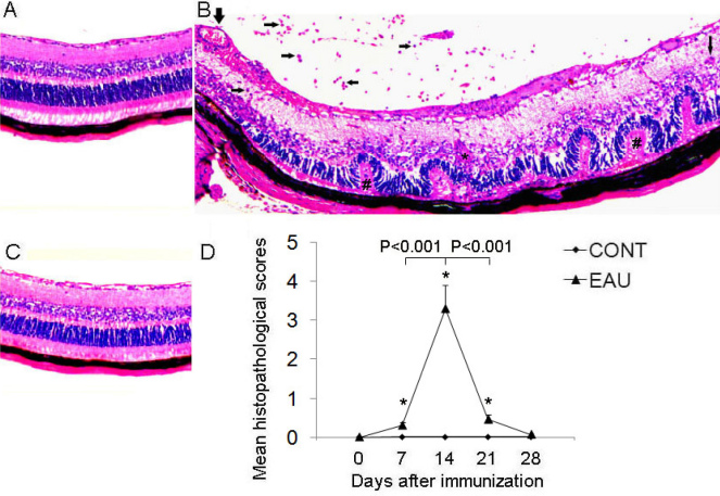
EAU was successfully induced in B10.RIII mice after immunization with 50 µg IRBP161–180 in CFA [52]. Immunohistochemistry revealed severe inflammation in the posterior segment and a massive influx of inflammatory cells infiltrating the vitreous body and retina, vitritis, vasculitis, granuloma formation, and retinal photoreceptor lesions (Figure 1B). Inflammation was initially identified on days 7–9 after immunization and reached its peak by day 14. Inflammation was then followed by a rapid resolution and recovery (Figure 1D). As published reports have shown, there was no apparent inflammation in the control (treated with CFA only) [50,52] and recovery groups (week 4–5 or more after IRBP immunization) [53] (Figure 1A,C).
Figure 1.
Histopathologic features of the eyes enucleated from experimental autoimmune uveitis (EAU), recovery phase and control mice. A: Eye of CFA controls. Normal retinal structure in a B10.RIII mouse. Hematoxylin-eosin (H and E) staining (magnification, 200×). B: Eye of EAU mice. An image from mice day 14 after IRBP immunization (at the peak of inflammation) shows inflammatory T lymphocytes (horizontal arrows) and macrophages (vertical thin arrow) infiltrating the vitreous and the retina, vasculitis (vertical bold arrow), damage to the retinal photoreceptor cell layer (pounds) and granuloma (asterisk). H and E staining (magnification, 200×). C: Eye of recovery phase mice. An image from mice week 4–5 or more after IRBP immunization shows no obvious inflammation in the retina. H and E staining (magnification, 200×). D: Mean histopathologic score during the development of EAU. * indicates p<0.001 when compared EAU with control mice. Each value represents the mean±SD (n=6).
Induction of EAU increases DR3 mRNA and protein expression in CD4+ T cells
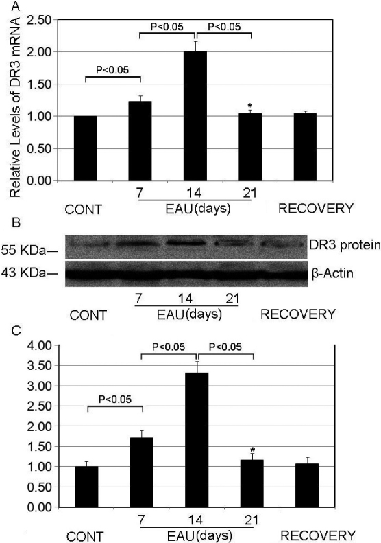
Since previous studies suggested that CD4+ T cells play a significant role in the development and maintenance of EAU [7,49], we used CD4 microbeads to sort CD4+ T cells from mouse lymphocytes obtained by draining LNs. The mRNA and total protein were extracted from CD4+ T cells for analysis with RT–PCR and WB. We found that the DR3 mRNA and protein levels increased in CD4+ T cells from the EAU group 7 or 14 days after immunization (Figure 2). However, there were no obvious differences in DR3 mRNA and protein levels between the controls, EAU (day 21), and recovery phase mice. We also found that the expression of DR3 in CD4+ T cells in EAU achieved the highest level at day 14, and then declined (Figure 2). These data raise the question of the role of DR3 upregulation in EAU.
Figure 2.
DR3 mRNA and protein levels in CD4+ T cells from experimental autoimmune uveitis (EAU) increased. A: Real-time PCR analysis of DR3 mRNA expression in CD4+ T cells isolated from the control, EAU, or recovery phase mice. GAPDH mRNA was used as a control to normalize the total mRNA levels. * indicates p<0.05 when compared day 7 with day 21 in the EAU group. B: western blotting analysis of DR3 protein expression in the CD4+ T cells. β-Actin was used as a loading control. C: Densitometry quantification of western-blotting results in panel B. * indicates p<0.05 when day 7 is compared with day 21 in the EAU group. Each value represents the mean±SD (n=6).
Secretion of IL-17 into CD4+ T cell culture medium is upregulated by the DR3 ligand, TL1A
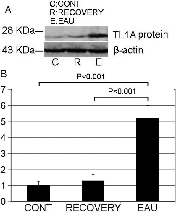
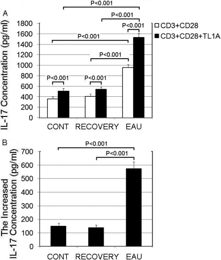
To determine the potential role for increased DR3 expression in EAU, we chose the cells of the most severe inflammatory mice (day 14) to represent the EAU [53] as shown in Figure 1 and Figure 2. At first, we detected the endogenous levels of TL1A in mice DLNs with WB and found that the concentration of TL1A in EAU was upregulated; then it decreased in the recovery phase mice (Figure 3). After that, we added recombinant TL1A into the medium of the CD4+ T cells. Since under conditions of suboptimal anti-CD3/CD28 stimulation CD4+ T cells are thoroughly activated [35], we included anti-CD3/CD28 in the medium. We then measured IL-17 in the culture supernatants with ELISA and found that TL1A interaction with DR3 clearly promoted IL-17 production by CD4+ T cells compared with the media without TL1A in the control, EAU, or recovery mice (Figure 4A). This effect was significantly more pronounced in CD4+ T cells obtained from the DLNs of the EAU mice. Then we calculated the increased concentration of IL-17 after stimulated by TL1A for 2 h, and found that the elevated IL-17 levels in the culture supernatant in the EAU group were much higher, about fourfold, than that in the other two groups (Figure 4B). However, the increased levels of IL-17 stimulated by TL1A in the control group were similar to those in the recovery group (Figure 4B).
Figure 3.
The mice endogenous level of TL1A was upregulated in experimental autoimmune uveitis (EAU). A: The concentration of TL1A in draining lymph nodes was detected with western blotting. B: Densitometry quantification of western-blotting results in panel A. Each value represents the mean±SD (n=6).
Figure 4.
TL1A (TNFSF15) promoted secretion of IL-17 by CD4+ T cells. A: CD4+ T cells from the control, experimental autoimmune uveitis (EAU), and recovery phase mice were cultured in the presence of anti-CD3 (1 µg/ml) and anti-CD28 (1 µg/ml), with or without recombinant TL1A (100 ng/ml) for 72 h. IL-17 levels in the media were then determined with ELISA. B: The increased concentration of IL-17 secreted by CD4+ T cell in each group after TL1A stimulation for 72 h. Each value represents the mean±SD (n=6).
Discussion
Uveitis is considered a typical T-cell mediating, organ-specific, autoimmune disease. EAU in B10.RIII mice induced by IRBP161–180, the best-studied model of uveitis recently shown to be IL-17 driven [23,44,55], has commonly been described as a monophasic disease [56], with a clinical peak about 2 weeks after immunization [37,57,58], then followed by remission and tolerance to reinduction; namely, EAU is a self-limited disease [1]. Similar to EAU, human posterior uveitis is also characterized by a bilateral granuloma, vasculitis, retinal lesions, and so on; however, human posterior uveitis often follows a relapsing and remitting course [59-61], of which the etiology and pathology are still elusive [62].
Previous studies showed that DR3 is expressed on T cells [28], and DR3:TL1A signaling has been associated with several autoinflammatory diseases, including EAE, lung inflammation [27], Crohn’s disease [29], and experimental arthritis [30,63].
To further investigate its function, we analyzed the role of DR3 in EAU. First, we examined the expression of DR3 in mice. The DR3 mRNA and protein levels in the CD4+ T cells of EAU mice were upregulated compared with the recovery phase mice and controls. Since DR3 expression coincides with the rapid induction of its ligand (TL1A) expression [29,32] and DR3 binding to TL1A regulates T-cell activation and expansion [27,63], we examined whether TL1A was also elevated in EAU. Results showed that the endogenous TL1A levels were significantly increased in EAU compared with the recovery phase mice and controls. These data imply that DR3 needs to be coupled with its ligand TL1A to execute its function.
Several studies show that Th17 cells or IL-17 are associated with ocular inflammatory diseases such as uveitis [60,64,65] and CD4+ T cells are capable of producing IL-17 [12]. Therefore, we studied the effect of DR3 on IL-17 production in CD4+ T cells and try to explain the mechanism of DR3 function in EAU. We found that DR3 interaction with TL1A could induce the increase of IL-17 production by CD4+ T cells. This is consistent with other findings that DR3-TL1A interaction regulates the Th17 cell function and IL-17-mediated autoimmune disease [35]. In summary, our study showed that increased DR3 production may be associated with the development of EAU in mice. These results indicate that stimulation of DR3 with TL1A could increase IL-17 production, with the suggestion that the DR3:TL1A signaling pathway may be involved in the pathogenesis of autoimmune uveitis.
In future research, we plan to determine how the interaction of TL1A with DR3 can increase the secretion of IL-17. Our studies will focus on elucidating cell signaling pathways that are activated in response to TL1A binding to DR3 and on the possibility of the production of cytokines other than IL-17. In addition, we want to knock out the DR3 gene in B10.RIII mice to observe whether the EAU model can still be induced. These studies will provide valuable new insights, and ultimately, we hope that elucidation of these mechanisms will enable the development of new therapeutic methods to treat human autoimmune and inflammatory diseases.
Acknowledgments
We thank Dr. Shasha Gao and the staff of Chongqing Eye Institute for excellent technical assistant.
References
- 1.Singh VK, Biswas S, Anand R, Agarwal SS. Experimental autoimmune uveitis as animal model for human posterior uveitis. Indian J Med Res. 1998;107:53–67. [PubMed] [Google Scholar]
- 2.Faure JP. Autoimmunity and the retina. Curr Top Eye Res. 1980;2:215–302. [PubMed] [Google Scholar]
- 3.Wacker WB, Donoso LA, Kalsow CM, Yankeelov JA, Jr, Organisciak DT. Experimental allergic uveitis. Isolation, characterization, and localization of a soluble uveitopathogenic antigen from bovine retina. J Immunol. 1977;119:1949–58. [PubMed] [Google Scholar]
- 4.Caspi RR, Roberge FG, McAllister CG, el-Saied M, Kuwabara T, Gery I, Hanna E, Nussenblatt RB. T cell lines mediating experimental autoimmune uveoretinitis (EAU) in the rat. J Immunol. 1986;136:928–33. [PubMed] [Google Scholar]
- 5.Sanui H, Redmond TM, Kotake S, Wiggert B, Hu LH, Margalit H, Berzofsky JA, Chader GJ, Gery I. Identification of an immunodominant and highly immunopathogenic determinant in the retinal interphotoreceptor retinoid-binding protein (IRBP). J Exp Med. 1989;169:1947–60. doi: 10.1084/jem.169.6.1947. [DOI] [PMC free article] [PubMed] [Google Scholar]
- 6.Fox GM, Redmond TM, Wiggert B, Kuwabara T, Chader GJ, Gery I. Dissociation between lymphocyte activation for proliferation and for the capacity to adoptively transfer uveoretinitis. J Immunol. 1987;138:3242–6. [PubMed] [Google Scholar]
- 7.Rizzo LV, Silver P, Wiggert B, Hakim F, Gazzinelli RT, Chan CC, Caspi RR. Establishment and characterization of a murine CD4+ T cell line and clone that induce experimental autoimmune uveoretinitis in B10.A mice. J Immunol. 1996;156:1654–60. [PubMed] [Google Scholar]
- 8.Rozenszajn LA, Muellenberg-Coulombre C, Gery I, el-Saied M, Kuwabara T, Mochizuki M, Lando Z, Nussenblatt RB. Induction of experimental autoimmune uveoretinitis by T-cell lines. Immunology. 1986;57:559–65. [PMC free article] [PubMed] [Google Scholar]
- 9.Caspi RR, Roberge FG, Chan CC, Wiggert B, Chader GJ, Rozenszajn LA, Lando Z, Nussenblatt RB. A new model of autoimmune disease. Experimental autoimmune uveoretinitis induced in mice with two different retinal antigens. J Immunol. 1988;140:1490–5. [PubMed] [Google Scholar]
- 10.Iwase K, Fujii Y, Nakashima I, Kato N, Fujino Y, Kawashima H, Mochizuki M. A new method for induction of experimental autoimmune uveoretinitis (EAU) in mice. Curr Eye Res. 1990;9:207–16. doi: 10.3109/02713689009044515. [DOI] [PubMed] [Google Scholar]
- 11.Sasamoto Y, Kotake S, Yoshikawa K, Wiggert B, Gery I, Matsuda H. Interphotoreceptor retinoid-binding protein derived peptide can induce experimental autoimmune uveoretinitis in various rat strains. Curr Eye Res. 1994;13:845–9. doi: 10.3109/02713689409025141. [DOI] [PubMed] [Google Scholar]
- 12.Peng Y, Han G, Shao H, Wang Y, Kaplan HJ, Sun D. Characterization of IL-17+ interphotoreceptor retinoid-binding protein-specific T cells in experimental autoimmune uveitis. Invest Ophthalmol Vis Sci. 2007;48:4153–61. doi: 10.1167/iovs.07-0251. [DOI] [PMC free article] [PubMed] [Google Scholar]
- 13.Chan CC, Nussenblatt RB, Wiggert B, Redmond TM, Fujikawa LS, Chader GJ, Gery I. Immunohistochemical analysis of experimental autoimmune uveoretinitis (EAU) induced by interphotoreceptor retinoid-binding protein (IRBP) in the rat. Immunol Invest. 1987;16:63–74. doi: 10.3109/08820138709055713. [DOI] [PubMed] [Google Scholar]
- 14.Gregerson DS, Fling SP, Obritsch WF, Merryman CF, Donoso LA. Identification of T cell recognition sites in S-antigen: dissociation of proliferative and pathogenic sites. Cell Immunol. 1989;123:427–40. doi: 10.1016/0008-8749(89)90302-x. [DOI] [PubMed] [Google Scholar]
- 15.Okunuki Y, Usui Y, Takeuchi M, Kezuka T, Hattori T, Masuko K, Nakamura H, Yudoh K, Usui M, Nishioka K, Kato T. Proteomic surveillance of autoimmunity in Behcet's disease with uveitis: selenium binding protein is a novel autoantigen in Behcet's disease. Exp Eye Res. 2007;84:823–31. doi: 10.1016/j.exer.2007.01.003. [DOI] [PubMed] [Google Scholar]
- 16.Okunuki Y, Usui Y, Kezuka T, Hattori T, Masuko K, Nakamura H, Yudoh K, Goto H, Usui M, Nishioka K, Kato T, Takeuchi M. Proteomic surveillance of retinal autoantigens in endogenous uveitis: implication of esterase D and brain-type creatine kinase as novel autoantigens. Mol Vis. 2008;14:1094–104. [PMC free article] [PubMed] [Google Scholar]
- 17.Caspi RR. A look at autoimmunity and inflammation in the eye. J Clin Invest. 2010;120:3073–83. doi: 10.1172/JCI42440. [DOI] [PMC free article] [PubMed] [Google Scholar]
- 18.Yoshimura T, Sonoda KH, Miyazaki Y, Iwakura Y, Ishibashi T, Yoshimura A, Yoshida H. Differential roles for IFN-gamma and IL-17 in experimental autoimmune uveoretinitis. Int Immunol. 2008;20:209–14. doi: 10.1093/intimm/dxm135. [DOI] [PubMed] [Google Scholar]
- 19.Romagnani S. T-cell subsets (Th1 versus Th2). Ann Allergy Asthma Immunol. 2000;85:9–18. doi: 10.1016/S1081-1206(10)62426-X. [DOI] [PubMed] [Google Scholar]
- 20.Hoey S, Grabowski PS, Ralston SH, Forrester JV, Liversidge J. Nitric oxide accelerates the onset and increases the severity of experimental autoimmune uveoretinitis through an IFN-gamma-dependent mechanism. J Immunol. 1997;159:5132–42. [PubMed] [Google Scholar]
- 21.Jones LS, Rizzo LV, Agarwal RK, Tarrant TK, Chan CC, Wiggert B, Caspi RR. IFN-gamma-deficient mice develop experimental autoimmune uveitis in the context of a deviant effector response. J Immunol. 1997;158:5997–6005. [PubMed] [Google Scholar]
- 22.Crane IJ, Xu H, Wallace C, Manivannan A, Mack M, Liversidge J, Marquez G, Sharp PF, Forrester JV. Involvement of CCR5 in the passage of Th1-type cells across the blood-retina barrier in experimental autoimmune uveitis. J Leukoc Biol. 2006;79:435–43. doi: 10.1189/jlb.0305130. [DOI] [PubMed] [Google Scholar]
- 23.Zhang R, Qian J, Guo J, Yuan YF, Xue K. Suppression of experimental autoimmune uveoretinitis by Anti-IL-17 antibody. Curr Eye Res. 2009;34:297–303. doi: 10.1080/02713680902741696. [DOI] [PubMed] [Google Scholar]
- 24.Langrish CL, Chen Y, Blumenschein WM, Mattson J, Basham B, Sedgwick JD, McClanahan T, Kastelein RA, Cua DJ. IL-23 drives a pathogenic T cell population that induces autoimmune inflammation. J Exp Med. 2005;201:233–40. doi: 10.1084/jem.20041257. [DOI] [PMC free article] [PubMed] [Google Scholar]
- 25.Commodaro AG, Bombardieri CR, Peron JP, Saito KC, Guedes PM, Hamassaki DE, Belfort RN, Rizzo LV, Belfort R, Jr, de Camargo MM. p38{alpha} MAP kinase controls IL-17 synthesis in vogt-koyanagi-harada syndrome and experimental autoimmune uveitis. Invest Ophthalmol Vis Sci. 2010;51:3567–74. doi: 10.1167/iovs.09-4393. [DOI] [PubMed] [Google Scholar]
- 26.Weaver CT, Harrington LE, Mangan PR, Gavrieli M, Murphy KM. Th17: an effector CD4 T cell lineage with regulatory T cell ties. Immunity. 2006;24:677–88. doi: 10.1016/j.immuni.2006.06.002. [DOI] [PubMed] [Google Scholar]
- 27.Meylan F, Davidson TS, Kahle E, Kinder M, Acharya K, Jankovic D, Bundoc V, Hodges M, Shevach EM, Keane-Myers A, Wang EC, Siegel RM. The TNF-family receptor DR3 is essential for diverse T cell-mediated inflammatory diseases. Immunity. 2008;29:79–89. doi: 10.1016/j.immuni.2008.04.021. [DOI] [PMC free article] [PubMed] [Google Scholar]
- 28.Migone TS, Zhang J, Luo X, Zhuang L, Chen C, Hu B, Hong JS, Perry JW, Chen SF, Zhou JX, Cho YH, Ullrich S, Kanakaraj P, Carrell J, Boyd E, Olsen HS, Hu G, Pukac L, Liu D, Ni J, Kim S, Gentz R, Feng P, Moore PA, Ruben SM, Wei P. TL1A is a TNF-like ligand for DR3 and TR6/DcR3 and functions as a T cell costimulator. Immunity. 2002;16:479–92. doi: 10.1016/s1074-7613(02)00283-2. [DOI] [PubMed] [Google Scholar]
- 29.Bamias G, Mishina M, Nyce M, Ross WG, Kollias G, Rivera-Nieves J, Pizarro TT, Cominelli F. Role of TL1A and its receptor DR3 in two models of chronic murine ileitis. Proc Natl Acad Sci USA. 2006;103:8441–6. doi: 10.1073/pnas.0510903103. [DOI] [PMC free article] [PubMed] [Google Scholar]
- 30.Bull MJ, Williams AS, Mecklenburgh Z, Calder CJ, Twohig JP, Elford C, Evans BA, Rowley TF, Slebioda TJ, Taraban VY, Al-Shamkhani A, Wang EC. The Death Receptor 3-TNF-like protein 1A pathway drives adverse bone pathology in inflammatory arthritis. J Exp Med. 2008;205:2457–64. doi: 10.1084/jem.20072378. [DOI] [PMC free article] [PubMed] [Google Scholar]
- 31.Bamias G, Martin C, 3rd, Marini M, Hoang S, Mishina M, Ross WG, Sachedina MA, Friel CM, Mize J, Bickston SJ, Pizarro TT, Wei P, Cominelli F. Expression, localization, and functional activity of TL1A, a novel Th1-polarizing cytokine in inflammatory bowel disease. J Immunol. 2003;171:4868–74. doi: 10.4049/jimmunol.171.9.4868. [DOI] [PubMed] [Google Scholar]
- 32.Kang YJ, Kim WJ, Bae HU, Kim DI, Park YB, Park JE, Kwon BS, Lee WH. Involvement of TL1A and DR3 in induction of pro-inflammatory cytokines and matrix metalloproteinase-9 in atherogenesis. Cytokine. 2005;29:229–35. doi: 10.1016/j.cyto.2004.12.001. [DOI] [PubMed] [Google Scholar]
- 33.Prehn JL, Mehdizadeh S, Landers CJ, Luo X, Cha SC, Wei P, Targan SR. Potential role for TL1A, the new TNF-family member and potent costimulator of IFN-gamma, in mucosal inflammation. Clin Immunol. 2004;112:66–77. doi: 10.1016/j.clim.2004.02.007. [DOI] [PubMed] [Google Scholar]
- 34.Prehn JL, Thomas LS, Landers CJ, Yu QT, Michelsen KS, Targan SR. The T cell costimulator TL1A is induced by FcgammaR signaling in human monocytes and dendritic cells. J Immunol. 2007;178:4033–8. doi: 10.4049/jimmunol.178.7.4033. [DOI] [PubMed] [Google Scholar]
- 35.Pappu BP, Borodovsky A, Zheng TS, Yang X, Wu P, Dong X, Weng S, Browning B, Scott ML, Ma L, Su L, Tian Q, Schneider P, Flavell RA, Dong C, Burkly LC. TL1A–DR3 interaction regulates Th17 cell function and Th17-mediated autoimmune disease. J Exp Med. 2008;205:1049–62. doi: 10.1084/jem.20071364. [DOI] [PMC free article] [PubMed] [Google Scholar]
- 36.Watts TH. TNF/TNFR family members in costimulation of T cell responses. Annu Rev Immunol. 2005;23:23–68. doi: 10.1146/annurev.immunol.23.021704.115839. [DOI] [PubMed] [Google Scholar]
- 37.Jiang HR, Lumsden L, Forrester JV. Macrophages and dendritic cells in IRBP-induced experimental autoimmune uveoretinitis in B10RIII mice. Invest Ophthalmol Vis Sci. 1999;40:3177–85. [PubMed] [Google Scholar]
- 38.Papadakis KA, Prehn JL, Landers C, Han Q, Luo X, Cha SC, Wei P, Targan SR. TL1A synergizes with IL-12 and IL-18 to enhance IFN-gamma production in human T cells and NK cells. J Immunol. 2004;172:7002–7. doi: 10.4049/jimmunol.172.11.7002. [DOI] [PubMed] [Google Scholar]
- 39.Papadakis KA, Zhu D, Prehn JL, Landers C, Avanesyan A, Lafkas G, Targan SR. Dominant role for TL1A/DR3 pathway in IL-12 plus IL-18-induced IFN-gamma production by peripheral blood and mucosal CCR9+ T lymphocytes. J Immunol. 2005;174:4985–90. doi: 10.4049/jimmunol.174.8.4985. [DOI] [PubMed] [Google Scholar]
- 40.Fang L, Adkins B, Deyev V, Podack ER. Essential role of TNF receptor superfamily 25 (TNFRSF25) in the development of allergic lung inflammation. J Exp Med. 2008;205:1037–48. doi: 10.1084/jem.20072528. [DOI] [PMC free article] [PubMed] [Google Scholar]
- 41.Takedatsu H, Michelsen KS, Wei B, Landers CJ, Thomas LS, Dhall D, Braun J, Targan SR. TL1A (TNFSF15) regulates the development of chronic colitis by modulating both T-helper 1 and T-helper 17 activation. Gastroenterology. 2008;135:552–67. doi: 10.1053/j.gastro.2008.04.037. [DOI] [PMC free article] [PubMed] [Google Scholar]
- 42.Schreiber TH, Wolf D, Tsai MS, Chirinos J, Deyev VV, Gonzalez L, Malek TR, Levy RB, Podack ER. Therapeutic Treg expansion in mice by TNFRSF25 prevents allergic lung inflammation. J Clin Invest. 2010;120:3629–40. doi: 10.1172/JCI42933. [DOI] [PMC free article] [PubMed] [Google Scholar]
- 43.Ferber IA, Brocke S, Taylor-Edwards C, Ridgway W, Dinisco C, Steinman L, Dalton D, Fathman CG. Mice with a disrupted IFN-gamma gene are susceptible to the induction of experimental autoimmune encephalomyelitis (EAE). J Immunol. 1996;156:5–7. [PubMed] [Google Scholar]
- 44.Tang J, Zhou R, Luger D, Zhu W, Silver PB, Grajewski RS, Su SB, Chan CC, Adorini L, Caspi RR. Calcitriol suppresses antiretinal autoimmunity through inhibitory effects on the Th17 effector response. J Immunol. 2009;182:4624–32. doi: 10.4049/jimmunol.0801543. [DOI] [PMC free article] [PubMed] [Google Scholar]
- 45.Komiyama Y, Nakae S, Matsuki T, Nambu A, Ishigame H, Kakuta S, Sudo K, Iwakura Y. IL-17 plays an important role in the development of experimental autoimmune encephalomyelitis. J Immunol. 2006;177:566–73. doi: 10.4049/jimmunol.177.1.566. [DOI] [PubMed] [Google Scholar]
- 46.Chen Y, Langrish CL, McKenzie B, Joyce-Shaikh B, Stumhofer JS, McClanahan T, Blumenschein W, Churakovsa T, Low J, Presta L, Hunter CA, Kastelein RA, Cua DJ. Anti-IL-23 therapy inhibits multiple inflammatory pathways and ameliorates autoimmune encephalomyelitis. J Clin Invest. 2006;116:1317–26. doi: 10.1172/JCI25308. [DOI] [PMC free article] [PubMed] [Google Scholar]
- 47.Bailey SL, Schreiner B, McMahon EJ, Miller SD. CNS myeloid DCs presenting endogenous myelin peptides 'preferentially' polarize CD4+ T(H)-17 cells in relapsing EAE. Nat Immunol. 2007;8:172–80. doi: 10.1038/ni1430. [DOI] [PubMed] [Google Scholar]
- 48.Kolls JK, Linden A. Interleukin-17 family members and inflammation. Immunity. 2004;21:467–76. doi: 10.1016/j.immuni.2004.08.018. [DOI] [PubMed] [Google Scholar]
- 49.Liu X, Lee YS, Yu CR, Egwuagu CE. Loss of STAT3 in CD4+ T cells prevents development of experimental autoimmune diseases. J Immunol. 2008;180:6070–6. doi: 10.4049/jimmunol.180.9.6070. [DOI] [PMC free article] [PubMed] [Google Scholar]
- 50.Chan CC, Caspi RR, Ni M, Leake WC, Wiggert B, Chader GJ, Nussenblatt RB. Pathology of experimental autoimmune uveoretinitis in mice. J Autoimmun. 1990;3:247–55. doi: 10.1016/0896-8411(90)90144-h. [DOI] [PubMed] [Google Scholar]
- 51.Prendergast RA, Iliff CE, Coskuncan NM, Caspi RR, Sartani G, Tarrant TK, Lutty GA, McLeod DS. T cell traffic and the inflammatory response in experimental autoimmune uveoretinitis. Invest Ophthalmol Vis Sci. 1998;39:754–62. [PubMed] [Google Scholar]
- 52.Sun M, Yang P, Du L, Zhou H, Ren X, Kijlstra A. Contribution of CD4+CD25+ T cells to the regression phase of experimental autoimmune uveoretinitis. Invest Ophthalmol Vis Sci. 2010;51:383–9. doi: 10.1167/iovs.09-3514. [DOI] [PubMed] [Google Scholar]
- 53.Liu L, Xu Y, Wang J, Li H. Upregulated IL-21 and IL-21 receptor expression is involved in experimental autoimmune uveitis (EAU). Mol Vis. 2009;15:2938–44. [PMC free article] [PubMed] [Google Scholar]
- 54.Zhang J, Wang X, Fahmi H, Wojcik S, Fikes J, Yu Y, Wu J, Luo H. Role of TL1A in the pathogenesis of rheumatoid arthritis. J Immunol. 2009;183:5350–7. doi: 10.4049/jimmunol.0802645. [DOI] [PubMed] [Google Scholar]
- 55.Caspi RR, Chan CC, Grubbs BG, Silver PB, Wiggert B, Parsa CF, Bahmanyar S, Billiau A, Heremans H. Endogenous systemic IFN-gamma has a protective role against ocular autoimmunity in mice. J Immunol. 1994;152:890–9. [PubMed] [Google Scholar]
- 56.de Kozak Y, Thillaye B, Renard G, Faure JP. Hyperacute form of experimental autoimmune uveo-retinitis in Lewis rats; electron microscopic study. Albrecht Von Graefes Arch Klin Exp Ophthalmol. 1978;208:135–42. doi: 10.1007/BF00406988. [DOI] [PubMed] [Google Scholar]
- 57.Kerr EC, Raveney BJ, Copland DA, Dick AD, Nicholson LB. Analysis of retinal cellular infiltrate in experimental autoimmune uveoretinitis reveals multiple regulatory cell populations. J Autoimmun. 2008;31:354–61. doi: 10.1016/j.jaut.2008.08.006. [DOI] [PubMed] [Google Scholar]
- 58.Silver PB, Chan CC, Wiggert B, Caspi RR. The requirement for pertussis to induce EAU is strain-dependent: B10.RIII, but not B10.A mice, develop EAU and Th1 responses to IRBP without pertussis treatment. Invest Ophthalmol Vis Sci. 1999;40:2898–905. [PubMed] [Google Scholar]
- 59.Dick AD. Immune mechanisms of uveitis: insights into disease pathogenesis and treatment. Int Ophthalmol Clin. 2000;40:1–18. doi: 10.1097/00004397-200004000-00003. [DOI] [PubMed] [Google Scholar]
- 60.Chi W, Yang P, Li B, Wu C, Jin H, Zhu X, Chen L, Zhou H, Huang X, Kijlstra A. IL-23 promotes CD4+ T cells to produce IL-17 in Vogt-Koyanagi-Harada disease. J Allergy Clin Immunol. 2007;119:1218–24. doi: 10.1016/j.jaci.2007.01.010. [DOI] [PubMed] [Google Scholar]
- 61.Forrester JV, Liversidge J, Dua HS, Towler H, McMenamin PG. Comparison of clinical and experimental uveitis. Curr Eye Res. 1990;9(Suppl):75–84. doi: 10.3109/02713689008999424. [DOI] [PubMed] [Google Scholar]
- 62.Sheu SJ. Update on uveomeningoencephalitides. Curr Opin Neurol. 2005;18:323–9. doi: 10.1097/01.wco.0000169753.31321.4e. [DOI] [PubMed] [Google Scholar]
- 63.Cassatella MA, Pereira-da-Silva G, Tinazzi I, Facchetti F, Scapini P, Calzetti F, Tamassia N, Wei P, Nardelli B, Roschke V, Vecchi A, Mantovani A, Bambara LM, Edwards SW, Carletto A. Soluble TNF-like cytokine (TL1A) production by immune complexes stimulated monocytes in rheumatoid arthritis. J Immunol. 2007;178:7325–33. doi: 10.4049/jimmunol.178.11.7325. [DOI] [PubMed] [Google Scholar]
- 64.Yoshimura T, Sonoda KH, Ohguro N, Ohsugi Y, Ishibashi T, Cua DJ, Kobayashi T, Yoshida H, Yoshimura A. Involvement of Th17 cells and the effect of anti-IL-6 therapy in autoimmune uveitis. Rheumatology (Oxford) 2009;48:347–54. doi: 10.1093/rheumatology/ken489. [DOI] [PMC free article] [PubMed] [Google Scholar]
- 65.Amadi-Obi A, Yu CR, Liu X, Mahdi RM, Clarke GL, Nussenblatt RB, Gery I, Lee YS, Egwuagu CE. TH17 cells contribute to uveitis and scleritis and are expanded by IL-2 and inhibited by IL-27/STAT1. Nat Med. 2007;13:711–8. doi: 10.1038/nm1585. [DOI] [PubMed] [Google Scholar]


