Abstract
Although the genetic component in the etiology of rheumatoid arthritis (RA) has been consistently suggested, many novel genetic loci remain to uncover. To identify RA risk loci, we performed a genome-wide association study (GWAS) with 100 RA cases and 600 controls using Affymetrix SNP array 5.0. The candidate risk locus (APOM gene) was re-sequenced to discover novel promoter and coding variants in a group of the subjects. Replication was performed with the independent case-control set comprising of 578 RAs and 711 controls. Through GWAS, we identified a novel SNP associated with RA at the APOM gene in the MHC class III region on 6p21.33 (rs805297, odds ratio (OR) = 2.28, P = 5.20 × 10⁻⁷). Three more polymorphisms were identified at the promoter region of the APOM by the re-sequencing. For the replication, we genotyped the four SNP loci in the independent case-control set. The association of rs805297 identified by GWAS was successfully replicated (OR = 1.40, P = 6.65 × 10⁻⁵). The association became more significant in the combined analysis of discovery and replication sets (OR = 1.56, P = 2.73 ± 10-10). The individuals with the rs805297 risk allele (A) at the promoter region showed a significantly lower level of APOM expression compared with those with the protective allele (C) homozygote. In the logistic regressions by the phenotype status, the homozygote risk genotype (A/A) consistently showed higher ORs than the heterozygote one (A/C) for the phenotype-positive RAs. These results indicate that APOM promoter polymorphisms are significantly associated with the susceptibility to RA.
Keywords: APOM protein, human; autoimmune diseases; genome-wide association study; polymorphism, single nucleotide; rheumatoid arthritis
Introduction
Rheumatoid arthritis (RA) is a common systemic autoimmune disease that is characterized by chronic inflammation of the synovium, which can lead to progressive joint destruction. It is a complex disease that is caused by multiple factors such as genetic, environmental, and hormonal contributions (Firestein, 2003). While the exact pathogenesis of RA is still unknown, multiple lines of evidence such as a higher concordance rate in monozygotic twins than in dizygotic twins and the higher risk in siblings of patients compared with that in a general population (MacGregor et al., 2000; Goronzy and Weyand, 2009), suggest the genetic component in the etiology of this disease.
As for the efforts to understand the genetic etiology of RA, various genome-wide association studies (GWAS) and meta-analyses have identified a number of risk loci including HLA-DRB1, PTPN22, CD40, STAT4, OLIG3, TNFAIP3, TNFRSF14, CTLA4, CCL2, PADI4 and TRAF1/C5 (Plenge et al., 2007a, 2007b; WTCCC, 2007; Julià et al., 2008; Raychaudhuri et al., 2008; Gregersen et al., 2009; Kochi et al., 2010; Stahl et al., 2010). Some of the significant SNPs in the candidate RA-associated genes have shown consistent significance across diverse ethnic groups, while some other SNPs have not. For example, the significant SNPs in HLA-DRB1, STAT4, OLIG3 and TNFAIP3 identified in Caucasians were consistently replicated in the Japanese population, while PTPN22 and CD40 were not replicated in Asians (Kochi et al., 2010; Stahl et al., 2010). When Lee et al examined whether the known genetic variants at 4q27, 6q23, CCL21, TRAF1/C5 and CD40 identified in Caucasians were also associated with RA in Koreans, those loci did not show any significant associations and some of them were not even polymorphic (Lee et al., 2009). Even within a similar ethnic group, the association of some candidate genetic markers to RA was differently reported. For example, polymorphisms in OLIG3 or TNFAIP3 genes were reported to be significantly associated with RA in Japanese (Kochi et al., 2010) but not in Korean population (Han et al., 2009). Recent meta-analysis of GWAS also suggested that only a small amount of the genetic component can be explained by the known RA risk alleles (Raychaudhuri et al., 2008; Stahl et al., 2010). These data imply that additional risk alleles remain to be identified.
Based on this inference, we attempted to find new risk loci for RA in Korean population. For this purpose, we performed a three step analysis. First, we identified the risk loci using GWAS analysis with Affymetrix SNP array 5.0 in the discovery set that consisted of 100 RA patients and 600 healthy controls. Second, after selecting the candidate risk loci, we screened the SNPs in the promoter region and in the entire exons, including the exon-intron boundaries of the candidate gene, by PCR-direct sequencing. Third, we performed a replication study with independent Korean samples of 578 RA patients and 711 healthy controls to verify the association. Haplotype analysis, qRT-PCT and reporter assay followed to refine our findings of the candidate markers.
Results
Genome-wide scan for RA
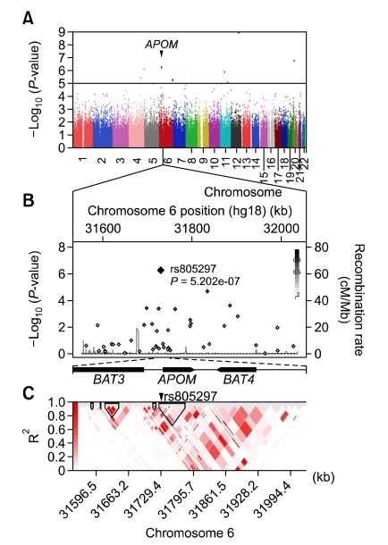
To identify the RA risk loci, we first performed whole-genome SNP genotyping for 100 RA cases and 600 normal controls using Affymetrix Human SNP array 5.0. After filtering the SNPs based on the threshold cutoff as described in the Methods section, we obtained 300,909 reliable SNPs. To check whether the process of SNP quality control effectively removed the false-associations, we drew the Quantile-Quantile plot (Q-Q plot) based on the P values from a logistic regression analysis (Supplemental Data Figure S1). Since we did not observe any notable divergence from the null hypothesis in the plot with the genomic inflation factor (λGC) = 1.03, we performed the GWAS with the 300,909 SNPs (Figure 1A). Eight SNPs were found to be significant with P values under 10-5 (unadjusted) (Table 1). Since it is thought that a SNP tends to be false positive if it is the solely significant SNP around the region, we examined the P-value profiles of the neighboring SNPs around the eight significant SNPs to minimize false positive results and improve the efficiency of replication (Supplemental Data Figure S2). Based on our strategy, we chose the one locus around rs805297 SNP in the promoter region of APOM gene on 6p21.33 for further analysis (OR = 2.28, 95% CI 1.65-3.14, P = 5.20 × 10⁻⁷), because it harbored a clusters of nearly significant SNPs (P = 10-3-10-5) (Figure 1B). After deciding the replication target, also to increase the statistical power, we performed the replication study with a larger set (578 cases and 711 controls). The details of the significance of the SNPs on 6p21.33 are available in Supplemental Data Table S1.
Figure 1.
GWAS results and plots in the region around APOM. (A) Manhattan plot displaying the -log10 (P-value) of SNPs in the GWAS for 100 Korean RA cases and 600 controls. APOM region is marked. (B) The -log10 (P-value) of SNPs around APOM region. SNPs are represented with diamond dots and the most strongly associated SNP (rs805297) was marked with the largest diamond. The strength of LD relationship (r2) between rs805297 and other SNPs is presented with red color intensities based on the JPT + CHB Hapmap data. The background recombination rate curve is drawn with the JPT + CHB Hapmap data (light blue curve). The location and orientation of APOM and neighboring genes are illustrated under the -log10 plot. (C) LD map of the APOM region. The r2 based LD map is drawn with the genotype data of the RA cases and controls using Golden helix SVS software version 7.3.0.
Table 1.
Results of genome-wide association study for RA in Korean population
*Significant SNPs with the smallest P-values were presented. †Based on NCBI human genome build 36.3 (hg18). ‡Unadjusted P-values from logistic regression. RA, Rheumatoid arthritis; OR, odds ratio of minor allele; CI, confidence interval.
Identification of SNPs in APOM gene in the Korean population
To expand the list of candidate SNPs, we screened the sequences of all six exons containing intronexon boundaries, a 5' flanking region and a 3' untranslated region (UTR) of APOM gene for more SNPs in 48 Koreans. In addition to the rs805297, three more SNPs (rs805296, rs1266078 and rs9404941) with MAF > 5% were identified in the promoter region (Table 2), while there was none in the exons. Linkage disequilibrium (LD) was not observed among the four SNPs (Supplemental Data Table S2).
Table 2.
Results of replication study between APOM polymorphisms and RA susceptibility in Korean population
*Logistic regression analysis was used to estimate odds ratio based on the additive model with age and gender as covariates. MAF, minor allele frequency; RA, rheumatoid arthritis; OR, odds ratios for minor allele; CI, confidence interval.
Replication of the association of APOM promoter polymorphism and RA
We performed the replication study with all four polymorphisms in the promoter region of APOM gene in the independent Korean RA case-control set (578 cases and 711 controls). The frequencies of all 4 SNP loci were in Hardy-Weinberg equilibrium in both the RA patients and the controls (P > 0.05, data not shown). The association of rs805297 polymorphism was successfully replicated in the replication set (OR = 1.40, 95% CI 1.19-1.65, P = 6.65 × 10⁻⁵) (Table 2). When we combined the discovery and replication sets, the association of rs805297 with RA became more significant (OR = 1.56, 95% CI 1.36-1.80, P = 2.73 × 10⁻¹⁰).
We found that rs805296, another polymorphism near the rs805297 in the promoter region, was also significantly associated with RA (OR = 1.52, 95% CI 1.08-2.14, P = 0.016), while the other two SNPs were not. LD was not observed between the two polymorphisms, suggesting that they may behave independently from each other. Indeed, multivariate analysis with the two significant polymorphisms (rs805296 and rs805297) revealed that both were independent indicators of RA risk (Hazard ratio [HR] = 1.76, 95% CI = 1.25-2.48, P = 0.001 and HR = 1.53, 95% CI = 1.29-1.80, P = 5.49 × 10⁻⁷, respectively). We also performed haplotype analysis based on the two significant SNPs (Table 3). All three haplotypes showed significant differences between the RA patients and the controls. Interestingly, Ht1 (C-T), which is composed of two non-risk alleles, indeed showed a significantly protective effect against RA (OR = 0.64, 95% CI 0.55-0.75, P = 3.56 × 10⁻⁸). On the other hand, Ht2 (A-T) and Ht3 (C-C), which are composed of one risk allele and one non-risk allele, were found to increase the risk of RA (OR = 1.44, 95% CI 1.23 -1.69, P = 7.01 × 10⁻⁶ and OR = 1.48, 95% CI 1.06-2.07, P = 0.02, respectively). There was no haplotype comprising two risk alleles in this study population.
Table 3.
Result of haplotype analysis with two SNPs in the promoter region of APOM gene
*Haplotype: rs805297-rs805296. †Haplotype frequency was constructed by the expectation maximization algorithm with genotyped SNPs. ‡Odds ratios (OR), 95% confidence intervals (95% CI) and P-values were obtained from chi-square (χ2) test. "Risk" or "Non-risk" presents the alleles more or less frequently occurred in RA cases than controls.
Associations between APOM polymorphisms and the various phenotypes of RA
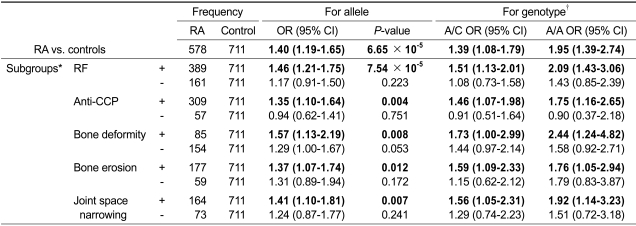
We then examined the relationship between the rs805297 polymorphism and the clinical phenotypes such as RF, anti-CCP antibodies, bone deformity, bone erosion and joint space narrowing in the independent Korean subjects (578 cases and 711 controls). We first assessed the associations among patients, but found no associations between any phenotypes and the rs805297 polymorphism (data not shown). Next, we performed the logistic regressions for the same clinical phenotypes, but used subgroups of patients based on each phenotype (e.g., RF positive patients vs. controls; RF negative patients vs. controls etc). The genotypes containing the A allele were significantly associated with the phenotype-positive cases, while not with the phenotype-negative cases (Table 4). When using the C/C genotype as reference (OR = 1), the homozygote risk genotype (A/A) consistently showed higher ORs than the heterozygote genotype (A/C) for all the phenotypes in the phenotype-positive cases. Although significant associations were not found in the phenotype-negative cases, the increasing trend across the genotypes remained.
Table 4.
Associations of rs805297 SNP in RA subgroups
*Positive (+) and negative (-) subgroup of each phenotype. †OR of the minor allele homozygote (A/A) and heterozygote (A/C) was calculated when using the C/C genotype as reference (OR = 1). Odds ratios (OR), 95% confidence intervals (95% CI) and P-values were obtained from logistic regression analysis with age and gender as covariate. RF, rheumatoid factor; Anti-CCP, anti-cyclic citrullinated peptide antibody.
The rs805297 polymorphism and APOM expression
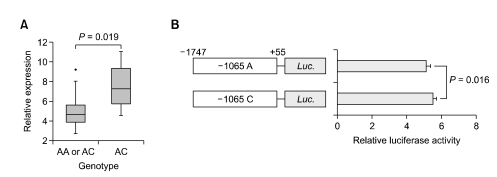
To assess the relationship between the rs805297 polymorphism and the mRNA expression of APOM, we performed qRT-PCR using RNA from 23 RA cases available for RNA extraction. The individuals with genotypes containing a risk allele (A/A or A/C, n = 19) showed a significantly lower level of the mRNA expression than the individuals with the protective allele homozygous genotype (C/C, n = 4) (median 4.6 [IQR 3.1-5.6] vs. 7.3 [IQR 5.7-9.3], P = 0.0186) (Figure 2A). Luciferase reporter assay was performed to further explore whether the C to A substitution at the promoter region of APOM affects the transcription activity. The luciferase activity of the rs805297-A construct was slightly decreased compared with that of the rs805297-C construct in the HEK293 cells (P = 0.016) (Figure 2B). However, in the in-silico prediction for transcription factor binding site based on the promoter sequence of APOM gene (http://www.cbrc.jp/research/db/TFSEARCH.html), we could not identify any binding site exceeding the threshold score (default: 85.0).
Figure 2.
Relationship between rs805297 genotypes and APOM expression. (A) APOM gene expression level by rs805297 genotypes. We performed qRT-PCR using RNA from 23 RA cases. The individuals with the genotypes containing the A allele showed significantly lower level of APOM expression than those with C/C homozygote genotype. (B) Effect of the rs805297C > A on the transcriptional activity of APOM promoter in 239HEK cells. The results are represented as mean ± SD of relative luciferase activities obtained from the triplicate experiments. P-values were determined by Mann-Whitney Test.
Discussion
In an effort to identify and characterize the polymorphisms associated with RA, we performed GWAS analysis using Affymetrix Genome-Wide Human SNP array 5.0 and identified 8 candidate SNP polymorphisms in the Korean population. Of the 8 candidate polymorphisms, we selected rs805297 for further replication based on its strong association with susceptibility to RA and the existence of a cluster of SNPs nearly significant SNPs (P = 10-3-10-5) around this locus. It is located on the promoter region of APOM gene in the MHC III region on chromosome 6p21.3. The MHC region is one of the well known loci that are strongly associated with RA. HLA-DRB1 locus in the MHC class II region has been known to be strongly associated with RA. However, in addition to the associations with the DRB1 alleles, additional genetic loci associated with RA are present within the MHC region (Newton et al., 2004). Several papers have reported the existence of genetic loci in the MHC I and III regions that are potentially associated with RA (Jawaheer et al., 2002; Newton et al., 2003; Harney et al., 2008). Apolipoprotein M (apoM) is high density lipoprotein (HDL)-associated apoprotein mainly expressed in the liver and kidney (Xu and Dahlback, 1999). A reduced level of serum HDL is commonly observed in untreated RAs (Park et al., 1999; Choy and Sattar, 2009). Recent evidence has indicated that apoM may contribute to the anti-inflammatory function of HDL (Burger and Dayer, 2002; Feingold et al., 2008) and apoM inhibited by siRNA resulted in decreased HDL level (Wolfrum et al., 2005). These findings suggest that decreased apoM expression may negatively affect anti-inflammatory function of HDL. Interestingly, there was a study reporting that displacement of apolipoprotein A-I, apolipoprotein family in HDL particle, led to a loss of anti-inflammatory activities of HDL (Khovidhunkit et al., 2004). We found that rs805297 (C > A) polymorphism in the APOM promoter decreased its transcription activity slightly, which may affect RA susceptibility through altering the HDL level.
In this study, the association of rs805297 polymorphism (C > A) at the promoter region of APOM was identified and successfully replicated in the independent validation set. A SNP (rs805296) located in APOM promoter region near rs805297 was also found to be significantly associated with RA. SNP rs805296 has been implicated in some diseases such as coronary artery disease and type 1 diabetes mellitus (T1DM), but not in RA (Jiao et al., 2007; Wu et al., 2009). Given that T1DM is an autoimmune disease and that there is a higher risk of atherosclerosis in RA patients than normal individuals, we could assume that APOM promoter polymorphism SNP rs805296 may also be involved in both autoimmune mediated- and inflammation mediated-diseases. According to the linkage and multivariate analysis, both SNPs seem to be independent indicators of RA risk.
We assessed the effect of the rs805297 on the phenotypes among patients, but found no significant associations. However, in the logistic regressions by the phenotype status, we found the significant associations of the rs805297 (C > A) polymorphism with the phenotype-positive groups, but not with the phenotype-negative ones. And the homozygote risk allele genotype consistently showed higher ORs than the heterozygote risk allele genotype in the phenotype-positive ones. It indirectly suggests the involvement of the APOM promoter polymorphism on the development of those phenotypes. Lower transcription activity and lower expression levels of APOM in the risk allele also supports this possibility. Taking our study results and known anti-inflammatory function of apoM into consideration, it is likely that rs805297 (C > A) polymorphism in the APOM promoter may decrease its transcription activity, which may affect RA susceptibility through altering the HDL level. However, although the promoter assay revealed slightly reduced transcription activity in the risk allele, we could not identify any potential transcription factor binding site in the promoter sequence of APOM including rs805297 site by in-silico prediction. Further functional assay will be required to assess the decreased APOM expression in the risk allele.
There are several limitations in this study. First, it is possible to miss potentially significant polymorphisms in the GWAS discovery due to the small size of the cases in the discovery set. However, the significance of APOM polymorphism was successfully replicated in the large independent set. This result suggests that our strategy, i.e., replication of the stringently selected SNPs from the discovery set, compensated the shortcomings of relatively small discovery set. Second, we could not find the direct associations between the rs805297 (C > A) polymorphism and the clinical phenotypes in the patient population. It is required to recruit a large number of patients to identify the effect of the polymorphism on the development of the phenotypes. Lastly, we did not examine the possibility of HLA-DRB1 shared epitope in this study. Further detailed HLA-DR genotyping in larger-scale RA case-control sets and linkage analysis will be required to verify this possibility.
In conclusion, we demonstrated that APOM promoter polymorphisms (rs805297 and rs805296) were significantly associated with RA. To the best of our knowledge, this is the first evidence that APOM promoter polymorphism is associated with the susceptibility to RA, and potentially with the clinical phenotypes.
Methods
Study population
This study was approved by the institutional review board of the Catholic University of Korea (CUMC09U034). All the participants were Korean and provided written informed consent. For the GWAS analysis, 100 RA patients (76 females and 24 males, mean age 54.1 ± 11.4 yr) were recruited from Eulji University Hospital, Korea who were diagnosed with RA according to the 1987 American College of Rheumatology (ACR; formerly, the American Rheumatism Association) classification criteria for RA (Arnett et al., 1988). As a normal control group, 600 healthy individuals (456 females and 144 males, mean age 52.4 ± 9.1 yr) were randomly selected from the Korean Genome Epidemiology Study (KoGES) (Yoo et al., 2005). We also recruited 578 RA patients (469 females and 109 males, mean age 53.2 ± 12.0 yr) and 711 healthy controls (452 females and 259 males, mean age 52.3 ± 12.9 yr) for replication of the candidate associations from St. Vincent Hospital, Suwon, Korea and Eulji University Hospital, Korea. Rheumatoid factor (RF) was measured by the latex fixation test using the Hitachi 7170S kit (Hitachi Co., Tokyo, Japan). Anti-cyclic citrullinated peptide antibodies (anti-CCP) were measured by an enzyme-linked immunosorbent assay (ELISA) using a Diastat anti-CCP kit (MBL Co., Nagoya, Japan). Of the 100 RA cases in GWAS, 72.0 and 63.5% were RF and anti-CCP positives, respectively. Similarly, of the 578 RA cases in replication study, 70.7 and 84.4% were RF and anti-CCP positives, respectively. Clinical characteristics of the RA cases are summarized in Supplemental Data Table S3. Genomic DNA was extracted from whole blood by using a DNeasy Blood & Tissue Kit (Qiagen, Valencia, CA) according to manufacturer's instructions.
SNP genotyping and GWAS
We genotyped the 100 RA cases and 600 controls using the Affymetrix Genome-Wide Human SNP array 5.0 platforms (Affymetrix, Santa Clara, CA). As a SNP genotyping algorithm, we used the Birdseed (version 2) (http://www.broadinstitute.org/mpg/birdsuite/birdseed.html). Based on the model previously established with known genotypes from 270 HapMap samples, the algorithm employs expectation-maximization algorithm to assign each SNP to the AA, AB or BB genotype clusters (Dempster et al., 1977). Using the PLINK software package (http://pngu.mgh.harvard.edu/~purcell/plink/), SNP quality control was performed for 503,590 SNPs and a total of 300,909 SNPs were selected to satisfy minor allele frequency > 5%, call rates > 95%, and HWE P > 0.001 as described previously (Sladek et al., 2007). Genome-wide association analysis was performed based on the PLINK. Logistic regression was applied for adjusting the effects of age and sex. P-values were obtained from the additive model analysis and P < 10-5 was considered significant at a genome-wide level.
Screening SNPs around APOM
To identify polymorphic sites in APOM gene, all six exons including of intron-exon boundaries, 2.30 kb of 5' flanking region, and 3' untranslated region (UTR) were amplified by PCR with the DNAs from 48 Koreans. PCR was performed using 50 ng of genomic DNA, Taq DNA polymerase (EF Taq, SolGent, Daejeon, Korea), and 0.5 pM of each primer under the following conditions: 30 cycles of PCR consisting of denaturation for 10 s at 98℃, annealing for 30 s at 65℃, extension for 2 min at 72℃, and a final extension for 10 min at 72℃ in a thermocycler (Gene Amp PCR system 9700, Applied Biosystems, Foster, CA). The primer formations for PCR and PCR-sequencing are available in Supplemental Data Table S4. The SNPs of APOM were detected by comparing the reference sequence of human chromosome 6 (GenBank accession NT_167249.1).
SNP genotyping for replication
Genotyping for replication was performed by TaqMan analysis. The primer formations for TaqMan assay are available in Supplemental Data Table S4. Primers and probes were designed by Applied Biosystems. The 10 µl of reaction mixture was made up of 225 nM of each primer, 50 nM of each probe, 5 µl of 2 × TaqMan Genotyping Master mix (Applied Biosystems), and 100 ng of genomic DNA. PCR conditions were as follows: 1 cycle at 95℃ for 10 min; 50 cycles at 95℃ for 15 s and 60℃ for 1 min. The PCR was carried out using the Rotor-Gene Thermal Cycler RG6000 (Corbett Research, Mortlake, NSW, Australia) and the products were read and analyzed using the software Rotor-Gene 1.7.40 (Corbett Research). Genotyping of two SNPs (rs9404941 and rs805296), for which TaqMan probes were not available, was conducted by a PCR-restriction fragment length polymorphism (PCR-RFLP). A total of 20 µl reaction mixture was used for PCR containing 50 ng of genomic DNA, Taq DNA polymerase, and 0.5 M of each primer under the following conditions: 30 cycles of PCR consisting of denaturation for 10 s at 98℃, annealing for 20 s at 65℃, extension for 40 s at 72℃, and a final extension for 10 min at 72℃ in a thermocycler (Gene Amp PCR system 9700, Applied Biosystems). The primer formations for PCR-RFLP are available in Supplemental Data Table S4. The PCR products were digested with each of 1 U of HaeIII and RsaI (Promega, Madison, WI) for 3 h at 37℃, respectively. After digestion, PCR fragments were examined on 4% agarose gel stained with ethidium bromide. The sizes of fragments are as follows; of rs9404941, 244 bp for TT, 138 bp and 106 bp for CC and 244 bp, 138 bp, and 106 bp for TC; of rs805296, 244 bp for TT, 183 bp and 61 pb for CC and 244 bp, 183 bp, and 61 bp for TC.
Quantitative RT-PCR (qRT-PCR) for APOM
Total RNA was extracted from the blood samples of 23 RA cases and 8 controls using TRIzol (Invitrogen, Carlsbad, CA) according to manufacturer's instructions. First-strand complementary DNA (cDNA) was synthesized using oligo-dT primer and superscript II reverse transcriptase (Invitrogen). Real-time qRT-PCR was performed using Mx3000P QPCR System and software MxPro Version 3.00 (Stratagene, La Jolla, CA). Reaction mixtures were composed of 1 × SYBR Green Tbr polymerase Master Mix (FINNZYMES, Vantaa, Finland), 0.5 × ROX, 20 pmol of each primer, and 10 ng of cDNA. Primers for APOM were: 5'-TTGTCTACTGCACAGGTTGTGGTC-3' (forward) and 5'-TAGACGCTGTACACACCGATGTCA-3' (reverse). GAPDH primers were used as internal control: 5'-GCGGGGCTCCAGAACATCAT-3' (forward) and 5'- CCAGCCCCAGCGTCAAGGTG-3' (reverse). The PCR cycle was as follows: denaturation at 95℃ for 5 min; 40 cycles of 95℃ for 30 s, 60℃ for 30 s, and 72℃ for 40 s followed by a 72℃ elongation step for 6 min. Relative quantification was performed by the ΔΔCT method as described elsewhere (Kim et al., 2008). All the experiments were repeated three times and the mean value of intensity ratios ± SD was plotted.
Plasmid construction for luciferase reporter assay
The fragment encompassing the promoter region of APOM, -1747 to +55, was amplified with primers containing restriction endonuclease sites. This region contains the four polymorphic sites examined in this study, rs805297, rs805296, rs1266078 and rs9404941. The primers used in the construction of plasmids were as follows: 5'-ggggtaccAGGCAGAGAATTGCTTTGAA-3' (forward) and 5'-ccaagctTGGTGCTCTGTG TGCCTTA-3' (reverse), containing the KpnI and HindIII linkers (underlined sequences), respectively. The PCR reaction was performed with 50 ng of genomic DNA, 10 pmol of the primers described above, and AccuPower™ HF PCR PreMix (Bioneer, Daejeon, Korea) in a 20-µl reaction mixture. The PCR cycle was as follows: denaturation at 94℃ for 5 min; 35 cycles of 94℃ for 30 s, 59℃ for 30 s, and 72℃ for 2 min followed by a 72℃ elongation step for 5 min. The amplified fragment was first ligated into pGEM®-T Easy vector (Promega) and then sub-cloned into the luciferase expression vector pGL3 (Promega) between KpnI and HindIII sites.
The rs805297 (C > A) polymorphic site was generated using the QuikChange® Site-Directed Mutagenesis Kit (Stratagene). The mutagenic primer sets for the rs805297 were as follows: 5'-gcaaatatatactttggaAttaccatgtgcaagtctt-3' (forward) and 5'-aagacttgcacatggtaaTtccaaagtatatatttgc-3' (reverse). All constructs used in this study were sequence-verified, and orientations were confirmed to be correct. Final construct was named pGL3-pAPOM-rs805297C and pGL3-pAPOM-rs805297A.
Luciferase reporter assay
293HEK was cultured in DMEM supplemented with 10% heat-inactivated fetal bovine serum (Hyclone, Waltham, MA), and were transfected between passages 5 and 10. Cultured cells (5 × 10⁵/well) were plated in 6-well plates and transfected with 1 µg of pGL3-pAPOM-rs805297C and -rs805297A constructs and 0.2 µg of pRL-TK vector (Promega) as internal control by using Fugene HD (Roche Applied Bioscience, Basel, Switzeland). After transfection, the 293HEK cells were incubated for 36 h. The preparation of cell lysate and assessment of luciferase activity were performed using the Dual-Glo Luciferase Assay System (Promega) according to manufacturer's protocol. Luciferase activity was measured using a Lumat LB 9507 luminometer (EG & G Berthhold, Bad Wildbad, Germany). Firefly luciferase activity, as AVPR1A promoter activity, was normalized using the activity of Renilla luciferase. Luciferase activity was expressed as relative light units (RLUs). The pGL3-control (pSV40-luc+) and pGL3-Basic (without promoter) vectors (Promega) were used as positive and negative controls, respectively. Three independent experiments were performed, and the results were represented graphically as a ratio of pGL3-Basic vector activity to controls.
Statistical analysis
Chi-square test was used to estimate the Hardy-Weinberg equilibrium (HWE). Pair-wise linkage disequilibrium (LD) analyses among four SNPs in APOM were performed with the SNPAnalyzer software (DYNACOM, Mobara, Japan). The haplotype frequencies were estimated using the expectation maximization (EM) algorithm with the Golden Helix SVS 7 software (http://www.goldenhelix.com/SNP_Variation/index.html). P-values < 0.05 were considered statistically significant. In the replication study, logistic regression analysis was performed to estimate odds ratio based on the additive model with age and gender as covariates. Gene expression levels for rs805297 genotypes (AA or AC vs. CC) were compared by Mann-Whitney test (SPSS Inc., Chicago, IL).
Supplemental data
Supplemental data include two figures and four tables and can be found with this article online at http://e-emm.or.kr/article/article_files/SP-43-11-02.pdf.
Acknowledgements
This study was supported by the Korea Research Foundation Grant (KRF-2008-220-E00025) and a grant from the Korea Health 21 R&D Project (A040002), Ministry of Health and Welfare, Republic of Korea. The authors would like to thank Mr. JH Kim who participated in the GWAS and logistic regression. The authors would like to thank Korea National Institute of Health (KNIH) for providing control genotyping data for GWAS.
Abbreviations
- APOM
apolipoprotein M
- GWAS
genome-wide association study
- RA
rheumatoid arthritis
- SNP
single nucleotide polymorphism
Supplementary Material
Supplemental Data
References
- 1.Arnett FC, Edworthy SM, Bloch DA, McShane DJ, Fries JF, Cooper NS, Healey LA, Kaplan SR, Liang MH, Luthra HS, et al. The American Rheumatism Association 1987 revised criteria for the classification of rheumatoid arthritis. Arthritis Rheum. 1988;31:315–324. doi: 10.1002/art.1780310302. [DOI] [PubMed] [Google Scholar]
- 2.Burger D, Dayer JM. High-density lipoprotein-associated apolipoprotein A-I: the missing link between infection, chronic inflammation? Autoimmun Rev. 2002;1:111–117. doi: 10.1016/s1568-9972(01)00018-0. [DOI] [PubMed] [Google Scholar]
- 3.Choy E, Sattar N. Interpreting lipid levels in the context of high-grade inflammatory states with a focus on rheumatoid arthritis: a challenge to conventional cardiovascular risk actions. Ann Rheum Dis. 2009;68:460–469. doi: 10.1136/ard.2008.101964. [DOI] [PubMed] [Google Scholar]
- 4.Dempster A, Laird N, Rubin D. Maximum likelihood from incomplete data via the EM algorithm. J R Stat Soc Series B Stat Methodol. 1977;39:1–38. [Google Scholar]
- 5.Feingold KR, Shigenaga JK, Chui LG, Moser A, Khovidhunkit W, Grunfeld C. Infection and inflammation decrease apolipoprotein M expression. Atherosclerosis. 2008;199:19–26. doi: 10.1016/j.atherosclerosis.2007.10.007. [DOI] [PubMed] [Google Scholar]
- 6.Firestein GS. Evolving concepts of rheumatoid arthritis. Nature. 2003;423:356–361. doi: 10.1038/nature01661. [DOI] [PubMed] [Google Scholar]
- 7.Goronzy JJ, Weyand CM. Developments in the scientific understanding of rheumatoid arthritis. Arthritis Res Ther. 2009;11:249. doi: 10.1186/ar2758. [DOI] [PMC free article] [PubMed] [Google Scholar]
- 8.Gregersen PK, Amos CI, Lee AT, Lu Y, Remmers EF, Kastner DL, Seldin MF, Criswell LA, Plenge RM, Holers VM, Mikuls TR, Sokka T, Moreland LW, Bridges SL, Jr, Xie G, Begovich AB, Siminovitch KA. REL, encoding a member of the NF-kappaB family of transcription factors, is a newly defined risk locus for rheumatoid arthritis. Nat Genet. 2009;41:820–823. doi: 10.1038/ng.395. [DOI] [PMC free article] [PubMed] [Google Scholar]
- 9.Han TU, Bang SY, Kang C, Bae SC. TRAF1 polymorphisms associated with rheumatoid arthritis susceptibility in Asians and in Caucasians. Arthritis Rheum. 2009;60:2577–2584. doi: 10.1002/art.24759. [DOI] [PubMed] [Google Scholar]
- 10.Harney SM, Vilarino-Guell C, Adamopoulos IE, Sims AM, Lawrence RW, Cardon LR, Newton JL, Meisel C, Pointon JJ, Darke C, Athanasou N, Wordsworth BP, Brown MA. Fine mapping of the MHC Class III region demonstrates association of AIF1 and rheumatoid arthritis. Rheumatology (Oxford) 2008;47:1761–1767. doi: 10.1093/rheumatology/ken376. [DOI] [PubMed] [Google Scholar]
- 11.Jawaheer D, Li W, Graham R, Chen W, Damle A, Xiao X, Monteiro J, Khalili H, Lee A, Lundsten R. Dissecting the genetic complexity of the association between human leukocyte antigens and rheumatoid arthritis. Am J Hum Genet. 2002;71:585–594. doi: 10.1086/342407. [DOI] [PMC free article] [PubMed] [Google Scholar]
- 12.Jiao G, Yuan Z, Xue Y, Yang C, Lu C. A prospective evaluation of apolipoprotein M gene T-778C polymorphism in relation to coronary artery disease in Han Chinese. Clin Biochem. 2007;40:1108–1112. doi: 10.1016/j.clinbiochem.2007.04.023. [DOI] [PubMed] [Google Scholar]
- 13.Julià A, Ballina J, Cañete J, Balsa A, Tornero Molina J, Naranjo A, Alperi López M, Erra A, Pascual Salcedo D, Barceló P. Genome wide association study of rheumatoid arthritis in the Spanish population: KLF12 as a risk locus for rheumatoid arthritis susceptibility. Arthritis Rheum. 2008;58:2275–2286. doi: 10.1002/art.23623. [DOI] [PubMed] [Google Scholar]
- 14.Khovidhunkit W, Kim MS, Memon RA, Shigenaga JK, Moser AH, Feingold KR, Grunfeld C. Effects of infection and inflammation on lipid and lipoprotein metabolism: mechanisms and consequences to the host. J Lipid Res. 2004;45:1169–1196. doi: 10.1194/jlr.R300019-JLR200. [DOI] [PubMed] [Google Scholar]
- 15.Kim TM, Yim SH, Shin SH, Xu HD, Jung YC, Park CK, Choi JY, Park WS, Kwon MS, Fiegler H, Carter NP, Rhyu MG, Chung YJ. Clinical implication of recurrent copy number alterations in hepatocellular carcinoma and putative oncogenes in recurrent gains on 1q. Int J Cancer. 2008;123:2808–2815. doi: 10.1002/ijc.23901. [DOI] [PMC free article] [PubMed] [Google Scholar]
- 16.Kochi Y, Okada Y, Suzuki A, Ikari K, Terao C, Takahashi A, Yamazaki K, Hosono N, Myouzen K, Tsunoda T, Kamatani N, Furuichi T, Ikegawa S, Ohmura K, Mimori T, Matsuda F, Iwamoto T, Momohara S, Yamanaka H, Yamada R, Kubo M, Nakamura Y, Yamamoto K. A regulatory variant in CCR6 is associated with rheumatoid arthritis susceptibility. Nat Genet. 2010;42:515–519. doi: 10.1038/ng.583. [DOI] [PubMed] [Google Scholar]
- 17.Lee H, Korman B, Le JM, Kastner DL, Remmers EF, Gregersen PK, Bae SC. Genetic risk factors for rheumatoid arthritis differ in Caucasian and Korean populations. Arthritis Rheum. 2009;60:364–371. doi: 10.1002/art.24245. [DOI] [PMC free article] [PubMed] [Google Scholar]
- 18.MacGregor AJ, Snieder H, Rigby AS, Koskenvuo M, Kaprio J, Aho K, Silman AJ. Characterizing the quantitative genetic contribution to rheumatoid arthritis using data from twins. Arthritis Rheum. 2000;43:30–37. doi: 10.1002/1529-0131(200001)43:1<30::AID-ANR5>3.0.CO;2-B. [DOI] [PubMed] [Google Scholar]
- 19.Newton J, Brown MA, Milicic A, Ackerman H, Darke C, Wilson JN, Wordsworth BP, Kwiatkowski D. The effect of HLA-DR on susceptibility to rheumatoid arthritis is influenced by the associated lymphotoxin alpha-tumor necrosis factor haplotype. Arthritis Rheum. 2003;48:90–96. doi: 10.1002/art.10719. [DOI] [PubMed] [Google Scholar]
- 20.Newton JL, Harney SM, Wordsworth BP, Brown MA. A review of the MHC genetics of rheumatoid arthritis. Genes Immun. 2004;5:151–157. doi: 10.1038/sj.gene.6364045. [DOI] [PubMed] [Google Scholar]
- 21.Park YB, Lee SK, Lee WK, Suh CH, Lee CW, Lee CH, Song CH, Lee J. Lipid profiles in untreated patients with rheumatoid arthritis. J Rheumatol. 1999;26:1701–1704. [PubMed] [Google Scholar]
- 22.Plenge RM, Cotsapas C, Davies L, Price AL, de Bakker PI, Maller J, Pe'er I, Burtt NP, Blumenstiel B, DeFelice M, Parkin M, Barry R, Winslow W, Healy C, Graham RR, Neale BM, Izmailova E, Roubenoff R, Parker AN, Glass R, Karlson EW, Maher N, Hafler DA, Lee DM, Seldin MF, Remmers EF, Lee AT, Padyukov L, Alfredsson L, Coblyn J, Weinblatt ME, Gabriel SB, Purcell S, Klareskog L, Gregersen PK, Shadick NA, Daly MJ, Altshuler D. Two independent alleles at 6q23 associated with risk of rheumatoid arthritis. Nat Genet. 2007a;39:1477–1482. doi: 10.1038/ng.2007.27. [DOI] [PMC free article] [PubMed] [Google Scholar]
- 23.Plenge RM, Seielstad M, Padyukov L, Lee AT, Remmers EF, Ding B, Liew A, Khalili H, Chandrasekaran A, Davies LR, Li W, Tan AK, Bonnard C, Ong RT, Thalamuthu A, Pettersson S, Liu C, Tian C, Chen WV, Carulli JP, Beckman EM, Altshuler D, Alfredsson L, Criswell LA, Amos CI, Seldin MF, Kastner DL, Klareskog L, Gregersen PK. TRAF1-C5 as a risk locus for rheumatoid arthritis-a genomewide study. N Engl J Med. 2007b;357:1199–1209. doi: 10.1056/NEJMoa073491. [DOI] [PMC free article] [PubMed] [Google Scholar]
- 24.Raychaudhuri S, Remmers E, Lee A, Hackett R, Guiducci C, Burtt N, Gianniny L, Korman B, Padyukov L, Kurreeman F. Common variants at CD40 and other loci confer risk of rheumatoid arthritis. Nat Genet. 2008;40:1216–1223. doi: 10.1038/ng.233. [DOI] [PMC free article] [PubMed] [Google Scholar]
- 25.Sladek R, Rocheleau G, Rung J, Dina C, Shen L, Serre D, Boutin P, Vincent D, Belisle A, Hadjadj S, Balkau B, Heude B, Charpentier G, Hudson TJ, Montpetit A, Pshezhetsky AV, Prentki M, Posner BI, Balding DJ, Meyre D, Polychronakos C, Froguel P. A genome-wide association study identifies novel risk loci for type 2 diabetes. Nature. 2007;445:881–885. doi: 10.1038/nature05616. [DOI] [PubMed] [Google Scholar]
- 26.Stahl EA, Raychaudhuri S, Remmers EF, Xie G, Eyre S, Thomson BP, Li Y, Kurreeman FA, Zhernakova A, Hinks A, Guiducci C, Chen R, Alfredsson L, Amos CI, Ardlie KG, Barton A, Bowes J, Brouwer E, Burtt NP, Catanese JJ, Coblyn J, Coenen MJ, Costenbader KH, Criswell LA, Crusius JB, Cui J, de Bakker PI, De Jager PL, Ding B, Emery P, Flynn E, Harrison P, Hocking LJ, Huizinga TW, Kastner DL, Ke X, Lee AT, Liu X, Martin P, Morgan AW, Padyukov L, Posthumus MD, Radstake TR, Reid DM, Seielstad M, Seldin MF, Shadick NA, Steer S, Tak PP, Thomson W, van der Helm-van Mil AH, van der Horst-Bruinsma IE, van der Schoot CE, van Riel PL, Weinblatt ME, Wilson AG, Wolbink GJ, Wordsworth BP, Wijmenga C, Karlson EW, Toes RE, de Vries N, Begovich AB, Worthington J, Siminovitch KA, Gregersen PK, Klareskog L, Plenge RM. Genome-wide association study meta-analysis identifies seven new rheumatoid arthritis risk loci. Nat Genet. 2010;42:508–514. doi: 10.1038/ng.582. [DOI] [PMC free article] [PubMed] [Google Scholar]
- 27.Wolfrum C, Poy MN, Stoffel M. Apolipoprotein M is required for prebeta-HDL formation and cholesterol efflux to HDL and protects against atherosclerosis. Nat Med. 2005;11:418–422. doi: 10.1038/nm1211. [DOI] [PubMed] [Google Scholar]
- 28.WTCCC. Genome-wide association study of 14,000 cases of seven common diseases and 3,000 shared controls. Nature. 2007;447:661–678. doi: 10.1038/nature05911. [DOI] [PMC free article] [PubMed] [Google Scholar]
- 29.Wu X, Niu N, Brismar K, Zhu X, Wang X, Efendic S, Du T, Liu Y, Gu HF. Apolipoprotein M promoter polymorphisms alter promoter activity and confer the susceptibility to the development of type 1 diabetes. Clin Biochem. 2009;42:17–21. doi: 10.1016/j.clinbiochem.2008.10.008. [DOI] [PubMed] [Google Scholar]
- 30.Xu N, Dahlback B. A novel human apolipoprotein (apoM) J Biol Chem. 1999;274:31286–31290. doi: 10.1074/jbc.274.44.31286. [DOI] [PubMed] [Google Scholar]
- 31.Yoo K, Shin H, Chang S, Choi B, Hong Y, Kim D, Kang D, Cho N, Shin C, Jin Y. Genomic epidemiology cohorts in Korea: present and the future. Asian Pac J Cancer Prev. 2005;6:238–243. [PubMed] [Google Scholar]
Associated Data
This section collects any data citations, data availability statements, or supplementary materials included in this article.
Supplementary Materials
Supplemental Data


