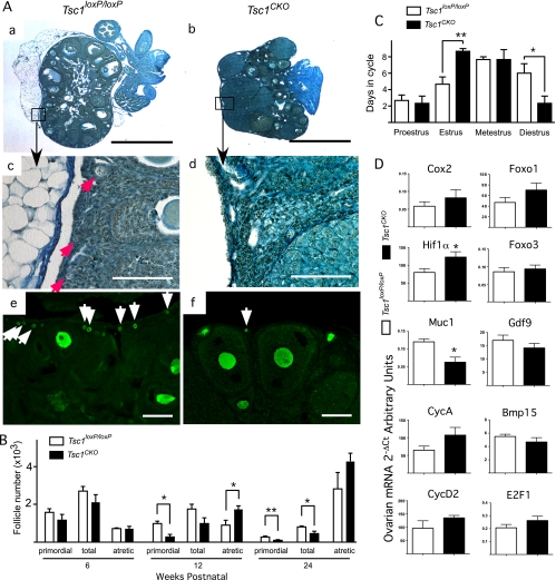
Fig. 2.
Ovarian function in Tsc1CKO mice. A, Hematoxylin-picric acid staining of sections from typical ovaries isolated from 12-wk-old control Tsc1loxP/loxP (a and c) and Tsc1CKO mice (b and d); boxed areas in a and b are shown at higher magnification in c and d, respectively; red arrows indicate primordial follicles (c); MVH immunofluorescence in control (e) and Tsc1CKO (f) ovaries; many small MVH-positive oocytes are seen in Tsc1loxP/loxP ovaries (white arrows in e), whereas few MVH-positive oocytes are seen in Tsc1CKO ovaries (white arrow in f). Bars, 100 μm except for a and b, which are 1 mm. Photos are representative of n = 3 for each group. B, Numbers of primordial, total immature (primordial, primary, and small preantral), and atretic follicles in mouse ovaries from Tsc1loxP/loxP and Tsc1CKO ovaries were counted, averaged, and plotted. C, Estrous cycling in 6-wk-old Tsc1loxP/loxP and Tsc1CKO mice was determined daily by vaginal lavage cytology over a period of 21 d and cumulatively plotted. D, Quantitative real-time PCR was performed with cDNA from control and mutant ovaries in estrus to determine the effect of TSC1 deletion on the expression of key genes involved in ovarian function. Graphs represent the mean ± sem; n = 3 for each group. *, P < 0.05; **, P < 0.01.

