Abstract
Background:
We have previously developed 11C-erlotinib as a new positron emission tomography (PET) tracer and shown that it accumulates in epidermal growth factor receptor (EGFR)-positive lung cancer xenografts in mice. Here, we present a study in patients with non-small cell lung cancer (NSCLC) investigating the feasibility of 11C-erlotinib PET as a potential method for the identification of lung tumours accumulating erlotinib.
Methods:
Thirteen patients with NSCLC destined for erlotinib treatment were examined by contrast-enhanced computed tomography (CT), 11C-erlotinib PET/low-dose CT and 18F-fluoro-2-deoxy-D-glucose (18F-FDG) PET/low-dose CT before start of the erlotinib treatment. After 12 weeks treatment, they were examined by 18F-FDG PET/contrast-enhanced CT for the assessment of clinical response.
Results:
Of the 13 patients included, 4 accumulated 11C-erlotinib in one or more of their lung tumours or lymph-node metastases. Moreover, 11C-erlotinib PET/CT identified lesions that were not visible on 18F-FDG PET/CT. Of the four patients with accumulation of 11C-erlotinib, one died before follow-up, whereas the other three showed a positive response to erlotinib treatment. Three of the nine patients with no accumulation died before follow-up, four showed progressive disease while two had stable disease after 12 weeks of treatment.
Conclusion:
Our data show a potential for 11C-erlotinib PET/CT for visualizing NSCLC lung tumours, including lymph nodes not identified by 18F-FDG PET/CT. Large clinical studies are now needed to explore to which extent pre-treatment 11C-erlotinib PET/CT can predict erlotinib treatment response.
Keywords: erlotinib, EGFR, tarceva, lung cancer, PET imaging
Lung cancer is one of the leading causes of cancer deaths worldwide (Parkin et al, 2005) and the treatment response and clinical outcome of the disease are difficult to predict. Recently, new treatment strategies targeting the epidermal growth factor receptor (EGFR) have been developed. The EGFR is one of the most frequently overexpressed proteins in various cancers including lung cancer, and signalling through this receptor is related to tumour progression and resistance to most treatments (Rusch et al, 1993; Fontanini et al, 1998; Ciardiello and Tortora, 2008). Therefore, the EGFR has become an attractive target for cancer treatment. The two most commonly used tyrosine kinase inhibitors targeting EGFR are gefitinib (Iressa, ZD1839) and erlotinib (Tarceva, OSI-774). Gefitinib and erlotinib are tailored drugs that compete with adenosine triphosphate (ATP) for the ATP binding site on the EGFR and thereby prevent phosphorylation and activation of downstream signalling molecules involved in cell proliferation and tumour growth. Gefitinib was the first EGFR inhibitor approved for treatment of advanced non-small cell lung cancer (NSCLC); however, clinical trials using gefitinib did not show significant improvement in survival (Comis, 2005). In contrast, trials with erlotinib have demonstrated prolonged progression-free survival and improved survival of patients with advanced NSCLC (Shepherd et al, 2004). Erlotinib was also superior to placebo with respect to quality of life (Cohen et al, 2005). Nevertheless, overall response rates have been relatively low in studies that have examined all NSCLC patients collectively (Shepherd et al, 2005), indicating that not all lung cancer patients are suitable for erlotinib treatment and that the treatment should only be given to selected patients. Various parameters have been used to classify patients who respond to erlotinib, such as type of tumour, smoking history, gender, and ethnicity, but none of these parameters had significant impact on survival (Fukuoka et al, 2003; Perez-Soler et al, 2004). Patients with tumours expressing high amounts of the EGFR had an improved response to treatment with erlotinib (Shepherd et al, 2005) and the presence of specific mutations around the ATP binding domain of the receptor was found to increase the response to gefitinib treatment (Lynch et al, 2004; Paez et al, 2004). Determination of the EGFR expression and the presence of mutations require a tumour biopsy, which is not possible to collect in all situations. Thus, non-invasive methods are needed that can identify the subset of patients who are most likely to benefit from erlotinib treatment.
Positron emission tomography (PET) is a 3-dimensional imaging technique that uses isotope-labelled tracers that decay with the emission of a positron, it is used for non-invasive assessment of biochemical and physiological processes in vivo. In the present study, we used 2-[18F]fluoro-2-deoxy-D-glucose (18F-FDG) for visualization of the higher glucose metabolism of tumour tissue compared with surrounding tissue (Gambhir, 2002; Jerusalem et al, 2003; Rohren et al, 2004) and 11C-labelled erlotinib for visualization of EGFRs. Labelling of gefitinib with 11C (Wang et al, 2006; Kawamura et al, 2009; Zhang et al, 2010) and 18F (Su et al, 2008) has been attempted but 18F-gefitinib showed a high non-specific cellular uptake both in vitro and in mice xenografted with human tumours. Furthermore, in this in-vivo model the 18F-gefitinib signal did not relate to EGFR expression (Su et al, 2008). In contrast, 11C-gefitinib showed enhanced accumulation in vitro in the cancer cells that had the highest EGFR expression (Zhang et al, 2010). In a recent micro-PET study, we reported the development of a new radiotracer, 11C-erlotinib, and its use in mice models of human lung cancer (Memon et al, 2009). Our results showed that 11C-erlotinib accumulated in xenografts that were sensitive to erlotinib treatment and expressed high levels of EGFR.
In the present study in patients with NSCLC, we examined the feasibility of using 11C-erlotinib PET combined with X-ray computed tomography (CT) for visualization of tumour tissue, metastases, and malignant lymph nodes.
Subjects and Methods
Subjects and recruitment
Thirteen patients with NSCLC were included between December 2008 and October 2009 before start on second-line treatment with erlotinib. According to clinical practice, patients with metastatic lung cancer were treated with first-line chemotherapy as a palliative treatment (n=10) and patients with locally advanced lung tumours received a potentially curative treatment with concomitant chemotherapy and radiotherapy (n=3). If the patients progressed within 6 months as assessed by the ‘Response evaluation criteria in solid tumours’ (RECIST) (Eisenhauer et al, 2009) using contrast-enhanced CT, treatment with erlotinib was offered as second-line treatment.
Patients were eligible if they were over 18 years of age, had normal liver and kidney function as judged from blood tests, were non-diabetic, and could lie in the PET/CT scanner for 90 min (Table 1). Patients were excluded if they were allergic to X-ray contrast agent or had marked dyspnoea at rest. The study was approved by the Central Denmark Region Committees on Biomedical Research Ethics (M-20080050) and the Danish Medical Association (2512-96464) and conducted in accordance with the Helsinki II Declaration.
Table 1. Clinical characteristics of patients with non-small lung cancer (NSCLC) at inclusion for 11C-erlotinib PET/CT.
| Patients (n) | 13 |
| Male | 4 |
| Female | 9 |
| Age (years) (median; range) | 63 (42–79) |
| Histology of NSCLC (n) | |
| Adenocarcinoma | 8 |
| Squamous cell carcinoma | 3 |
| Large-cell carcinoma | 1 |
| Unspecified | 1 |
| TNM classification (n) | |
| Stage | |
| Tx | 2 |
| T1 | 2 |
| T2 | 4 |
| T3–4 | 5 |
| Node | |
| Nx | 1 |
| N1 | 0 |
| N2 | 7 |
| N3 | 5 |
| Metastasis | |
| M0 | 2 |
| M1A | 0 |
| M1B | 11 |
Abbreviations: CT=computed tomography; PET=positron emission tomography; n=number of patients; TNM=tumour-node metastasis.
Study design
The patients were examined by contrast-enhanced CT before inclusion into the study, as mentioned above. After inclusion, patients underwent pre-treatment 11C-erlotinib PET and 18F-FDG PET examinations combined with low-dose CT scans (scan 1). Erlotinib treatment was started immediately hereafter. Twelve weeks after start of the treatment, 18F-FDG PET combined with contrast-enhanced CT (scan 2) was performed. In patient no. 12, who discontinued the treatment after 7 weeks, scan 2 was performed at this time. The primary end point was higher accumulation of 11C-erlotinib in localized foci than in surrounding tissues and the secondary end point was clinical response, as assessed after 12 weeks of erlotinib treatment by the RECIST criteria and 18F-FDG PET/CT scans, or death of the patient.
The evaluation of the CT images was performed by an experienced radiologist and a nuclear medicine specialist and the evaluation of the PET images was performed by two nuclear medicine specialists; all assessors were blinded for other imaging data and the clinical status of the patient.
Radiosynthesis of 11C-erlotinib
Erlotinib was labelled as described previously (Memon et al, 2009). Analytical HPLC showed the product to have >98% radiochemical purity with a specific activity of 5–200 GBq μmol−1; it contained no compounds except for the precursor and the product (0.1–0.2 μg ml−1) as determined by UV measurements. The product solution was clear and colourless with pH 5.5–6.5.
Contrast-enhanced CT
Contrast-enhanced CT of the thorax was performed using a Philips, Brilliance 64 CT scanner (Eindhoven, The Netherlands) with 2 ml kg−1 body weight of intravenous contrast agent (Visipaque) containing 270 mg of iodine per millilitre.
11C-erlotinib PET/CT and 18F-FDG PET/CT
The patients were asked to fast overnight but were free to drink water. The patient was placed on the back in a 40-slice Siemens Biograph TruePoint PET/CT camera with a 21-cm transaxial field-of-view (Siemens AG, Erlangen, Germany). A low-dose CT scan (50 effective mAs with CAREDose4D, 120 kV, pitch 0.8, slice thickness 5 mm) was performed for definition of anatomical structures and attenuation correction of the PET recordings.
For the 11C-erlotinib PET/CT study, 500 MBq±10% 11C-erlotinib was administered intravenously and immediately followed by a dynamic PET recording of the thorax for 90 min using list-mode data acquisition. Two hours after the 11C-erlotinib administration (six times the radioactive half-life for 11C of 20 min), 400 MBq±10% 18F-FDG was administered intravenously and after 1 h, a static PET recording was performed from head to groin using list-mode acquisition. Images were reconstructed by an iterative algorithm (6 iterations, 14 subsets) resulting in 3-dimensional images consisting of 168 × 168 × 73 voxels of 4.0 × 4.0 × 3.0 mm3 followed by a post-reconstruction smoothing Gaussian filter (5-mm full-width at half-maximum).
Data from the dynamic 11C-erlotinib PET recordings following the initial 5 min vascular phase were summed to give images of the average radioactivity concentrations. Foci with radioactivity concentrations higher than in the surrounding tissue were defined as hotspots of lung tumours, lymph-node metastases, or distant metastases. Volumes-of-interest were drawn in the hotspots and in the normal lung and muscle tissue for extraction of time courses of radioactivity concentrations.
For 18F-FDG PET/CT, hotspots were defined as foci with increased activity concentration compared with the surrounding tissues as previously described (Fischer et al, 2009).
Assessment of treatment response
Treatment response was assessed by comparison of pre-treatment (13–34 days before start of treatment, median 20 days) and post-treatment (12 weeks after start of treatment) contrast-enhanced CT images, using the CT-based tumour-node metastasis (TNM) staging system for lung cancer (Sobin and Fleming, 1997), the RECIST criteria, and 18F-FDG PET/CT. Patients having stable disease at the evaluation were classified as responders (n=5), whereas the non-responders consisted of the patients with progressive disease and the patients who died before the final assessment (n=8).
Results
Of the 13 patients included, 5 patients had stable disease at the post-treatment assessment, 4 patients had progressive disease, and 4 patients had died (Table 2). Among the five patients with stable disease, three had 11C-erlotinib hotspots (Table 2). One patient (patient no. 7) with erlotinib hotspot died 5 days after the start of treatment due to liver failure. Patient no. 12 discontinued treatment after 7 weeks due to severe side effects (fatigue and diarrhoea, grade 3 based on Common Terminology Criteria for Adverse Events v3.0 (Cancer Therapy Evaluation Program, Common Terminology Criteria for Adverse Events, Version 3.0, DCTD, NCI, NIH, DHHS (31 March 2003) (http://ctep.cancer.gov), 9 August 2006)). Among the nine patients with no 11C-erlotinib hotspots, three died before evaluation, four showed progressive disease, while two had stable disease at the post-treatment assessment (Table 2).
Table 2. Clinical parameters related to erlotinib treatment, status of 11C-erlotinib PET/CT, and clinical response after 12 weeks erlotinib treatment.
| Patient no.a | Gender | Smoking status | Histology | 11C-erlotinib hotspot | Treatment response |
|---|---|---|---|---|---|
| 6 | F | Never smoked | Adeno | Yes | SD |
| 8 | F | Never smoked | Adeno | Yes | SD |
| 12 | F | Never smoked | Adeno | Yes | SDb |
| 7 | M | Never smoked | Adeno | Yes | —c,d |
| 5 | F | Former smoker | SCC | No | SD |
| 11 | F | Former smoker | Adeno | No | SD |
| 2 | F | Former smoker | Adeno | No | PD |
| 3 | F | Former smoker | Not specified | No | PD |
| 9 | M | Former smoker | Adeno | No | PD |
| 10 | M | Active smoker | SCC | No | PD |
| 1 | M | Never smoked | Adeno | No | —d |
| 4 | F | Former smoker | Large-cell carcinoma | No | —d |
| 13 | F | Former smoker | SCC | No | —d |
Abbreviations: Adeno=adenocarcinoma; CT=computed tomography; PD=progressive disease; PET=positron emission tomography; SCC=squamous-cell carcinoma; SD=stable disease.
No. indicates the ID of the patients and was assigned as the patient was included in the study.
Patient discontinued treatment after 7 weeks because of severe side effects.
Patient died 5 days after start of erlotinib treatment.
Patient died within study period.
Figure 1 exemplifies CT, 18F-FDG PET/CT, and 11C-erlotinib PET/CT of a metastasis located in the sternum in a patient (no. 12) who responded to the treatment.
Figure 1.
Accumulation of 11C-erlotinib in a bone metastasis from a NSCLC. Left: transaxial slices of contrast-enhanced CT; middle: 18F-FDG PET/low-dose CT; right: 11C-erlotinib PET/low-dose CT. A 79-year-old patient (no. 12) had NSCLC with metastasis to the sternum as shown on CT (arrow left figure). Both 18F-FDG and 11C-erlotinib accumulated in the metastatic lesion (arrows middle and right figures).
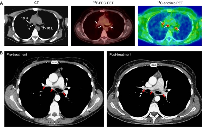
Patient no. 6, who also responded to the treatment, CT showed non-enlarged (<10 mm) hilar lymph nodes (positions 10R and 10L) and an enlarged (>10 mm) subcarinal lymph node (position 7) (Figure 2A). There was no accumulation of 18F-FDG in any of these lymph nodes, whereas 11C-erlotinib PET/CT showed accumulation in both the enlarged and the non-enlarged lymph nodes (Figure 2A). The patient continued treatment after end of the study and at follow-up 1 year later, 18F-FDG PET/CT was negative and contrast-enhanced CT showed no changes in the size of these three lymph nodes (Figure 2B).
Figure 2.
11C-erlotinib accumulation in lymph nodes that were negative on 18F-FDG PET/CT in a 42-year-old patient (no. 6). (A) Left: transaxial slices of contrast-enhanced CT; middle: 18F-FDG PET/low-dose CT; right: 11C-erlotinib PET/low-dose CT. CT (left figure) showed an enlarged lymph node (>10 mm) at position 7 (arrow) and non-enlarged lymph nodes (<10 mm) at positions 10R and 10L (arrows). None of these lymph nodes were visualized by 18F-FDG PET/CT (arrows, middle figure), whereas both enlarged and non-enlarged lymph nodes were visualized by 11C-erlotinib PET/CT (arrows, right figure). The ratio between 11C-erlotinib average radioactivity concentrations in the lymph nodes and that in surrounding lung tissue was 2. (B) Comparison of pre-treatment and 1-year post-treatment CT scans showed no significant change in the size of any of these lymph nodes.
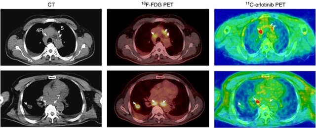
Patient no. 7 showed different tumour foci that differed with regard to accumulation of 11C-erlotinib as illustrated in Figure 3. Enlarged lymph nodes in the mediastinum at positions 4R and 5 (CT, arrows) showed hotspots on 18F-FDG PET/CT and on 11C-erlotinib PET/CT (upper panel, arrows) whereas the lung tumour and one of the enlarged lymph nodes at position 7 did not accumulate 11C-erlotinib (lower panel, arrows). Time courses of the radioactivity concentrations of 11C-erlotinib in this patient (Figure 4) show higher initial distribution in lung tissue and in the lung tumour than in the subcarinal lymph node and muscle tissue (Figure 4, left), which can be ascribed to differences in the blood perfusion. Ten minutes after the 11C-erlotinib administration and throughout the remaining 80-min measurement period, the radioactivity concentration was higher in the lymph node and the lung tumour than in the non-malignant lung and muscle tissue (Figure 4, right). The ratio between radioactivity concentrations lung tumour/lung tissue was around 1.3 and that of the lymph node/lung tissue around 2.0.
Figure 3.
11C-erlotinib PET/CT demonstrates the heterogeneous nature of advanced lung cancer. Two transaxial slices of (left) contrast-enhanced CT; (middle) 18F-FDG PET/low-dose CT, and (right) 11C-erlotinib PET/low-dose CT. A 48-year-old patient (no. 7) with NSCLC in the right lung and enlarged mediastinal lymph nodes (upper and lower panel left figure, arrows). Both 18F-FDG and 11C-erlotinib accumulated in lymph nodes at positions 4R and 5 (upper panel, arrows). The tumour in the right lung and one of the lymph nodes at position 7 showed only a weak accumulation of 11C-erlotinib (arrows). The ratio between the 11C-erlotinib average radioactivity concentrations in the lymph node metastasis and that in surrounding lung tissue was 2 (see Figure 4).
Figure 4.
Time courses of tissue radioactivity of 11C-erlotinib (patient no. 7, see Figure 3) for the right-sided lung tumour, metastatic lymph node 4R, lung tissue, and muscle tissue. Left: initial time courses and right: final time courses. The final accumulation of 11C-erlotinib was higher in the tumour and the metastatic lymph node than in lung and muscle tissue.
Discussion
In total, four of the thirteen patients examined showed 11C-erlotinib accumulation in one or more tumour foci and 11C-erlotinib accumulated in non-enlarged 18F-FDG PET/CT-negative lymph nodes. Our results also showed variation in 11C-erlotinib accumulation between different malignant lesions in the same patient.
Erlotinib is a targeted drug that inhibits signalling through the EGFR and thereby prolongs survival of a subgroup of lung cancer patients treated with this drug. Clinical parameters and mutational status of EGFR are considered helpful but insufficient to predict treatment response; and therefore, additional methods are required to improve the selection of patients for erlotinib treatment. We previously showed that 11C-erlotinib could be used to identify tumours overexpressing the EGFR in animal models (Memon et al, 2009), and we have presented a case report showing accumulation of 11C-erlotinib in brain metastasis of a patient with NSCLC (Weber et al, 2011). Here, we investigated the use of 11C-erlotinib PET/CT as a non-invasive method to identify tumours accumulating erlotinib and have established a method that may prove useful for the selection of patients suitable for erlotinib treatment. Our results showed that normal lung tissue accumulated no or minimal 11C-erlotinib, whereas some malignant lesions and metastases did accumulate the tracer.
Because tumour material was not available from all lesions, the EGFR status in 11C-erlotinib accumulating and non-accumulating tumour lesions is not known. However, in the present study, we observed different accumulation of 11C-erlotinib in the primary tumour and metastatic lesions in the same patient. This suggests that it may be of limited value to know the EGFR status in a biopsy from just one tumour lesion.
Variation in the accumulation of 11C-erlotinib between different tumours in the same patient is in agreement with the molecular evolution of the disease in these patients. The majority of lung cancer patients who initially respond to treatment eventually have a relapse (Sequist et al, 2007). Some of the known causes of these relapses are mutations in EGFR causing erlotinib resistance, amplification of the met proto-oncogene (MET) (Engelman et al, 2007), activation of other receptor tyrosine kinases (insulin-like growth factor 1) (Morgillo et al, 2007), and KRAS mutations (Eberhard et al, 2005). Our observations of variation in the uptake of 11C-erlotinib further suggest that not all tumours in the same patient are driven by EGFR signalling, and therefore may not respond to erlotinib treatment.
Staging of lung cancer is crucial when deciding treatment options and the prognosis also differs significantly according to stage. The TNM preoperative staging system employing CT is widely used; however, lymph-node staging (N staging) of hilar and mediastinal lymph nodes is still a challenge and CT scanning results in a significant amount of false positive and false negative results (Al-Sarraf et al, 2008). Another method for staging is 18F-FDG PET/CT (Fischer et al, 2009). The major obstacle with this method is the difficulty in distinguishing between benign and metastatic lymph nodes (Love et al, 2005). According to standard CT criteria, lymph nodes with a diameter >10 mm (enlarged) are classified as malignant and lymph nodes with a diameter <10 mm (non-enlarged) are classified as non-malignant. However, recent studies suggest that non-enlarged lymph nodes can also be metastatic even in the absence of a positive 18F-FDG PET/CT and small lymph nodes can therefore not be identified as non-malignant until other characteristics are considered (Liu et al, 2009). Interestingly, 11C-erlotinib PET/CT identified both enlarged and non-enlarged lymph nodes, which were negative on 18F-FDG PET/CT. These results indicate that it is possible that non-enlarged lymph nodes in cancer patients may harbour tumour cells expressing EGFR. We monitored these lymph nodes during erlotinib treatment in patient no. 6, and both enlarged and non-enlarged lymph nodes remained stable according to the RECIST criteria for more than a year and were also negative on 18F-FDG PET/CT. If these findings are confirmed in more patients, then this observation could have clinical significance as it may change the criteria for staging of lung cancer and thereby the treatment strategy for the patients.
Conclusions
This study showed a potential for 11C-erlotinib in PET/CT for visualizing NSCLC lung tumours, including lymph nodes not identified by 18F-FDG PET/CT. Moreover, 11C-erlotinib in PET/CT could be a useful tool to identify molecular heterogeneity between tumours in the same patient. Large clinical studies are now needed to explore to which extent pre-treatment 11C-erlotinib PET/CT can predict response to erlotinib treatment.
Acknowledgments
We thank the patients who participated. This work was supported by the Danish Research Medical Council, Danish Cancer Society, Danish Cancer Foundation, and Novo Nordisk Foundation.
Footnotes
This work is published under the standard license to publish agreement. After 12 months the work will become freely available and the license terms will switch to a Creative Commons Attribution-NonCommercial-Share Alike 3.0 Unported License.
The authors declare no conflict of interest.
References
- Al-Sarraf N, Aziz R, Gately K, Lucey J, Wilson L, McGovern E, Young V (2008) Pattern and predictors of occult mediastinal lymph node involvement in non-small cell lung cancer patients with negative mediastinal uptake on positron emission tomography. Eur J Cardiothorac Surg 33: 104–109 [DOI] [PubMed] [Google Scholar]
- Ciardiello F, Tortora G (2008) EGFR antagonists in cancer treatment. N Engl J Med 358: 1160–1174 [DOI] [PubMed] [Google Scholar]
- Cohen MH, Johnson JR, Chen YF, Sridhara R, Pazdur R (2005) FDA drug approval summary: erlotinib (Tarceva) tablets. Oncologist 10: 461–466 [DOI] [PubMed] [Google Scholar]
- Comis RL (2005) The current situation: erlotinib (Tarceva) and gefitinib (Iressa) in non-small cell lung cancer. Oncologist 10: 467–470 [DOI] [PubMed] [Google Scholar]
- Eberhard DA, Johnson BE, Amler LC, Goddard AD, Heldens SL, Herbst RS, Ince WL, Janne PA, Januario T, Johnson DH, Klein P, Miller VA, Ostland MA, Ramies DA, Sebisanovic D, Stinson JA, Zhang YR, Seshagiri S, Hillan KJ (2005) Mutations in the epidermal growth factor receptor and in KRAS are predictive and prognostic indicators in patients with non-small-cell lung cancer treated with chemotherapy alone and in combination with erlotinib. J Clin Oncol 23: 5900–5909 [DOI] [PubMed] [Google Scholar]
- Eisenhauer EA, Therasse P, Bogaerts J, Schwartz LH, Sargent D, Ford R, Dancey J, Arbuck S, Gwyther S, Mooney M, Rubinstein L, Shankar L, Dodd L, Kaplan R, Lacombe D, Verweij J (2009) New response evaluation criteria in solid tumours: revised RECIST guideline (version 1.1). Eur J Cancer 45(2): 228–247 [DOI] [PubMed] [Google Scholar]
- Engelman JA, Zejnullahu K, Mitsudomi T, Song Y, Hyland C, Park JO, Lindeman N, Gale CM, Zhao X, Christensen J, Kosaka T, Holmes AJ, Rogers AM, Cappuzzo F, Mok T, Lee C, Johnson BE, Cantley LC, Janne PA (2007) MET amplification leads to gefitinib resistance in lung cancer by activating ERBB3 signaling. Science 316: 1039–1043 [DOI] [PubMed] [Google Scholar]
- Fischer B, Lassen U, Mortensen J, Larsen S, Loft A, Bertelsen A, Ravn J, Clementsen P, Hogholm A, Larsen K, Rasmussen T, Keiding S, Dirksen A, Gerke O, Skov B, Steffensen I, Hansen H, Vilmann P, Jacobsen G, Backer V, Maltbaek N, Pedersen J, Madsen H, Nielsen H, Hojgaard L (2009) Preoperative staging of lung cancer with combined PET-CT. N Engl J Med 361: 32–39 [DOI] [PubMed] [Google Scholar]
- Fontanini G, De Laurentiis M, Vignati S, Chine S, Lucchi M, Silvestri V, Mussi A, De Placido S, Tortora G, Bianco AR, Gullick W, Angeletti CA, Bevilacqua G, Ciardiello F (1998) Evaluation of epidermal growth factor-related growth factors and receptors and of neoangiogenesis in completely resected stage I-IIIA non-small-cell lung cancer: amphiregulin and microvessel count are independent prognostic indicators of survival. Clin Cancer Res 4: 241–249 [PubMed] [Google Scholar]
- Fukuoka M, Yano S, Giaccone G, Tamura T, Nakagawa K, Douillard JY, Nishiwaki Y, Vansteenkiste J, Kudoh S, Rischin D, Eek R, Horai T, Noda K, Takata I, Smit E, Averbuch S, Macleod A, Feyereislova A, Dong RP, Baselga J (2003) Multi-institutional randomized phase II trial of gefitinib for previously treated patients with advanced non-small-cell lung cancer. J Clin Oncol 21: 2237–2246 [DOI] [PubMed] [Google Scholar]
- Gambhir SS (2002) Molecular imaging of cancer with positron emission tomography. Nat Rev Cancer 2: 683–693 [DOI] [PubMed] [Google Scholar]
- Jerusalem G, Hustinx R, Beguin Y, Fillet G (2003) PET scan imaging in oncology. Eur J Cancer 39: 1525–1534 [DOI] [PubMed] [Google Scholar]
- Kawamura K, Yamasaki T, Yui J, Hatori A, Konno F, Kumata K, Irie T, Fukumura T, Suzuki K, Kanno I, Zhang MR (2009) In vivo evaluation of P-glycoprotein and breast cancer resistance protein modulation in the brain using [11C]gefitinib. Nucl Med Biol 36: 239–246 [DOI] [PubMed] [Google Scholar]
- Liu BJ, Dong JC, Xu CQ, Zuo CT, Le JJ, Guan YH, Zhao J, Wu JF, Duan XH, Cao YX (2009) Accuracy of 18F-FDG PET/CT for lymph node staging in non-small-cell lung cancers. Chin Med J (Engl) 122: 1749–1754 [PubMed] [Google Scholar]
- Love C, Tomas MB, Tronco GG, Palestro CJ (2005) FDG PET of infection and inflammation. Radiographics 25: 1357–1368 [DOI] [PubMed] [Google Scholar]
- Lynch TJ, Bell DW, Sordella R, Gurubhagavatula S, Okimoto RA, Brannigan BW, Harris PL, Haserlat SM, Supko JG, Haluska FG, Louis DN, Christiani DC, Settleman J, Haber DA (2004) Activating mutations in the epidermal growth factor receptor underlying responsiveness of non-small-cell lung cancer to gefitinib. N Engl J Med 350: 2129–2139 [DOI] [PubMed] [Google Scholar]
- Memon AA, Jakobsen S, Dagnaes-Hansen F, Sorensen BS, Keiding S, Nexo E (2009) Positron emission tomography (PET) imaging with [11C]-labeled erlotinib: a micro-PET study on mice with lung tumor xenografts. Cancer Res 69: 873–878 [DOI] [PubMed] [Google Scholar]
- Morgillo F, Kim WY, Kim ES, Ciardiello F, Hong WK, Lee HY (2007) Implication of the insulin-like growth factor-IR pathway in the resistance of non-small cell lung cancer cells to treatment with gefitinib. Clin Cancer Res 13: 2795–2803 [DOI] [PubMed] [Google Scholar]
- Paez JG, Janne PA, Lee JC, Tracy S, Greulich H, Gabriel S, Herman P, Kaye FJ, Lindeman N, Boggon TJ, Naoki K, Sasaki H, Fujii Y, Eck MJ, Sellers WR, Johnson BE, Meyerson M (2004) EGFR mutations in lung cancer: correlation with clinical response to gefitinib therapy. Science 304: 1497–1500 [DOI] [PubMed] [Google Scholar]
- Parkin DM, Bray F, Ferlay J, Pisani P (2005) Global cancer statistics, 2002. CA Cancer J Clin 55: 74–108 [DOI] [PubMed] [Google Scholar]
- Perez-Soler R, Chachoua A, Hammond LA, Rowinsky EK, Huberman M, Karp D, Rigas J, Clark GM, Santabarbara P, Bonomi P (2004) Determinants of tumor response and survival with erlotinib in patients with non-small-cell lung cancer. J Clin Oncol 22: 3238–3247 [DOI] [PubMed] [Google Scholar]
- Rohren EM, Turkington TG, Coleman RE (2004) Clinical applications of PET in oncology. Radiology 231: 305–332 [DOI] [PubMed] [Google Scholar]
- Rusch V, Baselga J, Cordon-Cardo C, Orazem J, Zaman M, Hoda S, McIntosh J, Kurie J, Dmitrovsky E (1993) Differential expression of the epidermal growth factor receptor and its ligands in primary non-small cell lung cancers and adjacent benign lung. Cancer Res 53: 2379–2385 [PubMed] [Google Scholar]
- Sequist LV, Bell DW, Lynch TJ, Haber DA (2007) Molecular predictors of response to epidermal growth factor receptor antagonists in non-small-cell lung cancer. J Clin Oncol 25: 587–595 [DOI] [PubMed] [Google Scholar]
- Shepherd FA, Pereira J, Ciuleanu TE, Tan EH, Hirsh V, Thongprasert S, Bezjak A, Tu D, Santabarbara P, Seymour L (2004) A randomized placebo-controlled trial of erlotinib in patients with advanced non-small cell lung cancer (NSCLC) following failure of 1(st) line or 2(nd) line chemotherapy. A National Cancer Institute of Canada Clinical Trials Group (NCIC CTG) trial. J Clin Oncol 22: 622S [Google Scholar]
- Shepherd FA, Pereira JR, Ciuleanu T, Tan EH, Hirsh V, Thongprasert S, Campos D, Maoleekoonpiroj S, Smylie M, Martins R, van Kooten M, Dediu M, Findlay B, Tu DS, Johnston D, Bezjak A, Clark G, Santabarbara P, Seymour L (2005) Erlotinib in previously treated non-small-cell lung cancer. N Engl J Med 353: 123–132 [DOI] [PubMed] [Google Scholar]
- Sobin LH, Fleming ID (1997) TNM classification of malignant tumors, fifth edition (1997). Union Internationale Contre le Cancer and the American Joint Committee on Cancer. Cancer 80: 1803–1804 [DOI] [PubMed] [Google Scholar]
- Su H, Seimbille Y, Ferl GZ, Bodenstein C, Fueger B, Kim KJ, Hsu YT, Dubinett SM, Phelps ME, Czernin J, Weber WA (2008) Evaluation of [18F]gefitinib as a molecular imaging probe for the assessment of the epidermal growth factor receptor status in malignant tumors. Eur J Nucl Med Mol Imaging 35: 1089–1099 [DOI] [PubMed] [Google Scholar]
- Wang JQ, Gao M, Miller KD, Sledge GW, Zheng QH (2006) Synthesis of [11C]Iressa as a new potential PET cancer imaging agent for epidermal growth factor receptor tyrosine kinase. Bioorg Med Chem Lett 16: 4102–4106 [DOI] [PubMed] [Google Scholar]
- Weber B, Winterdahl M, Memon A, Sorensen BS, Keiding S, Sorensen L, Nexo E, Meldgaard P (2011) Erlotinib accumulation in brain metastases from non-small cell lung cancer: visualization by positron emission tomography in a patient harboring a mutation in the epidermal growth factor receptor. J Thorac Oncol 6: 1287–1289 [DOI] [PubMed] [Google Scholar]
- Zhang MR, Kumata K, Hatori A, Takai N, Toyohara J, Yamasaki T, Yanamoto K, Yui J, Kawamura K, Koike S, Ando K, Suzuki K (2010) [11C]Gefitinib ([11C]Iressa): radiosynthesis, in vitro uptake, and in vivo imaging of intact murine fibrosarcoma. Mol Imaging Biol 12: 181–191 [DOI] [PubMed] [Google Scholar]


