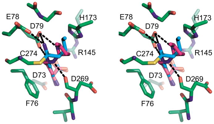
Figure 5.
Comparison of 4 complexed to wild type and C274S DDAH-1. In this divergent stereo image, the protein is shown with green carbon backbone. 4 in the wild type is shown as cyan sticks, while 4 in the mutant is shown in magenta. Hydrogen bonds are shown as black dashed lines; for clarity, only bonds to the C-alkyl amidine moiety are shown and those to the amino acid portion of the inhibitor have been omitted.

