Abstract
We report the case of a female patient with incomplete distal renal tubular acidosis with nephrocalcinosis. She was admitted to the hospital because of acute pyelonephritis. Imaging studies showed dual medullary nephrocalcinosis. Subsequent evaluations revealed hypokalemia, hypocalcemia, hypercalciuria, and hypocitraturia with normal acid-base status. A modified tubular acidification test with NH4Cl confirmed a defect of urine acidification, which is compatible with incomplete distal tubular acidosis. We treated our patient with potassium citrate, which corrects hypokalemia and prevents further deposition of calcium salts.
Keywords: Renal tubular acidosis, Nephrocalcinosis, Kidney
THE CASE: WHAT IS THE CAUSE OF NEPHROCALCINOSIS?
A 25-year-old female presented to Chonnam National University Hospital complaining of dysuria for 3 days with a fever of 38.3℃, chills, and right flank pain. Over the previous 5 years she had recurrent urinary tract infections. On admission, she had mild hypotension, blood pressure 100/60 mmHg, and her pulse rate was 96/minute. Physical examination showed a soft abdomen with severe costovertebral angle tenderness. The diagnosis of acute pyelonephritis was made and empirical antibiotic treatment with fluid resuscitation was started.
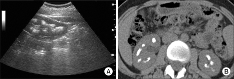
Laboratory studies revealed blood urea nitrogen 19.0 mg/dl and serum creatinine 1.0 mg/dl. The electrolytes were sodium 138, chloride 104, and potassium 3.4 mEq/L. The venous blood gas analysis was as follows: pH 7.39, PO2 38.7 mmHg, PCO2 38.7 mmHg, HCO3- 23.7 mmol/L. Calcium was 7.7 mg/dl and phosphorus was 3.1 mg/dl. Serum albumin and total protein were within normal ranges. Urinalysis showed a specific gravity of 1.005, pH 7.0, negative protein, 2+ white blood cells, and 1+ red blood cells. The renal ultrasound showed medullary nephrocalcinosis with total involvement of the medullary pyramids (Fig. 1A), which was confirmed by abdominal computed tomography (Fig. 1B).
FIG. 1.
(A) Renal ultrasound showed a hyperechogenic region in the medulla and postacoustic shadowing beyond the region. (B) Abdominal computed tomography confirmed medullary calcification.
The 24-hour electrolyte excretion was sodium 160 mEq (normal 40-220), potassium 66.5 mEq (normal 25-125), and calcium 440 mg (normal 100-300). The transtubular gradient for potassium [(Osmolality serum * K+ urine)/(K+ serum * Osmolality urine)] was 4.84, which was inappropriately high. Serum parathyroid hormone and vitamin D (calcidiol) were normal, 50 pg/ml and 14.9 ng/ml, respectively. A test for 24-hour urinary citric acid was ordered, and the level was 13.8 mg, which was markedly low.
The aforementioned investigations revealed hypokalemia, hypocalcemia, hypercalciuria, and hypocitraturia. The patient's metabolic acid-base status was normal and imaging study revealed medullary nephrocalcinosis of both kidneys. What is the cause of these findings?
THE DIAGNOSIS: INCOMPLETE DISTAL RENAL TUBULAR ACIDOSIS
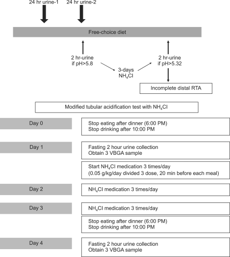
Because urine pH was sustained between 6.5 and 7.0 for the entire hospital stay, a modified tubular acidification test with NH4Cl was performed (Fig. 2).1 Before the challenge of NH4Cl, venous pH was 7.40, venous HCO3- was 26.5 mmol/L, and urine pH was 7.0. After an oral challenge of NH4Cl at a dosage of 0.05 g/kg/day for 3 days, venous HCO3- decreased to 19.7 mmol/L, but urine pH was 6.1, which strongly suggested the presence of a urine acidification defect. These results confirmed the diagnosis of incomplete distal renal tubular acidosis (RTA), which leads to hypokalemia, hypocalciuria, hypocitraturia, and nephrocalcinosis. In addition, it was considered that the nephrocalcinosis had been the cause of the recurrent urinary tract infections.
FIG. 2.
Protocol for a modified tubular acidification test with NH4Cl.
Nephrocalcinosis can occur in some hypercalcemic disorders, such as hyperparathyroidism, vitamin D intoxication, and sarcoidosis.2 However, in the present case, the patient had hypocalcemia and an inappropriately high 24-hour calcium excretion, which strongly suggests renal tubulopathy, such as Bartter syndrome, furosemide abuse, or distal RTA. The lower urinary citrate excretion and absence of metabolic alkalosis indicate the possibility of distal RTA. For that reason, we conducted a modified tubular acidification test and confirmed incomplete distal RTA.
Distal RTA is a syndrome of systemic hyperchloremic acidosis with alkaline urine pH, hypocitraturia, and hypercalciuria due to reduced secretion of H+ ions by the cells of the collecting tubules.3 In contrast, incomplete distal RTA shows normal acid-base status under the basal condition, but increased protein intake or catabolic stress may cause transient positive acid loads, which lead to decreased tubular reabsorption of calcium and increased reabsorption of citrate by intracellular acidosis and hypokalemia.4 Subsequently, hypercalciuria, alkaline urine, and a lower urinary level of citrate lead to renal deposition of calcium salts. We treated our patient with potassium citrate. Administration of potassium citrate not only provides potassium, but also corrects the acidosis because citrate is converted to bicarbonate after absorption. Furthermore, potassium citrate lowers urinary calcium excretion and promotes citrate excretion, hence preventing further deposition of calcium salts.
In conclusion, we described our diagnostic approach in patients with nephrocalcinosis. Incomplete distal RTA should be considered in patients with nephrocalcinosis, who show hypokalemia, sustained alkaline urine, and normal acid-base status under the baseline condition. In such cases, a tubular acidification test with NH4Cl will help to clarify the underlying defect of urine acidification.
References
- 1.Arampatzis S, Röpke-Rieben B, Lippuner K, Hess B. Prevalence and densitometric characteristics of incomplete distal renal tubular acidosis in men with recurrent calcium nephrolithiasis. Urol Res. 2011 doi: 10.1007/s00240-011-0397-3. [Epub ahead of print] [DOI] [PubMed] [Google Scholar]
- 2.Alon US. Nephrocalcinosis. Curr Opin Pediatr. 1997;9:160–165. [PubMed] [Google Scholar]
- 3.Kitiyakara C, Yamwong S, Cheepudomwit S, Domrongkitchaiporn S, Unkurapinun N, Pakpeankitvatana V, et al. The metabolic syndrome and chronic kidney disease in a Southeast Asian cohort. Kidney Int. 2007;71:693–700. doi: 10.1038/sj.ki.5002128. [DOI] [PubMed] [Google Scholar]
- 4.Weger W, Kotanko P, Weger M, Deutschmann H, Skrabal F. Prevalence and characterization of renal tubular acidosis in patients with osteopenia and osteoporosis and in non-porotic controls. Nephrol Dial Transplant. 2000;15:975–980. doi: 10.1093/ndt/15.7.975. [DOI] [PubMed] [Google Scholar]


