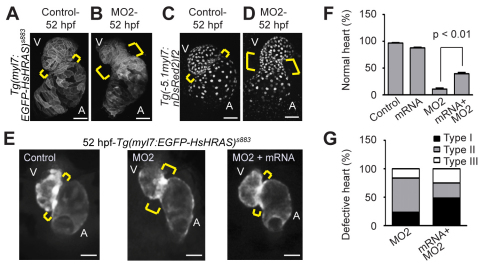
Fig. 3.
Npnt knockdown causes extension of the AV canal. (A-D) Projections of confocal images of hearts from control (A,C) and MO2-injected (B,D) embryos from transgenic Tg(myl7:EGFP-HsHRAS)s883 (A,B) and Tg(–5.1myl7:nDsRed2)f2 (C,D) zebrafish at 52 hpf, suggesting an extension of the AV canal (brackets) in npnt morphants. (E) Representative images of hearts from control, MO2-injected and MO2 + npnt mRNA-injected embryos from transgenic Tg(myl7:EGFP-HsHRAS)s883 zebrafish at 52 hpf. Brackets indicate the AV boundary. (F) Quantitative analysis (n>160 from three independent experiments; mean ± s.e.m.). Note that injection of npnt mRNA rescued the MO2-mediated AV canal extension. (G) Quantitative analysis scoring of type I (mild extension of the AV canal), type II (obvious extension of the AV canal) and type III (straight heart) AV canal defects (mean ± s.e.m.). A, atrium; V, ventricle. Scale bars: 50 μm.

