Abstract
We measured changes in maximal voluntary and electrically evoked torque and rate of torque development because of limb unloading. We investigated whether these changes during single joint isometric muscle contractions were related to changes in jump performance involving dynamic muscle contractions and several joints. Six healthy male subjects (21 ± 1 years) underwent 3 weeks of unilateral lower limb suspension (ULLS) of the right limb. Plantar flexor and knee extensor maximal voluntary contraction (MVC) torque and maximal rate of torque development (MRTD), voluntary activation, and maximal triplet torque (thigh; 3 pulses at 300 Hz) were measured next to squat jump height before and after ULLS. MVC of plantar flexors and knee extensors (MVCke) and triplet torque decreased by 12% (P = 0.012), 21% (P = 0.001) and 11% (P = 0.016), respectively. Voluntary activation did not change (P = 0.192). Absolute MRTD during voluntary contractions decreased for plantar flexors (by 17%, P = 0.027) but not for knee extensors (P = 0.154). Absolute triplet MRTD decreased by 17% (P = 0.048). The reduction in MRTD disappeared following normalization to MVC. Jump height with the previously unloaded leg decreased significantly by 28%. No significant relationships were found between any muscle variable and jump height (r < 0.48), but decreases in torque were (triplet, r = 0.83, P = 0.04) or tended to be (MVCke r = 0.71, P = 0.11) related to decreases in jump height. Thus, reductions in isometric muscle torque following 3 weeks of limb unloading were significantly related to decreases in the more complex jump task, although torque in itself (without intervention) was not related to jump performance.
Keywords: Unloading, Muscle, Torque, Knee extensor, Limb suspension
Introduction
Unloading, for instance, during prolonged bed rest or during space flight, is known to decrease muscle cross-sectional area (CSA) (Alkner and Tesch 2004; Reeves et al. 2002; Shackelford et al. 2004) and strength (Dudley et al. 1992; Berg et al. 1991; LeBlanc and Schneider 1992; de Boer et al. 2007; Mulder et al. 2009). Gravitational muscles, like the knee extensors and the plantar flexors, are exposed to altered patterns of activity because of unloading and therefore their contractile properties change (Trappe et al. 2004; Fitts et al. 2007). In addition, a reduced neural drive capacity may be expected after unloading (Duchateau 1995; Koryak 2001; Ruegg et al. 2003), which could further decrease the ability to generate maximal voluntary torque (Gondin et al. 2004; Deschenes et al. 2002) and fast torque development (Mulder et al. 2009; Suetta et al. 2007; de Boer et al. 2007). Although the consequences of muscle unloading for contractile properties of individual muscle groups is well documented (Duchateau and Hainaut 1990; Berg and Tesch 1996), the consequences for more complex whole body functioning are less well studied. In the present study, we aimed to relate changes in static muscle contractile properties and voluntary activation of single muscle groups, to dynamic multi-joint squat jump performance.
The capacity to activate a muscle as fast as possible is important during daily life, since the time allowed to build up muscle torque is limited (Kakihana and Suzuki 2001) during balance corrections and fast powerful tasks such as jumping. Maximal rate of isometric torque development (MRTD) in vivo is dependent on properties of the muscle tendon complex and on neural activation. It is indicated that contractions in which torque needs to be developed as forcefully and rapidly as possible require higher levels of neural activation compared with maximal contractions of several seconds or longer (de Ruiter et al. 2004; Duchateau 1995; de Boer et al. 2007; Kubo et al. 2000; Bamman et al. 1998). Maximal rate of torque development (MRTD) has been related to fall prevention (Pijnappels et al. 2008) and jump performance (de Ruiter et al. 2007).
Jump height is thought to be a more relevant measure of leg-extension power than isolated force responses. It is shown that very good jumpers are stronger than bad jumpers (Vanezis and Lees 2005; Kraska et al. 2009), but for the quadriceps muscles, correlations between maximal voluntary knee extension torque and jump height have only been found in heterogeneous subject groups that for example included both men and women (Hakkinen 1991). Researchers have hypothesized that jump performance would be positively related to either maximal knee-extension torque and/or measure of contractile speed. However, in light of the logical basis of these hypotheses, the results of such studies have in general been unclear. Most studies did not find relationships between isometric torque and jump performance (Andersen and Aagaard 2006; Hakkinen 1991; Jaric et al. 2002; Young et al. 1999).
Also correlations between muscle contractile speed during fast voluntary contractions and jump performance were found to be poor (Marcora and Miller 2000; Paasuke et al. 2001; Wilson and Murphy 1995) or absent (Marcora and Miller 2000; Baker et al. 1994; Driss et al. 1998; Matavulj et al. 2001; de Ruiter et al. 2007) in cross-sectional studies. In longitudinal studies, which are not obscured by inter individual differences, such as in body composition and jumping technique, positive effects of muscle strength training on complex whole body performance such as jumping have been reported (Cormie et al. 2010; Lamont et al. 2008; Kalapotharakos et al. 2005). Therefore, it is expected that interventions which would negatively affect contractile properties of individual muscle groups would have a detrimental effect on more complex dynamic tasks such as jumping. The present study investigated whether changes in maximal torque and MRTD because of unloading were related to changes in jump height. We used unilateral lower limb suspension (ULLS), a model promoting disuse with intact joint mobility, relatively low costs and only moderate encroachment on daily physical activities. To our knowledge, the present study is the first to relate changes in maximal torque and MRTD, due to unloading, during single-joint isometric knee extensor and plantar flexor torque development, to changes in a more natural whole body movement like jumping. We hypothesized that without an intervention, the relationships between contractile properties of individual muscle groups (knee extensors and plantar flexors) and jump performance would be weak or absent, but that decreases in muscle force and speed following unilateral lower limb suspension would be related to decreases in jump performance.
Methods
Subjects
Ten healthy young men with a minimum age of 18 years (22 ± 2 years, 1.88 ± 0.06 m and 77 ± 8 kg) volunteered in this study. Exclusion criteria were more than 5 h physical activity per week; hypertension; contraindication for maximal exercise or electrical stimulation; recent bone fracture; heart failure; muscle, skin, metabolic and bone diseases; functional limitations in upper or lower limbs; use of drugs with hemodynamic effects and any medical or surgical history that could influence the study outcome.
The project carried the approval of the Medical Ethics Committee of the Radboud University Nijmegen Medical Centre. All subjects signed an informed consent before participation. One week prior to the baseline measurements, subjects were familiarized with all testing procedures. Unfortunately, two subjects withdrew because of poor tolerance of electrical stimulation and another two for personal reasons unrelated to the study. Therefore, six subjects (21 ± 1 years, 1.87 ± 0.06 m and 79 ± 9 kg) completed the 3 weeks of ULSS.
Unilateral lower limb suspension
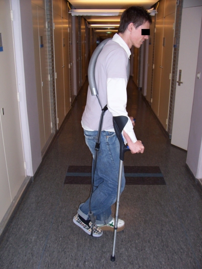
The Unilateral lower limb suspension (ULLS) model used in this study was described by Berg et al. (1991) and Seynnes et al. (2010). In brief, the subjects were wearing a left shoe with a 7.5 cm thick sole and performed all daily activities on crutches. The right leg was unloaded by means of a shoulder strap running around the foot and the ankle (Fig. 1), which was worn during all locomotor activities. The length of the strap was adjusted to prevent the foot touching the floor and to maintain the ankle angle 90° (instead of a more plantar flexion position), so that no additional loss of muscle mass occurred because of short muscle length (Tabary et al. 1972; Williams and Goldspink 1973). Subjects were instructed not to load the suspended limb (e.g. no touching the ground, no driving, etc.). They were constrained to wear support stockings in order to limit the risk of deep venous thrombosis. Compliance with the suspension was monitored through regularly (personal, by telephone or via email) communication with the subjects and they kept a diary.
Fig. 1.
Model of unilateral lower limb suspension, with a left shoe with thick sole and the shoulder strap running around foot and ankle to unload the right foot
Experimental design
Maximal voluntary isometric knee extensions and plantar flexions and fast voluntary contractions of both muscle groups were performed. Moreover, electrically evoked contractions (supramaximal triplets, see below) were used for the knee extensors to establish voluntary activation and MRTD during maximal electrical activation. Squat jump height on one leg (both left and right) and on two legs was determined. The experiments were performed on the same day, starting with the jumps, followed by measurements of the contractile properties of the plantar flexors and that of the knee extensors, with at least 1.5 h of rest in between these three tests. After the measurements at baseline (pre), the subjects started walking on crutches and 3 weeks later, this period ended with the performance of the same measurements done prior to the ULLS (post).
Torque measurements
Knee extensors
Maximal voluntary isometric forces of the knee extensors of the right limb were measured on a custom built dynamometer (VU University Amsterdam, The Netherlands) at a knee angle of 120° (180° corresponds with fully extended knee), at which angle usually the highest torque is produced (de Ruiter et al. 2004). Subjects were tightened with a hip and trunk belt to avoid changes in hip and knee angle during isometric contractions. The lower leg was strapped tightly to a force transducer (KAP, E/200 Hz, Bienfait B.V. Haarlem, The Netherlands, range 0–2 kN) just above the ankle by means of a cuff. The knee angle of 120° was determined with a handheld goniometer (model G300, Whitehall Manufacturing, California, USA) using the greater trochanter, the lateral epicondyle of the femur and the lateral malleolus of the fibula as references (Kooistra et al. 2005), while the subject produced at voluntary contraction at about 50% MVC (dynamometer) or when he was standing in the start position prior to the jump test. The distance between the lateral femur epicondyle and the force transducer in the dynamometer was taken as the external moment arm which was multiplied with the measured forces to obtain torques.
Plantar flexors
Maximal voluntary isometric forces of the plantar flexors were measured using another custom made dynamometer (VU University Amsterdam, The Netherlands). Subjects were seated, hip and knee angle were 90° and ankle angle was 15° dorsal flexion position. In this position, also the gastrocnemius muscles and not only the soleus muscles have a substantial contribution to the triceps surae torque (Sale et al. 1982; Martin et al. 1994). Subjects were tightened with a hip and trunk belt and their right foot was strapped into a standard shoe with a sturdy flat sole that was in turn tightly strapped to a force transducer (K.A.S., A.S.T., Wolnzach, Germany, range 0–10 kN). The leg was fixed tightly by a top restraining bar that was secured on the thigh, just proximal to the knee joint to minimize the movement of the leg and to avoid changes in knee and ankle angles during force generation. The distance between the middle of the medial malleolus and a fixed point at the shoe (at the height of the ball of the foot) was measured representing the external moment arm used to calculate plantar flexion torque.
Experimental procedures
Familiarization session
The measurements of the familiarization session were performed with the right leg 1 week before the baseline measurements. Dynamometer adjustments and shoe sizes were noted. After a warming-up, maximal voluntary contractions and fast voluntary contractions were practiced. Thereafter, subjects were familiarized with electrical stimulation for the quadriceps muscle, by increasing current during triplets (train of three 200 μs pulses at 300 Hz) at a relaxed muscle. Finally, supramaximal triplets were superimposed on a maximal voluntary isometric contraction.
Muscle torque
On the pre and post-ULLS test days, the subjects were asked to generate maximal isometric knee extensions and plantar flexions for 3–4 s to determine maximal voluntary contraction torque (MVC). Two to maximally four attempts were allowed, separated by 2 min of rest. MVC was taken as the highest value of these attempts, which did not exceed preceding attempts by >10%. Real-time force production was visible on a computer screen. Subjects were vigorously encouraged to exceed their maximal value, which was also displayed.
Voluntary activation
Volitional tests rely heavily on the subject’s motivation and the ability to maximally recruit his muscles and are often not an accurate reflection of the maximal torque generating capacity of the muscle. Electrically evoked contractions are independent of the subject’s effort. Therefore, a modified super-imposed stimulation technique was used in which electrically evoked triplets (pulse train of three rectangular 200 μs pulses applied at 300 Hz) were used to establish the subjects’ capacity to voluntarily activate their knee extensor muscles (Kooistra et al. 2005). After explanation of the procedure, the skin of the thigh of the subject was shaved and a pair of self-adhesive surface electrodes (13 × 8 cm, Schwa-Medico, The Netherlands) was placed over the proximal and distal part of the anterior thigh. The knee extensors were electrically stimulated using a computer-controlled constant current stimulator (Digitimer DSH7, Digitimer Ltd., Welwyn Garden City, UK). First, stimulation current was increased until torque measured in response to a triplet leveled off. The current was then increased by a further 20 mA to ensure supramaximal stimulation. It was assumed that at this point all muscle fibres of the knee extensors were activated. These high frequency stimulations (maximal triplet torque) result in maximal muscle activation with peak torques for about 30% MVC (de Haan 1998). Triplet stimulation was preferred to twitches because triplet torque is less sensitive to, for instance, length-dependent changes in calcium sensitivity and post-tetanic potentiation and improves the signal-to-noise ratio. Finally, subjects underwent measurements consisting of a triplet superimposed on the plateau of the force signal of the MVC.
Contraction speed
Maximal rates of isometric torque development (MRTD) were assessed as indicators of contractile speed using voluntary contractions. For this purpose, fast voluntary isometric contractions were performed. Subjects were asked to perform knee extensions and plantar flexions as fast as possible without a countermovement. Subjects performed these fast contractions until they had two attempts without countermovement or an enhanced pretension (both indicating anticipation of the subject and influencing the MRTD) just before the fast extension (de Ruiter et al. 2004) with a maximum of five attempts to avoid fatigue. After each attempt, subjects received feedback and were encouraged to exceed previous values.
For the knee extensors, maximal rates of torque development were also established during electrically evoked contractions, independent of voluntary activation, using the supramaximal triplets.
Jump height
The jump protocol of De Ruiter et al. (2007) was used in the present study. In brief, after a warming up of 12 squat jumps with increasing height, squat jumps were performed with both hands gripped together behind the back to avoid arm support for standardization. The subjects were instructed to hold their trunk as upright as possible. Squat jumps were started from 120° knee angle (the same angle as during the isometric knee extension measurements (180° was fully extended knee), which was set manually with a hand-held goniometer using as anatomical landmarks the greater trochanter, the lateral epicondyle and the lateral malleolus, as used during the isometric knee extensions. During the one-leg jumps, the other leg was held a bit forward, so that it was unable to support the jump. No countermovement (>1 cm) was allowed. Jump height was measured with a tape measure around the hips of the subject, which slid between two small messing plates between the feet in the floor when subjects jumped (de Ruiter et al. 2010). If the subject landed more than 2 cm forward (backward was impossible due to the horizontal bar) compared to the starting position, the jump was not used for data analysis. The reproducibility of this jump method is high (ICC = 0.94) (de Ruiter et al. 2010). The sequence of two-leg, right and left leg squat jumps was randomly chosen, but was the same per subject during the pre and post-ULLS measurements. Between consecutive jumps there were 2 min of rest and between the sets (one-leg right, one-leg left and two-leg) 5 min. In each of the three sets, three good jumps (i.e. no countermovement, no arm support, landing within 2 cm of the starting position), were performed with a maximum of six jumps.
Data analysis
Real-time force applied to the force transducer was digitally stored (1 kHz) on computer disc. Force signals were automatically corrected for gravity of the leg: average force applied by the limb weight to the transducer during the first 50 ms after start of a recording, with the subject seated in a relaxed manner, was set to zero by the computer program. All force signals were low-pass filtered (4th order, 150 Hz, Butterworth). MVC torque (Nm) was defined as the peak force from the force plateau multiplied by the external moment arm.
MRTD was defined as the steepest slope of torque development during both fast voluntary contractions (vol) (de Ruiter et al. 2004) and during the triplets at 300 Hz (stim). The MRTD as such not only depends on speed properties of the muscle but also on its maximal torque generating capacity. Therefore, to get a relevant comparison of the contractile speed of muscles between the different subjects independent of absolute maximal torques, MRTDvol was normalized to MVC torque and MRTDstim to triplet torque.
The ICC’s (intraclass correlation coefficient for test–retest reliability) for MVC, MRTD, voluntary activation (VA), and squat jump were shown to be, respectively, 0.97, 0.85, 0.82 and 0.90 (P < 0.05) [ICC’s found in our group with the same apparatuses (De Ruiter et al. 2007)]. De Ruiter et al. (2010) showed that CVs [coefficient of variation = (SD/mean) × 100%] for MVC were between 3 and 6%. Although the CVs for MRTD were quite high, the relative differences among the subjects were greater (MRTD varies substantially, even in healthy subjects with a high level of voluntary activation (De Ruiter et al. 2004), which resulted in good ICCs for the MRTDs.
Voluntary activation was defined as the completeness of skeletal muscle activation during voluntary contractions and was calculated by means of a modified interpolated twitch technique (Kooistra et al. 2005):
Here, the superimposed triplet is the force increment during a maximal contraction at the time of stimulation and the control triplet is that evoked in the relaxed muscle (Shield and Zhou 2004). (Supra maximal) triplet torque of the relaxed muscle was used as a measure for the maximal (intrinsic) torque capacity of the knee extensors, independent of voluntary activation.
Statistics
All results are presented as mean ± SD. Paired sample t tests were used to compare pre and post-ULLS values of MVCs, voluntary activation, triplet torque and jump heights. Pearson’s correlation coefficient was used to establish significance of correlation between (changes in) muscle variables (MVC of the knee extensors (MVCke), MVC of the plantar flexors (MVCpf), triplet torque, voluntary activation, MRTDvol and MRTDstim) and (changes in) one-leg (right) and two-leg jump height. In each statistical analysis, the level of significance was set at P < 0.05.
Results
Torque
Table 1 shows the results of the muscle variables before and after limb suspension. The MVC of the plantar flexors significantly decreased by 12.1 ± 5.9% (P = 0.01) after ULLS and the MVC of the knee extensors by 20.5 ± 4.6% (P = 0.001). In addition, triplet torque of the knee extensors decreased significantly (by 11.3 ± 8.7%, P = 0.02), whereas activation did not show a significant change (P = 0.19).
Table 1.
Values of the muscle variables pre and post-unilateral lower limb suspension
| Pre | Post | |
|---|---|---|
| MVC plantar flexors (n = 6) (Nm) | 242 ± 54 | 211 ± 36* |
| MVC knee extensors (n = 6) (Nm) | 308 ± 57 | 246 ± 42* |
| Triplet torque knee extensors (n = 6) (Nm) | 76 ± 12 | 68 ± 15* |
| Voluntary activation (n = 6) (%) | 88.3 ± 6.8 | 84.7 ± 9.1 |
| MRTDvol calf (n = 6) (Nm s−1) | 8,256 ± 7,076 | 7,076 ± 1,213* |
| MRTDvol thigh (n = 5) (Nm s−1) | 13,676 ± 4,100 | 11,214 ± 1,589 |
| MRTDstim thigh (n = 6) (Nm s−1) | 10,201 ± 2,903 | 8,271 ± 1,993* |
| Norm. MRTDvol calf (n = 6) (% ms−1) | 0.45 ± 0.08 | 0.44 ± 0.06 |
| Norm. MRTDvol thigh (n = 5) (% ms−1) | 1.30 ± 0.59 | 1.20 ± 0.39 |
| Norm. MRTDstim thigh (n = 6) (% ms−1) | 3.48 ± 0.77 | 3.16 ± 0.33 |
MVC maximal voluntary torque, norm. MRTD normalized maximal rate of torque development, vol voluntary, stim stimulated
* Significantly different from pre-ULLS values (P < 0.05)
Maximal rate of torque development
Absolute maximal rate of torque development (MRTD) during voluntary contractions decreased by 16.9 ± 11.4% (from 0.92 ± 0.17 to 0.76 ± 0.13 kNm s−1) for the plantar flexors (P = 0.03, n = 6) and 15.6 ± 32.4% (i.e. from 2.68 ± 0.66 to 2.11 ± 0.33 kNm s−1) for the knee extensors (being not significant; P = 0.15, n = 5). Absolute MRTD during electrically evoked contractions in the quadriceps muscles significantly decreased with 17.0 ± 17.7% (i.e. from 2.65 ± 0.75 to 2.15 ± 0.52 kNm s−1) (P = 0.048, n = 6).
The values for MRTD normalized for maximal torques are also shown in Table 1. Normalized voluntary MRTD of the plantar flexors (P = 0.683) and knee extensors (P = 0.434) or normalized electrically evoked MRTD of the knee extensors (P = 0.296, n = 6) changed significantly after ULLS. Three weeks of suspension did not affect the ratio of voluntary over stimulated MRTD in the knee extensor muscles for normalized (1.17 ± 0.46 pre and 1.06 ± 0.47 post; P = 0.862) or absolute values (0.38 ± 0.14 pre and 0.39 ± 0.14 post; P = 0.39).
Jump height
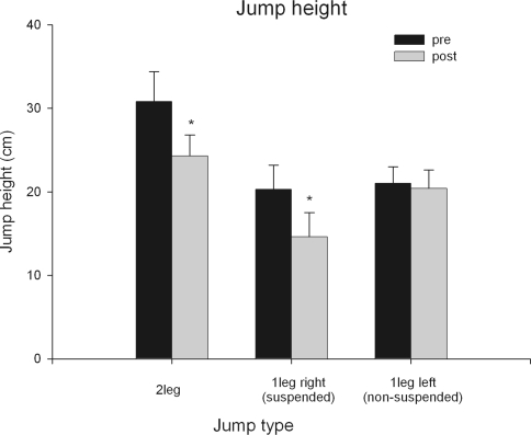
Figure 2 shows that two-leg jump height decreased significantly (P = 0.02) by 20 ± 13%. One-leg jump height with the right (suspended) leg decreased significantly (P = 0.006) with 28 ± 13%, whereas jump height with the left (non-suspended) leg did not change (P = 0.48).
Fig. 2.
Jump height pre and post-unilateral lower limb suspension for two-leg squat jumps and one-leg squat jump with the right (suspended) and left (non-suspended) leg. *Significantly different from pre-ULLS
Relationships between torque variables and jump height
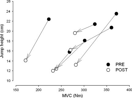
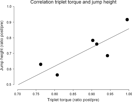
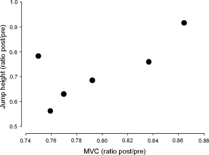
In line with our expectations, no correlations were found between any of the muscle variables and jump height before limb suspension or after limb suspension. Figure 3 shows that in all subjects both jump height and MVCke were lower post-unloading. The relationship between reductions in maximal triplet torque and one-leg (right) jump height was significant (r = 0.83, P = 0.04) (Fig. 4). The relationship between the reductions in jump height and MVCke (r = 0.71, P = 0.11, Fig. 5) tended to be significant. No significant correlations were found between changes in MVCpf and one-leg jump height (r = 0.63, P = 0.18).
Fig. 3.
Individual subject pre and post-unloading maximal voluntary knee extension contraction (MVC) torque and jump height values
Fig. 4.
Relationship between changes (ratio post/pre unloading value) in knee extensor triplet torque and jump height
Fig. 5.
Relationship between change (post/pre unloading value) in knee extensor MVC and jump height
Discussion
As hypothesized, no relationships were found between any of the muscle variables and jump height before ULLS. As also expected, maximal voluntary torque, absolute MRTD during voluntary contractions of plantar flexor muscles and electrically evoked MRTD in the knee extensor muscles significantly decreased after 3 weeks of unloading. However, maximal voluntary activation of the knee extensors was not affected by ULLS. When normalized for maximal torque, MRTD did not change after unloading, indicating that the reduction in MRTD may be accounted for by the reduction in maximal torque. Two-leg jump height and jump height of the suspended leg decreased significantly. Most importantly, the decreases in isometric knee extensor torque were significantly related (involuntary by triplet) or tended to be related (voluntary as MVCke) to the decreases in jump height.
Torque
Our study showed a significant decrease of 12% in the MVC of the plantar flexors after ULLS and the MVC of the knee extensors decreased with 21%, which is well in accordance with the 21% decrease in knee extensor torque found by de Boer et al. (2007) after 23 days of ULLS. The decreased maximal voluntary torque with unchanged, maximal voluntary activation (discussed below) suggests that the loss of muscle mass (decrease in CSA) was the dominating factor. This was confirmed by the decrease in thigh triplet torque of the suspended leg following ULLS.
Voluntary activation
We did not find a change in activation capacity from pre (88.3 ± 6.8%) to post-ULLS (84.7 ± 9.1%), which is in accordance with De Boer et al. (2007) who also did not find changes in voluntary activation of the knee extensors after 23 days of ULLS, using the Central Activation Ratio (98.4 ± 0.7% pre suspension and 97.8 ± 1.3% post-suspension). Mulder et al. (2006) did not find changes in maximal voluntary activation level of the knee extensors after 8 weeks of horizontal bed rest (values ranging between 88 and 99%), whereas Kawakami et al. (2001) showed that neural activation was decreased after 20 days of bed rest from 86.0 ± 5.3 to 80.2 ± 1.9%, which is in the same range as our activation values. Others also found decreased activation, but they used maximal integrated or rectified EMG of the knee extensors after longer periods of unloading, 6 weeks of bed rest (Berg et al. 1997) or 6 weeks of ULLS (Dudley et al. 1992).
Contraction speed
Absolute MRTD during fast voluntary contractions in the calf was significantly reduced by 17% after 3 weeks of ULLS, which is in line with the results of Duchateau (1995) who showed that MRTD of the calf during voluntary contractions was reduced by 24% after 5 weeks of bed rest. However, if we normalize the MRTD to maximal voluntary torque, we did not observe differences pre and post-ULLS, which is also in line with the findings of Duchateau (1995) and Mulder et al. (2009). This suggests that the reduction in MRTD may be accounted for by the reduction in maximal torque and that muscle fibre contractile speed was not significantly increased, which could have occurred if unloading had lead to a shift of fibre types towards fast twitch fibres and potentially increased muscle fibre maximum shortening velocity after unloading would attenuate the reduction in function, reported by others (Larsson et al. 1996; Widrick et al. 1997; Trappe et al. 2004; Shigematsu et al. 2006). However, care should be taken with interpretation of the lack of change in normalized MRTD, since other factors such as an unloading induced decrease in tendon stiffness, changes in muscle architecture (de Boer et al. 2007) or altered calcium movements and sarcoplasmatic reticulum function (Witzmann et al. 1982) potentially could have had the opposite effect (decrease) on MRTD.
We did not find significant decreases in absolute MRTD during voluntary contractions of the knee extensors (P = 0.15). A possible reason could be the fact that we had five instead of six values for knee extensor MRTD because one subject was not able to perform a valid voluntary MRTD attempt without countermovement or pre-tension after the suspension period.
Jump height
As expected, jump height of the non-suspended leg did not change, whereas the jump height of the suspended leg and the two-legged squat performance deteriorated significantly after unloading with 28 and 21%, respectively. To our knowledge, there are no other studies in which the effects of unloading on jump performance, or other dynamic whole body tasks, in correlation with changes in muscle variables were investigated. As stated before, relationships between MVC torque and MRTD of isolated muscles groups and jump height are often weak or absent without an experimental intervention. In addition, in the present study, both before and after ULLS this was the case. However, on the individual level the decreases in maximal torque of were accompanied by decreases in jump performance. Despite the small sample size of the present study, the relationship between reduction in triplet torque and decrease in jump height was significant and the decrease in MVC of the knee extensors tended to be related to the reduction in jump height after ULLS. Response diversity among subjects following unloading with respect to the muscle mass reduction and force decline has been found in other studies and probably is genetically determined. The present findings indicate that the extent of muscle force losses have direct effects for more complex whole body motor tasks such as jumping.
The fact that torque decreases after ULLS and therefore jump height also decreases may seem to be logical, but the apparent linear relation between both factors is surprising. Muscle activation patterns differ considerably between both tasks, and a fast onset of muscle activation has been related to jump performance (de Ruiter et al. 2007). However, the present findings suggest that changes in muscle activation is not the most important factor, but that the loss of intrinsic torque of the muscle fibers themselves seems to be the main factor accounting for the decline in jump performance.
Conclusions
Whereas cross-sectional studies and our separate pre and post-ULLS data indicated only poor relationships between maximal muscle torque (and speed) and jump height, the reduction in triplet torque (torque independent of voluntary activation) after ULLS was significantly related to the reduction in jump height. Thus, although muscle torque in itself was not related to jump performance, ULLS induced changes in torque production during static contractions of a single muscle group had clear consequences for dynamic multi-joint squat jump performance. Furthermore, the present findings indicate that when comparing subjects, the different effects of ULLS on the decrease of maximal muscle force determine to an important extent the decline in jump performance.
Open Access
This article is distributed under the terms of the Creative Commons Attribution Noncommercial License which permits any noncommercial use, distribution, and reproduction in any medium, provided the original author(s) and source are credited.
References
- Alkner BA, Tesch PA. Knee extensor and plantar flexor muscle size and function following 90 days of bed rest with or without resistance exercise. Eur J Appl Physiol. 2004;93(3):294–305. doi: 10.1007/s00421-004-1172-8. [DOI] [PubMed] [Google Scholar]
- Andersen LL, Aagaard P. Influence of maximal muscle strength and intrinsic muscle contractile properties on contractile rate of force development. Eur J Appl Physiol. 2006;96(1):46–52. doi: 10.1007/s00421-005-0070-z. [DOI] [PubMed] [Google Scholar]
- Baker D, Wilson G, Carlyon B. Generality versus specificity: a comparison of dynamic and isometric measures of strength and speed-strength. Eur J Appl Physiol Occup Physiol. 1994;68(4):350–355. doi: 10.1007/BF00571456. [DOI] [PubMed] [Google Scholar]
- Bamman MM, Clarke MS, Feeback DL, Talmadge RJ, Stevens BR, Lieberman SA, Greenisen MC. Impact of resistance exercise during bed rest on skeletal muscle sarcopenia and myosin isoform distribution. J Appl Physiol. 1998;84(1):157–163. doi: 10.1152/jappl.1998.84.1.157. [DOI] [PubMed] [Google Scholar]
- Berg HE, Tesch PA. Changes in muscle function in response to 10 days of lower limb unloading in humans. Acta Physiol Scand. 1996;157(1):63–70. doi: 10.1046/j.1365-201X.1996.476217000.x. [DOI] [PubMed] [Google Scholar]
- Berg HE, Dudley GA, Haggmark T, Ohlsen H, Tesch PA. Effects of lower limb unloading on skeletal muscle mass and function in humans. J Appl Physiol. 1991;70(4):1882–1885. doi: 10.1152/jappl.1991.70.4.1882. [DOI] [PubMed] [Google Scholar]
- Berg HE, Larsson L, Tesch PA. Lower limb skeletal muscle function after 6 wk of bed rest. J Appl Physiol. 1997;82(1):182–188. doi: 10.1063/1.365796. [DOI] [PubMed] [Google Scholar]
- Cormie P, McGuigan MR, Newton RU (2010) Adaptations in athletic performance after ballistic power versus strength training. Med Sci Sports Exerc 42(8):1582–1598 [DOI] [PubMed]
- de Boer MD, Maganaris CN, Seynnes OR, Rennie MJ, Narici MV. Time course of muscular, neural and tendinous adaptations to 23 day unilateral lower-limb suspension in young men. J Physiol. 2007;583(Pt 3):1079–1091. doi: 10.1113/jphysiol.2007.135392. [DOI] [PMC free article] [PubMed] [Google Scholar]
- de Haan A. The influence of stimulation frequency on force-velocity characteristics of in situ rat medial gastrocnemius muscle. Exp Physiol. 1998;83(1):77–84. doi: 10.1113/expphysiol.1998.sp004093. [DOI] [PubMed] [Google Scholar]
- de Ruiter CJ, Kooistra RD, Paalman MI, de Haan A. Initial phase of maximal voluntary and electrically stimulated knee extension torque development at different knee angles. J Appl Physiol. 2004;97(5):1693–1701. doi: 10.1152/japplphysiol.00230.2004. [DOI] [PubMed] [Google Scholar]
- de Ruiter CJ, Vermeulen G, Toussaint HM, de Haan A. Isometric knee-extensor torque development and jump height in volleyball players. Med Sci Sports Exerc. 2007;39(8):1336–1346. doi: 10.1097/mss.0b013e318063c719. [DOI] [PubMed] [Google Scholar]
- de Ruiter CJ, de Korte A, Schreven S, de Haan A. Leg dominancy in relation to fast isometric torque production and squat jump height. Eur J Appl Physiol. 2010;108(2):247–255. doi: 10.1007/s00421-009-1209-0. [DOI] [PMC free article] [PubMed] [Google Scholar]
- Deschenes MR, Giles JA, McCoy RW, Volek JS, Gomez AL, Kraemer WJ. Neural factors account for strength decrements observed after short-term muscle unloading. Am J Physiol Regul Integr Comp Physiol. 2002;282(2):R578–R583. doi: 10.1152/ajpregu.00386.2001. [DOI] [PubMed] [Google Scholar]
- Driss T, Vandewalle H, Monod H. Maximal power and force-velocity relationships during cycling and cranking exercises in volleyball players. Correlation with the vertical jump test. J Sports Med Phys Fit. 1998;38(4):286–293. [PubMed] [Google Scholar]
- Duchateau J. Bed rest induces neural and contractile adaptations in triceps surae. Med Sci Sports Exerc. 1995;27(12):1581–1589. [PubMed] [Google Scholar]
- Duchateau J, Hainaut K. Effects of immobilization on contractile properties, recruitment and firing rates of human motor units. J Physiol. 1990;422:55–65. doi: 10.1113/jphysiol.1990.sp017972. [DOI] [PMC free article] [PubMed] [Google Scholar]
- Dudley GA, Duvoisin MR, Adams GR, Meyer RA, Belew AH, Buchanan P. Adaptations to unilateral lower limb suspension in humans. Aviat Space Environ Med. 1992;63(8):678–683. [PubMed] [Google Scholar]
- Fitts RH, Romatowski JG, Peters JR, Paddon-Jones D, Wolfe RR, Ferrando AA. The deleterious effects of bed rest on human skeletal muscle fibers are exacerbated by hypercortisolemia and ameliorated by dietary supplementation. Am J Physiol Cell Physiol. 2007;293(1):C313–C320. doi: 10.1152/ajpcell.00573.2006. [DOI] [PubMed] [Google Scholar]
- Gondin J, Guette M, Maffiuletti NA, Martin A. Neural activation of the triceps surae is impaired following 2 weeks of immobilization. Eur J Appl Physiol. 2004;93(3):359–365. doi: 10.1007/s00421-004-1225-z. [DOI] [PubMed] [Google Scholar]
- Hakkinen K. Force production characteristics of leg extensor, trunk flexor and extensor muscles in male and female basketball players. J Sports Med Phys Fit. 1991;31(3):325–331. [PubMed] [Google Scholar]
- Jaric S, Ugarkovic D, Kukolj M. Evaluation of methods for normalizing muscle strength in elite and young athletes. J Sports Med Phys Fit. 2002;42(2):141–151. [PubMed] [Google Scholar]
- Kakihana W, Suzuki S. The EMG activity and mechanics of the running jump as a function of takeoff angle. J Electromyogr Kinesiol. 2001;11(5):365–372. doi: 10.1016/S1050-6411(01)00008-6. [DOI] [PubMed] [Google Scholar]
- Kalapotharakos VI, Tokmakidis SP, Smilios I, Michalopoulos M, Gliatis J, Godolias G. Resistance training in older women: effect on vertical jump and functional performance. J Sports Med Phys Fit. 2005;45(4):570–575. [PubMed] [Google Scholar]
- Kawakami Y, Akima H, Kubo K, Muraoka Y, Hasegawa H, Kouzaki M, Imai M, Suzuki Y, Gunji A, Kanehisa H, Fukunaga T. Changes in muscle size, architecture, and neural activation after 20 days of bed rest with and without resistance exercise. Eur J Appl Physiol. 2001;84(1–2):7–12. doi: 10.1007/s004210000330. [DOI] [PubMed] [Google Scholar]
- Kooistra RD, de Ruiter CJ, de Haan A. Muscle activation and blood flow do not explain the muscle length-dependent variation in quadriceps isometric endurance. J Appl Physiol. 2005;98(3):810–816. doi: 10.1152/japplphysiol.00712.2004. [DOI] [PubMed] [Google Scholar]
- Koryak YU. Electrically evoked and voluntary properties of the human triceps surae muscle: effects of long-term spaceflights. Acta Physiol Pharmacol Bulg. 2001;26(1–2):21–27. [PubMed] [Google Scholar]
- Kraska JM, Ramsey MW, Haff GG, Fethke N, Sands WA, Stone ME, Stone MH. Relationship between strength characteristics and unweighted and weighted vertical jump height. Int J Sports Physiol Perform. 2009;4(4):461–473. doi: 10.1123/ijspp.4.4.461. [DOI] [PubMed] [Google Scholar]
- Kubo K, Akima H, Kouzaki M, Ito M, Kawakami Y, Kanehisa H, Fukunaga T. Changes in the elastic properties of tendon structures following 20 days bed-rest in humans. Eur J Appl Physiol. 2000;83(6):463–468. doi: 10.1007/s004210000309. [DOI] [PubMed] [Google Scholar]
- Lamont HS, Cramer JT, Bemben DA, Shehab RL, Anderson MA, Bemben MG. Effects of 6 weeks of periodized squat training with or without whole-body vibration on short-term adaptations in jump performance within recreationally resistance trained men. J Strength Cond Res. 2008;22(6):1882–1893. doi: 10.1519/JSC.0b013e3181821a1a. [DOI] [PubMed] [Google Scholar]
- Larsson L, Li X, Berg HE, Frontera WR. Effects of removal of weight-bearing function on contractility and myosin isoform composition in single human skeletal muscle cells. Pflugers Arch. 1996;432(2):320–328. doi: 10.1007/s004240050139. [DOI] [PubMed] [Google Scholar]
- LeBlanc A, Schneider V. Countermeasures against space flight related bone loss. Acta Astronaut. 1992;27:89–92. doi: 10.1016/0094-5765(92)90182-I. [DOI] [PubMed] [Google Scholar]
- Marcora S, Miller MK. The effect of knee angle on the external validity of isometric measures of lower body neuromuscular function. J Sports Sci. 2000;18(5):313–319. doi: 10.1080/026404100402377. [DOI] [PubMed] [Google Scholar]
- Martin L, Cometti G, Pousson M, Morlon B. The influence of electrostimulation on mechanical and morphological characteristics of the triceps surae. J Sports Sci. 1994;12(4):377–381. doi: 10.1080/02640419408732184. [DOI] [PubMed] [Google Scholar]
- Matavulj D, Kukolj M, Ugarkovic D, Tihanyi J, Jaric S. Effects of plyometric training on jumping performance in junior basketball players. J Sports Med Phys Fit. 2001;41(2):159–164. [PubMed] [Google Scholar]
- Mulder ER, Stegeman DF, Gerrits KH, Paalman MI, Rittweger J, Felsenberg D, de Haan A. Strength, size and activation of knee extensors followed during 8 weeks of horizontal bed rest and the influence of a countermeasure. Eur J Appl Physiol. 2006;97(6):706–715. doi: 10.1007/s00421-006-0241-6. [DOI] [PubMed] [Google Scholar]
- Mulder ER, Horstman AM, Stegeman DF, de Haan A, Belavý DL, Miokovic T, Armbrecht G, Felsenberg D, Gerrits KH (2009) Influence of vibration resistance training on knee extensor and plantar flexor size, strength, and contractile speed characteristics after 60 days of bed rest. J Appl Physiol 107(6):1789–1798 [DOI] [PubMed]
- Paasuke M, Ereline J, Gapeyeva H. Knee extension strength and vertical jumping performance in nordic combined athletes. J Sports Med Phys Fit. 2001;41(3):354–361. [PubMed] [Google Scholar]
- Pijnappels M, Reeves ND, Maganaris CN, van Dieen JH. Tripping without falling; lower limb strength, a limitation for balance recovery and a target for training in the elderly. J Electromyogr Kinesiol. 2008;18(2):188–196. doi: 10.1016/j.jelekin.2007.06.004. [DOI] [PubMed] [Google Scholar]
- Reeves NJ, Maganaris CN, Ferretti G, Narici MV. Influence of simulated microgravity on human skeletal muscle architecture and function. J Gravit Physiol. 2002;9(1):P153–P154. [PubMed] [Google Scholar]
- Ruegg DG, Kakebeeke TH, Gabriel JP, Bennefeld M. Conduction velocity of nerve and muscle fiber action potentials after a space mission or a bed rest. Clin Neurophysiol. 2003;114(1):86–93. doi: 10.1016/S1388-2457(02)00329-2. [DOI] [PubMed] [Google Scholar]
- Sale D, Quinlan J, Marsh E, McComas AJ, Belanger AY. Influence of joint position on ankle plantarflexion in humans. J Appl Physiol. 1982;52(6):1636–1642. doi: 10.1152/jappl.1982.52.6.1636. [DOI] [PubMed] [Google Scholar]
- Seynnes OR, Maffiuletti NA, Horstman AM, Narici MV (2010) Increased h-reflex excitability is not accompanied by changes in neural drive following 24 days of unilateral lower limb suspension. Muscle Nerve 42(5):749–755 [DOI] [PubMed]
- Shackelford LC, LeBlanc AD, Driscoll TB, Evans HJ, Rianon NJ, Smith SM, Spector E, Feeback DL, Lai D. Resistance exercise as a countermeasure to disuse-induced bone loss. J Appl Physiol. 2004;97(1):119–129. doi: 10.1152/japplphysiol.00741.2003. [DOI] [PubMed] [Google Scholar]
- Shield A, Zhou S. Assessing voluntary muscle activation with the twitch interpolation technique. Sports Med. 2004;34(4):253–267. doi: 10.2165/00007256-200434040-00005. [DOI] [PubMed] [Google Scholar]
- Shigematsu R, Rantanen T, Saari P, Sakari-Rantala R, Kauppinen M, Sipila S, Heikkinen E. Motor speed and lower extremity strength as predictors of fall-related bone fractures in elderly individuals. Aging Clin Exp Res. 2006;18(4):320–324. doi: 10.1007/BF03324666. [DOI] [PubMed] [Google Scholar]
- Suetta C, Aagaard P, Magnusson SP, Andersen LL, Sipila S, Rosted A, Jakobsen AK, Duus B, Kjaer M. Muscle size, neuromuscular activation, and rapid force characteristics in elderly men and women: effects of unilateral long-term disuse due to hip-osteoarthritis. J Appl Physiol. 2007;102(3):942–948. doi: 10.1152/japplphysiol.00067.2006. [DOI] [PubMed] [Google Scholar]
- Tabary JC, Tabary C, Tardieu C, Tardieu G, Goldspink G. Physiological and structural changes in the cat’s soleus muscle due to immobilization at different lengths by plaster casts. J Physiol. 1972;224(1):231–244. doi: 10.1113/jphysiol.1972.sp009891. [DOI] [PMC free article] [PubMed] [Google Scholar]
- Trappe S, Trappe T, Gallagher P, Harber M, Alkner B, Tesch P. Human single muscle fibre function with 84 day bed-rest and resistance exercise. J Physiol. 2004;557(Pt 2):501–513. doi: 10.1113/jphysiol.2004.062166. [DOI] [PMC free article] [PubMed] [Google Scholar]
- Vanezis A, Lees A. A biomechanical analysis of good and poor performers of the vertical jump. Ergonomics. 2005;48(11–14):1594–1603. doi: 10.1080/00140130500101262. [DOI] [PubMed] [Google Scholar]
- Widrick JJ, Romatowski JG, Bain JL, Trappe SW, Trappe TA, Thompson JL, Costill DL, Riley DA, Fitts RH. Effect of 17 days of bed rest on peak isometric force and unloaded shortening velocity of human soleus fibers. Am J Physiol. 1997;273(5 Pt 1):C1690–C1699. doi: 10.1152/ajpcell.1997.273.5.c1690. [DOI] [PubMed] [Google Scholar]
- Williams PE, Goldspink G. The effect of immobilization on the longitudinal growth of striated muscle fibres. J Anat. 1973;116(Pt 1):45–55. [PMC free article] [PubMed] [Google Scholar]
- Wilson G, Murphy A. The efficacy of isokinetic, isometric and vertical jump tests in exercise science. Aust J Sci Med Sport. 1995;27(1):20–24. [PubMed] [Google Scholar]
- Witzmann FA, Kim DH, Fitts RH. Hindlimb immobilization: length-tension and contractile properties of skeletal muscle. J Appl Physiol. 1982;53(2):335–345. doi: 10.1152/jappl.1982.53.2.335. [DOI] [PubMed] [Google Scholar]
- Young W, Wilson G, Byrne C. Relationship between strength qualities and performance in standing and run-up vertical jumps. J Sports Med Phys Fitness. 1999;39(4):285–293. [PubMed] [Google Scholar]


