Abstract
Down syndrome (DS) is mainly caused by the presence of an extra copy of human chromosome 21 (Hsa21) and is a leading genetic cause for developmental cognitive disabilities in humans. The mouse is a premier model organism for DS because the regions on Hsa21 are syntenically conserved with three regions in the mouse genome, which are located on mouse chromosome 10 (Mmu10), Mmu16 and Mmu17. With the advance of chromosomal manipulation technologies, new mouse mutants have been generated to mimic DS at both the genotypic and phenotypic levels. Further mouse-based molecular genetic studies in the future may lead to the unraveling of the mechanisms underlying DS-associated developmental cognitive disabilities, which would lay the groundwork for developing effective treatments for this phenotypic manifestation. In this review, we will discuss recent progress and future challenges in modeling DS-associated developmental cognitive disability in mice with an emphasis on hippocampus-related phenotypes.
Key Words: Down syndrome, Human trisomy 21, Developmental cognitive disabilities, Mouse models, Targeted chromosome manipulation
Introduction
Down syndrome (DS), associated with an extra copy of human chromosome 21 (Hsa21) or a fragment of Hsa21, is a leading genetic cause of developmental cognitive disabilities [Epstein, 1986; Pulsifer, 1996; Pennington et al., 2003; Roizen and Patterson, 2003; Antonarakis et al., 2004; Antonarakis and Epstein, 2006]. The average IQ of individuals with DS is significantly lower than of individuals without DS [Pulsifer, 1996; Chapman and Hesketh, 2000]. Cognitive deficits include impairment in spatial memory and long-term memory as well as difficulties in acquiring new skills [Haxby, 1989; Nadel, 1999; Pennington et al., 2003]. Neuropsychological tests have shown that adolescents with DS exhibit deficits in hippocampal functions [Uecker et al., 1993; Pennington et al., 2003]. Studies of the brains of people with DS have shown changes in neuronal density in the cortex, especially of granular neurons [Ross et al., 1984; Schmidt-Sidor et al., 1990; Wisniewski, 1990], but these findings are inconsistent [Cragg, 1975]. In contrast, changes have been found consistently in the structure of dendritic spines in the cortex and hippocampus [Marin-Padilla, 1972; Purpura, 1975; Suetsugu and Mehraein, 1980; Takashima et al., 1981; Ferrer and Gullotta, 1990; Takashima et al., 1994; for a review see Fiala et al., 2002]. Marin-Padilla [1976] described the abnormalities associated with dendrites and spines, including spines with very large heads on dendrites with decreased numbers of spines. Ferrer and Gullotta [1990] found a 15% decrease in spines in the CA1 and CA2–CA3 areas of the hippocampus in adult individuals with DS but without Alzheimer's disease. Since dendritic spines are the principal sites of synapse formation [Gray, 1959; DeFelipe and Farinas, 1992], abnormalities in the structure of spines suggest the possibility of abnormal synaptic function. Neuropsychological tests have also shown that adolescents with DS exhibit deficits in hippocampal functions [Uecker et al., 1993; Pennington et al., 2003]. Thus, morphological and behavioral data point to hippocampal involvement in the cognitive and memory impairment of children and young people with DS.
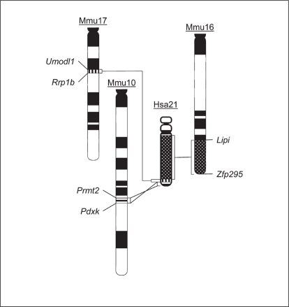
Relatively little is known about the molecular mechanism underlying DS-associated developmental cognitive disabilities and no treatment has yet proved effective. Therefore, animal models are essential to understand the molecular pathophysiology and therapeutic interventions. Since phenotypes of DS are likely to be the result of the allelic gene expression levels on Hsa21 and its interaction with the gene expression and allelic variation of the rest of the genome [Cowles et al., 2002; Wade et al., 2002; Waterston et al., 2002; Antonarakis et al., 2004; Kahlem et al., 2004; Lyle et al., 2004; Dimas et al., 2009; Altshuler et al., 2010; Montgomery et al., 2010; Pickrell et al., 2010], any animal model system will have some limitations and will not mimic exactly the situation in humans. This is partly because the control of gene expression is somewhat different between a model organism and humans, and the conservation of regulatory and other functional genomic elements is variable. Thus, one does not expect that all phenotypes in DS will be reproducible in a model organism. If solely based on evolutionary closeness, nonhuman primates would be the most desirable animal models of DS. Autosomal trisomies have been reported in nonhuman primates [McClure et al., 1969; McClure, 1972; Andrle et al., 1979; Ruppenthal et al., 1983, 1986; de Waal et al., 1996] and transgenic nonhuman primates have also been generated for modeling human neurological disorders [Yang et al., 2008]. Rat models could also have some desirable features since this species has been used extensively in neuroscience research. Gene knockouts have recently been generated in rats using newly established rat embryonic stem (ES) cells [Buehr et al., 2008; Li et al., 2008; Tong et al., 2010] or zinc finger nucleases [Geurts et al., 2009]. However, DS modeling has been almost exclusively carried out in mice so far because of the combination of several advantages. First, there are syntenic regions highly conserved between Hsa21 and the mouse genome (fig. 1) (www.ensembl.org). Second, there have been several widely used mouse models, particularly Ts65Dn, based on their segmental trisomies of the Hsa21 syntenic regions on Mmu16 (fig. 2). Third, the development of the Cre/loxP-mediated mouse chromosome engineering technology enables the generation of new DS mouse models carrying desired chromosomal rearrangements with predetermined endpoints [Yu and Bradley, 2001]. In the following sections, we will discuss the current status of modeling DS-associated developmental cognitive disabilities as well as the future perspectives of molecular genetic studies of this DS phenotype in mouse models. Extensive evidence has indicated that cognitive impairments in mouse models of DS are due to dysfunctions of different brain regions, including the hippocampus and forebrain [Chakrabarti et al., 2007; Chakrabarti et al., 2010]. Here we mainly focused on hippocampus-related phenotypes.
Fig. 1.
Hsa21 and the mouse syntenic regions. There are 3 regions in the mouse genome that are syntenically conserved with Hsa21. The endpoints of these syntenic regions are shown. All the Hsa21 orthologs of the mouse genes in the 3 syntenic regions are located on human 21q.
Fig. 2.
Hsa21, the mouse syntenic regions and the segmentally trisomic mouse models. The endpoints of the syntenic regions and the segmental trisomies in the mouse models are shown.
Current Status of Modeling DS
Segmentally Trisomic Mouse Models
Ts65Dn is currently the most widely used mouse model for DS. This strain is the chromosomally unbalanced progeny of a mouse mutant carrying a balanced translocation; the genetic alteration was randomly induced by irradiation at Muriel Davisson's laboratory [Davisson et al., 1990; Reeves et al., 1995]. The unbalanced derivative chromosome in Ts65Dn consists of a genomic fragment of approximately 13 Mb extending from Mrpl39 to the telomere on Mmu16 with approximately 49.2% of the syntenic regions and approximately 55% of the Hsa21 gene orthologs triplicated (fig. 2). Ts65Dn mice also carry a trisomic segment for a >5.8-Mb subcentromeric region of Mmu17 that is not syntenic to a region on Hsa21 [Davisson et al., 1990; Akeson et al., 2001; Li et al., 2007]. Similar to individuals with DS, the density of dendritic spines was decreased and spine heads were enlarged in Ts65Dn mice [Belichenko et al., 2004, 2007, 2009b; Popov et al., 2011]. Ts65Dn mice consistently exhibit impairments of hippocampal-mediated behaviors. Significantly severe impairment of hippocampal long-term potentiation (LTP), particularly in the dentate gyrus, was observed in Ts65Dn mice [Siarey et al., 1997; Kleschevnikov et al., 2004; Belichenko et al., 2007, 2009b]. Excess inhibition has been proposed to explain this abnormal synaptic plasticity, which is supported by the following evidence. (1) Immunocytochemical analysis of synapse-associated proteins showed no change in the overall density of inhibitory and excitatory synapses in Ts65Dn mice in the dentate gyrus. However, there was a marked increase in colocalization of synaptophysin with the inhibitory presynaptic proteins GAD65 and VGAT (table 1) [Belichenko et al., 2007, 2009b]. (2) At the electron microscopic level, the synaptic apposition length of inhibitory synapses (i.e., symmetric synapses) was significantly increased, with no change in excitatory (i.e., asymmetric) synapses [Belichenko et al., 2009b]. (3) Blocking GABA(A) receptor-mediated inhibitory neurotransmission with picrotoxin restored LTP in the dentate gyrus [Kleschevnikov et al., 2004; Belichenko et al., 2007]. Interestingly, the application of another GABAergic inhibitor, PTZ, not only has restored hippocampal LTP but also has enhanced hippocampus-mediated learning in Ts65Dn mice [Fernandez et al., 2007; Heller et al., 2009].
Table 1.
Alterations of the protein markers in hippocampal synapses in Ts65Dn mice
| Marker for all synapses | Markers for inhibitory synapses | Marker for excitatory synapses | |
|---|---|---|---|
| SYP (p38) | GAD65, VGAT | VGLUT1 | |
| Overall density (puncta/μm2) | no change | no change | no change |
| Colocalization with SYP in the molecular layer of the dentate gyms | N/A | ↑ | no change |
Ts1Cje is the unbalanced progeny of a mouse mutant carrying a balanced translocation, t(12;16), which was induced by gene-targeting in Charles Epstein's laboratory [Sago et al., 1998, 2000]. The unbalanced derivative chromosome in Ts1Cje consists of a genomic fragment of approximately 8.1 Mb from Sod1 to the telomere on Mmu16, with the Sod1 gene inactivated [Sago et al., 1998, 2000] (fig. 2). Ts1Cje mice harbor 3 copies of approximately 67% of the Hsa21 gene orthologs triplicated in Ts65Dn mice [Olson et al., 2004b]. A 2-Mb heterozygous deletion on Mmu12 was reported in Ts1Cje mice in a recent study [Laffaire et al., 2009]. There are important similarities between Ts1Cje and Ts65Dn mice with regard to synaptic and cognitive phenotypes [Olson et al., 2004b; Belichenko et al., 2007]. For instance, widespread enlargement of dendritic spines and decreased density of spines in the dentate gyrus were found in both strains [Belichenko et al., 2007]. Ms1Ts65Dn mice are produced by crossing Ts65Dn mice with t(12;16)1Cje mice [Sago et al., 1998; Sago et al., 2000]; these mice are segmentally trisomic for the genetic segment from Mrpl39 to Sod1. No significant cognitive impairment was found in the Morris water maze test for this mutant [Sago et al., 2000].
Engineered in Roger Reeves’ laboratory, Ts1Rhr is trisomic for the Cbr1-Fam3b region [Olson et al., 2004a; Aldridge et al., 2007] and syntenic to the so-called DS critical chromosomal region on Hsa21 (fig. 2) [Delabar et al., 1993; Sinet et al., 1993; Korenberg et al., 1994]. Cognitively relevant phenotypes of this mutant have been extensively characterized and significant abnormalities in synaptic structure and functions as well as cognitive behaviors were detected [Belichenko et al., 2009a]. Ms1Ts1Rhr mice were generated by crossing Ts65Dn mice with a mutant carrying a deletion in the Cbr1-Fam3b region [Olson et al., 2004a], thus resulting in the reduction of the Cbr1-Fam3b segment to 2 copies in Ts65Dn mice. A reported phenotype of Ms1Ts1Rhr mice was an approximately 18% reduction in the brain volume [Aldridge et al., 2007].
Ts1Yah mice that carry a 0.59-Mb duplication between Abcg1 and U2af1 in the Hsa21 syntenic region on Mmu17 were generated recently in Yann Herault's laboratory (fig. 2) [Pereira et al., 2009]. Interestingly, this duplication apparently led to increased hippocampal LTP in the mutant mice, providing the first evidence of possible genetic interaction between different mouse syntenic regions underlying altered synaptic plasticity associated with DS.
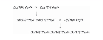
To further understand the impact of the different syntenic regions on developmental cognitive disabilities, the laboratory of Eugene Yu has recently generated the mouse mutants Dp(10)1Yey/+, Dp(16)1Yey/+ and Dp(17)1Yey/+, carrying the duplications spanning the entire Hsa21 syntenic regions on Mmu10, Mmu16 and Mmu17, respectively (fig. 2) [Li et al., 2007; Yu et al., 2010b]. The phenotypic results show that, while the genotype of Dp(17)1Yey/+ results in increased hippocampal LTP, the genotype of Dp(16)1Yey/+ leads to decreased hippocampal LTP and impaired cognitive behaviors. Surprisingly, no significant abnormalities have been detected in Dp(10)1Yey/+ mice based on Morris water maze tests, contextual fear conditioning test and hippocampal LTP analysis even though some genes in the duplicated region have been implicated in neurological disorder, such as S100b [Yu et al., 2010c]. This is consistent with a recent report from Yann Herault's laboratory that the 2.2-Mb heterozygous deletion of the Hsa21 syntenic region on Mmu10 did not alter cognitive deficiency in Tc1 mice [Duchon et al., 2010]. However, it remains possible that some cognitively relevant phenotypes could be detected in Dp(10)1Yey/+ mice if different phenotyping approaches are used. To assess the effect of the simultaneous presence of all the segmental trisomies, Dp(10)1Yey/+;Dp(16)1Yey/+; Dp(17)1Yey/+ mice were generated by crossing the mutants carrying individual duplications, which represent all the evolutionarily conserved genetic alterations and interactions of DS in mice (fig. 3). We showed that these mutant mice exhibited abnormal cognitively relevant phenotypes: a significant decrease in hippocampal LTP and a significant impairment in cognitive behaviors that are based on the Morris water maze and contextual fear conditioning tests [Yu et al., 2010b]. Because of their desirable genotypes and phenotypes, these mouse models offer a new platform for further understanding DS. Unlike Ts65Dn and Tc1 models, Dp(1)1Yey/+, Dp(16)1Yey/+ and Dp(17)1Yey/+ mice can be maintained in 129S5 background and they are also viable and fertile after backcrossing to C57BL/6J mice for five generations. Compound mutants can also be generated in these backgrounds. Therefore, these new models can be used to alleviate the effects of heterogeneous strain backgrounds. In addition, the inbred and congenic backgrounds of the models can facilitate the analysis to identify the genetic modifiers for mutant phenotypes, including cognitively relevant phenotypes.
Fig. 3.
Schematic representation of a breeding strategy to generate a mouse model of DS trisomic for all Hsa21 syntenic regions by crossing mouse mutants carrying individual duplications.
Transchromosomic Mouse Models
Extensive efforts to develop transchromosomic mice by introducing Hsa21 into mouse ES cells using microcell-mediated chromosome transfer [Hernandez et al., 1999; Inoue et al., 2000; Shinohara et al., 2001; O'Doherty et al., 2005] have led to the establishment of the Tc1 mouse model, which is a major achievement in DS modeling. The genotyping result has shown that the transchromosome contains almost the entire Hsa21 with only an approximately 4.9-Mb deletion that contains approximately 19 genes [O'Doherty et al., 2005]. Tc1 mice have been characterized by a number of laboratories and several major DS-related phenotypes were observed in this model, including abnormal developmental cognitive phenotypes, despite a subset of cells in Tc1 mice that did not carry the transchromosome [O'Doherty et al., 2005]. The behavioral experiments, including novel object recognition and Morris water maze tests, showed that Tc1 mice are impaired in learning and memory. Analysis of the dentate gyrus of hippocampal slices isolated from Tc1 mice showed decreased LTP within 60 min after induction [O'Doherty et al., 2005; Morice et al., 2008]. However, additional behavioral experiments and analysis of the dentate gyrus of freely moving mice showed that long-term memory and synaptic plasticity are preserved in Tc1 mice [Morice et al., 2008].
Transgenic Models
Transgenic mice are powerful tools in the functional characterizations of genes. However, many transgenic mouse mutants may not be appropriate for analyzing the impact of overexpression of Hsa21 orthologous genes on DS-related phenotypes because the regulatory elements of the transgenes were not derived from the endogenous loci so the expression levels as well as the spatial and temporal expression patterns might be different from those of the endogenous genes. Nevertheless, BAC or YAC transgenic mice may be useful for unraveling the consequences of the dosage increase for Hsa21 gene orthologs because these types of transgenes may retain all the endogenous regulatory elements for the associated genes [Smith et al., 1995, 1997; Chabert et al., 2004; Roubertoux and Carlier, 2010]. As an interesting example of such BAC transgenic mice, mice harboring a single copy of DYRK1A BAC showed impaired cognitive behaviors but, surprisingly, showed increasedhippocampal LTP [Ahn et al., 2006]. In contrast, the mouse models of DS carrying 3 copies of large segments of Mmu16 syntenic to Hsa21 (i.e., Ts65Dn, Ts1Cje and Dp(16)1Yey/+, all of which contain 3 copies of the mouse Dryk1a gene) exhibited decreased hippocampal LTP [Siarey et al., 1997; Kleschevnikov et al., 2004; Belichenko et al., 2007; Yu et al., 2010c]. One interpretation of this discrepancy is that the triplication(s) of (an)other Hsa21 gene ortholog(s) in Ts65Dn, Ts1Cje and Dp(16)1Yey/+ mice is/are responsible for the decrease in hippocampal LTP and that triplication of the Dyrk1a ortholog may actually help to reduce the impact of the causative genes [Ahn et al., 2006]. The more compelling conclusion is that analyses confined solely to examining the impact of an individual gene without the context of an optimal reference model trisomic for Hsa21 syntenic regions may be insufficient to unravel the details of the contribution of the gene to a DS phenotype.
Future Prospects of Further Molecular Genetic Studies of DS-Associated Developmental Cognitive Disabilities in Mice
Constitutional Transchromosomic Mouse Models
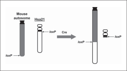
Being segmentally trisomic for all the Hsa21 syntenic regions, Dp(10)1Yey/+;Dp(16)1Yey/+;Dp(17)1Yey/+ mice, despite the complexity and difficulty in their generation, is an optimized genetic model for DS. Transchromosomic models are an ideal alternative for assessing the impact of Hsa21 genes as the third copy of the orthologs. The significance of such models has been well illustrated by Tc1 mice. However, the human chromosome in Tc1 mice is not present in all the cells [O'Doherty et al., 2005]. Therefore, it will be critical to understand the mechanism for the loss of the human chromosome in mouse cells during the developmental process. Since all the Hsa21 orthologs of the mouse genes in the 3 syntenic regions are located on human 21q, it may be possible to overcome mosaicism and establish a constitutional transchromosomic model by targeting human 21q to a mouse chromosome via Cre/loxP-mediated recombination (fig. 4) [Kazuki et al., 2003].
Fig. 4.
A strategy to generate a constitutional transchromosomic mouse model [Kazuki et al., 2003]. A loxP is inserted into the telomere region of a mouse autosome in mouse ES cells while another loxP is inserted into the centromere region of human 21q. The human chromosome can be delivered to the mouse ES cells using microcell-mediated chromosome transfer. Cre/loxP-mediated chromosomal rearrangement in the ES cells can lead to transferring of human 21q to the targeted autosome. The resultant ES cells can be used to generate germ line chimeras by injecting them into wild-type mouse blastocysts.
Besides human-specific genes on Hsa21 that have no mouse orthologs in the mouse syntenic regions, there are mouse-specific genes in the mouse syntenic regions that have no human orthologs on Hsa21 [Yu et al., 2010c; Gardiner et al., 2003]. Therefore, to assess the impact of the triplications of the human-specific Hsa21 genes, it will be desirable to generate and analyze a mouse model carrying 3 copies of human 21q. To avoid the presence of 4 or 5 copies of human 21q gene orthologs and eliminate the contributions of mouse-specific genes in Hsa21 syntenic regions, it will be necessary to remove the mouse syntenic regions from such a model. We and others have established mouse mutants carrying a heterozygous deletion of a partial or entire Hsa21 syntenic region on Mmu10 or Mmu17 [Besson et al., 2007; Duchon et al., 2010; Yu et al., 2010a]. Therefore, it may be possible to delete both copies of these syntenic regions in a model carrying 3 copies of human 21q.
Further Genetic Analysis of DS-Associated Developmental Cognitive Phenotypes in Mice
The true mechanisms underlying the major DS phenotypes remain largely unknown. Recent evidence supports the possibility that some of the DS phenotypes may be associated with the overexpression or underexpression of large chromosomal domains throughout the entire genome [Antonarakis et al., unpubl. results; Gardiner et al., 2010]. The experimental data have also demonstrated that some DS phenotypes are causally associated with the dosage increase of specific Hsa21 gene orthologs, such as App [Salehi et al., 2006]. The mouse-based genetic analysis could be used systemically to identify the dosage-sensitive genes associated with DS-related phenotypes, including developmental cognitive disabilities. To narrow down the genomic regions associated with these phenotypes, new mouse mutants carrying smaller deletions and duplications could be generated. A compound mutant carrying (a) large duplication(s) and a smaller deletion can be used in a subtractive strategy, while smaller duplications can be used in an additive strategy. The importance of the combination of these two strategies is based on the following related possibilities: (1) that the impact of the gene may only be manifested in the context of the action of the other genes present in 3 copies, and that demonstrating a role for a gene will therefore be most evident when that gene is selectively deleted; (2) that the effect of single-gene deletion may not fully abrogate the phenotype but nevertheless may significantly alleviate the severity of the phenotype, and (3) that triplication of just a single gene of interest onto the euploid background may not recapitulate the phenotype. The subtractive and additive strategies are ideal for performing genetic dissection in such situations because they allow for each of the aforementioned possibilities to be explored. When the critical genomic region for a specific phenotype is narrowed down by using duplications and deletions, single-gene knockouts could be incorporated in the subtractive strategy. In those experiments, a compound mutant could be generated to carry a null allele of the gene and a duplication. The contribution of the gene to the phenotype could be established based on elimination or significant alleviation of a DS-related phenotype observed in a mouse mutant carrying the duplication alone [Cataldo et al., 2003; Salehi et al., 2006; Sussan et al., 2008; Baek et al., 2009; Chakrabarti et al., 2010]. Such efforts to identify critical genes are benefitting tremendously from the global mouse gene knockout projects [Austin et al., 2004; Wurst, 2005].
Although the mouse is presently the most important and useful animal model for DS, the introduction and use of other models may in the future further facilitate research that will enable us to understand the molecular mechanisms of trisomy 21; yeast [Moldrich, 2007], Caenorhabditis elegans [Deplancke et al., 2006], Drosophila [Finelli et al., 2004], and zebrafish [North and Zon, 2003], each one, with their own advantages and limitations, may contribute to the understanding of some aspects of the gene expression networks, neuropathology and developmental dysregulation in DS. With the recent progress, major obstacles to efficiently performing mouse-based molecular genetic analysis of DS have been surmounted. Therefore, we can expect the molecular mechanisms underlying this genomic disorder to be gradually unraveled in the coming years, which should in turn significantly enhance our ability to rationally design novel therapeutic interventions for DS-associated clinical phenotypes, including developmental cognitive disabilities.
Acknowledgements
We thank the anonymous reviewers for their helpful suggestions on the manuscript. Projects in the authors’ laboratories are supported in part by grants from the Children's Guild Foundation, the Jerome Lejeune Foundation, the Larry L. Hillblom Foundation, the Down Syndrome Research and Treatment Foundation, the NIH (R01HL91519, R01NS55371 and R01NS66072), the Swiss National Science Foundation, NCCR ‘Frontiers in Genetics’ and the European Union (AnEUploidy Grant LSHG-CT-2006- 037627).
References
- Ahn KJ, Jeong HK, Choi HS, Ryoo SR, Kim YJ, Goo JS, Choi SY, Han JS, Ha I, Song WJ. DYRK1A BAC transgenic mice show altered synaptic plasticity with learning and memory defects. Neurobiol Dis. 2006;22:463–472. doi: 10.1016/j.nbd.2005.12.006. [DOI] [PubMed] [Google Scholar]
- Akeson EC, Lambert JP, Narayanswami S, Gardiner K, Bechtel LJ, Davisson MT. Ts65Dn – localization of the translocation breakpoint and trisomic gene content in a mouse model for Down syndrome. Cytogenet Cell Genet. 2001;93:270–276. doi: 10.1159/000056997. [DOI] [PubMed] [Google Scholar]
- Aldridge K, Reeves RH, Olson LE, Richtsmeier JT. Differential effects of trisomy on brain shape and volume in related aneuploid mouse models. Am J Med Genet A. 2007;143A:1060–1070. doi: 10.1002/ajmg.a.31721. [DOI] [PMC free article] [PubMed] [Google Scholar]
- Altshuler DM, Gibbs RA, Peltonen L, Dermitzakis E, Schaffner SF, Yu F, Bonnen PE, de Bakker PI, Deloukas P, Gabriel SB, Gwilliam R, Hunt S, Inouye M, Jia X, Palotie A, Parkin M, Whittaker P, Chang K, Hawes A, Lewis LR, Ren Y, Wheeler D, Muzny DM, Barnes C, Darvishi K, Hurles M, Korn JM, Kristiansson K, Lee C, McCarrol SA, Nemesh J, Keinan A, Montgomery SB, Pollack S, Price AL, Soranzo N, Gonzaga-Jauregui C, Anttila V, Brodeur W, Daly MJ, Leslie S, McVean G, Moutsianas L, Nguyen H, Zhang Q, Ghori MJ, McGinnis R, McLaren W, Takeuchi F, Grossman SR, Shlyakhter I, Hostetter EB, Sabeti PC, Adebamowo CA, Foster MW, Gordon DR, Licinio J, Manca MC, Marshall PA, Matsuda I, Ngare D, Wang VO, Reddy D, Rotimi CN, Royal CD, Sharp RR, Zeng C, Brooks LD, McEwen JE. Integrating common and rare genetic variation in diverse human populations. Nature. 2010;467:52–58. doi: 10.1038/nature09298. [DOI] [PMC free article] [PubMed] [Google Scholar]
- Andrle M, Fiedler W, Rett A, Ambros P, Schweizer D. A case of trisomy 22 in Pongo pygmaeus. Cytogenet Cell Genet. 1979;24:1–6. doi: 10.1159/000131350. [DOI] [PubMed] [Google Scholar]
- Antonarakis SE, Epstein CJ. The challenge of Down syndrome. Trends Mol Med. 2006;12:473–479. doi: 10.1016/j.molmed.2006.08.005. [DOI] [PubMed] [Google Scholar]
- Antonarakis SE, Lyle R, Dermitzakis ET, Reymond A, Deutsch S. Chromosome 21 and down syndrome: from genomics to pathophysiology. Nat Rev Genet. 2004;5:725–738. doi: 10.1038/nrg1448. [DOI] [PubMed] [Google Scholar]
- Austin CP, Battey JF, Bradley A, Bucan M, Capecchi M, Collins FS, Dove WF, Duyk G, Dymecki S, Eppig JT, Grieder FB, Heintz N, Hicks G, Insel TR, Joyner A, Koller BH, Lloyd KC, Magnuson T, Moore MW, Nagy A, Pollock JD, Roses AD, Sands AT, Seed B, Skarnes WC, Snoddy J, Soriano P, Stewart DJ, Stewart F, Stillman B, Varmus H, Varticovski L, Verma IM, Vogt TF, von Melchner H, Witkowski J, Woychik RP, Wurst W, Yancopoulos GD, Young SG, Zambrowicz B. The knockout mouse project. Nat Genet. 2004;36:921–924. doi: 10.1038/ng0904-921. [DOI] [PMC free article] [PubMed] [Google Scholar]
- Baek KH, Zaslavsky A, Lynch RC, Britt C, Okada Y, Siarey RJ, Lensch MW, Park IH, Yoon SS, Minami T, Korenberg JR, Folkman J, Daley GQ, Aird WC, Galdzicki Z, Ryeom S. Down's syndrome suppression of tumour growth and the role of the calcineurin inhibitor DSCR1. Nature. 2009;459:1126–1130. doi: 10.1038/nature08062. [DOI] [PMC free article] [PubMed] [Google Scholar]
- Belichenko NP, Belichenko PV, Kleschevnikov AM, Salehi A, Reeves RH, Mobley WC. The ‘Down syndrome critical region’ is sufficient in the mouse model to confer behavioral, neurophysiological, and synaptic phenotypes characteristic of Down syndrome. J Neurosci. 2009a;29:5938–5948. doi: 10.1523/JNEUROSCI.1547-09.2009. [DOI] [PMC free article] [PubMed] [Google Scholar]
- Belichenko PV, Kleschevnikov AM, Masliah E, Wu C, Takimoto-Kimura R, Salehi A, Mobley WC. Excitatory-inhibitory relationship in the fascia dentata in the Ts65Dn mouse model of Down syndrome. J Comp Neurol. 2009b;512:453–466. doi: 10.1002/cne.21895. [DOI] [PMC free article] [PubMed] [Google Scholar]
- Belichenko PV, Kleschevnikov AM, Salehi A, Epstein CJ, Mobley WC. Synaptic and cognitive abnormalities in mouse models of Down syndrome: exploring genotype-phenotype relationships. J Comp Neurol. 2007;504:329–345. doi: 10.1002/cne.21433. [DOI] [PubMed] [Google Scholar]
- Belichenko PV, Masliah E, Kleschevnikov AM, Villar AJ, Epstein CJ, Salehi A, Mobley WC. Synaptic structural abnormalities in the Ts65Dn mouse model of Down syndrome. J Comp Neurol. 2004;480:281–298. doi: 10.1002/cne.20337. [DOI] [PubMed] [Google Scholar]
- Besson V, Brault V, Duchon A, Togbe D, Bizot JC, Quesniaux VF, Ryffel B, Herault Y. Modeling the monosomy for the telomeric part of human chromosome 21 reveals haploinsufficient genes modulating the inflammatory and airway responses. Hum Mol Genet. 2007;16:2040–2052. doi: 10.1093/hmg/ddm152. [DOI] [PubMed] [Google Scholar]
- Buehr M, Meek S, Blair K, Yang J, Ure J, Silva J, McLay R, Hall J, Ying QL, Smith A. Capture of authentic embryonic stem cells from rat blastocysts. Cell. 2008;135:1287–1298. doi: 10.1016/j.cell.2008.12.007. [DOI] [PubMed] [Google Scholar]
- Cataldo AM, Petanceska S, Peterhoff CM, Terio NB, Epstein CJ, Villar A, Carlson EJ, Staufenbiel M, Nixon RA. App gene dosage modulates endosomal abnormalities of Alzheimer's disease in a segmental trisomy 16 mouse model of Down syndrome. J Neurosci. 2003;23:6788–6792. doi: 10.1523/JNEUROSCI.23-17-06788.2003. [DOI] [PMC free article] [PubMed] [Google Scholar]
- Chabert C, Jamon M, Cherfouh A, Duquenne V, Smith DJ, Rubin E, Roubertoux PL. Functional analysis of genes implicated in Down syndrome. 1. Cognitive abilities in mice transpolygenic for Down Syndrome Chromosomal Region-1 (DCR-1) Behav Genet. 2004;34:559–569. doi: 10.1007/s10519-004-5584-3. [DOI] [PubMed] [Google Scholar]
- Chakrabarti L, Best TK, Cramer NP, Carney RS, Isaac JT, Galdzicki Z, Haydar TF. Olig1 and Olig2 triplication causes developmental brain defects in Down syndrome. Nat Neurosci. 2010;13:927–934. doi: 10.1038/nn.2600. [DOI] [PMC free article] [PubMed] [Google Scholar]
- Chakrabarti L, Galdzicki Z, Haydar TF. Defects in embryonic neurogenesis and initial synapse formation in the forebrain of the Ts65Dn mouse model of Down syndrome. J Neurosci. 2007;27:11483–11495. doi: 10.1523/JNEUROSCI.3406-07.2007. [DOI] [PMC free article] [PubMed] [Google Scholar]
- Chapman RS, Hesketh LJ. Behavioral phenotype of individuals with Down syndrome. Ment Retard Dev Disabil Res Rev. 2000;6:84–95. doi: 10.1002/1098-2779(2000)6:2<84::AID-MRDD2>3.0.CO;2-P. [DOI] [PubMed] [Google Scholar]
- Cowles CR, Hirschhorn JN, Altshuler D, Lander ES. Detection of regulatory variation in mouse genes. Nat Genet. 2002;32:432–437. doi: 10.1038/ng992. [DOI] [PubMed] [Google Scholar]
- Cragg BG. The density of synapses and neurons in normal, mentally defective ageing human brains. Brain. 1975;98:81–90. doi: 10.1093/brain/98.1.81. [DOI] [PubMed] [Google Scholar]
- Davisson MT, Schmidt C, Akeson EC. Segmental trisomy of murine chromosome 16: a new model system for studying Down syndrome. Prog Clin Biol Res. 1990;360:263–280. [PubMed] [Google Scholar]
- DeFelipe J, Farinas I. The pyramidal neuron of the cerebral cortex: morphological and chemical characteristics of the synaptic inputs. Prog Neurobiol. 1992;39:563–607. doi: 10.1016/0301-0082(92)90015-7. [DOI] [PubMed] [Google Scholar]
- Delabar JM, Theophile D, Rahmani Z, Chettouh Z, Blouin JL, Prieur M, Noel B, Sinet PM. Molecular mapping of twenty-four features of Down syndrome on chromosome 21. Eur J Hum Genet. 1993;1:114–124. doi: 10.1159/000472398. [DOI] [PubMed] [Google Scholar]
- Deplancke B, Mukhopadhyay A, Ao W, Elewa AM, Grove CA, Martinez NJ, Sequerra R, Doucette-Stamm L, Reece-Hoyes JS, Hope IA, Tissenbaum HA, Mango SE, Walhout AJ. A gene-centered C. elegans protein-DNA interaction network. Cell. 2006;125:1193–1205. doi: 10.1016/j.cell.2006.04.038. [DOI] [PubMed] [Google Scholar]
- de Waal FB, Uno H, Luttrell LM, Meisner LF, Jeannotte LA. Behavioral retardation in a macaque with autosomal trisomy and aging mother. Am J Ment Retard. 1996;100:378–390. [PubMed] [Google Scholar]
- Dimas AS, Deutsch S, Stranger BE, Montgomery SB, Borel C, Attar-Cohen H, Ingle C, Beazley C, Gutierrez Arcelus M, Sekowska M, Gagnebin M, Nisbett J, Deloukas P, Dermitzakis ET, Antonarakis SE. Common regulatory variation impacts gene expression in a cell type-dependent manner. Science. 2009;325:1246–1250. doi: 10.1126/science.1174148. [DOI] [PMC free article] [PubMed] [Google Scholar]
- Duchon A, Pothion S, Brault V, Sharp AJ, Tybulewicz VL, Fisher EM, Herault Y. The telomeric part of the human chromosome 21 from Cstb to Prmt2 is not necessarry for the locomotor and short-term memory deficits observed in the Tc1 mouse model of Down syndrome. Behav Brain Res. 2010;217:271–281. doi: 10.1016/j.bbr.2010.10.023. [DOI] [PMC free article] [PubMed] [Google Scholar]
- Epstein CJ. The Consequences of Chromosome Imbalance: Principles, Mechanism and Models. New York: Cambridge University Press; 1986. [Google Scholar]
- Fernandez F, Morishita W, Zuniga E, Nguyen J, Blank M, Malenka RC, Garner CC. Pharmacotherapy for cognitive impairment in a mouse model of Down syndrome. Nat Neurosci. 2007;10:411–413. doi: 10.1038/nn1860. [DOI] [PubMed] [Google Scholar]
- Ferrer I, Gullotta F. Down's syndrome and Alzheimer's disease: dendritic spine counts in the hippocampus. Acta Neuropathol. 1990;79:680–685. doi: 10.1007/BF00294247. [DOI] [PubMed] [Google Scholar]
- Fiala JC, Spacek J, Harris KM. Dendritic spine pathology: cause or consequence of neurological disorders? Brain Res Brain Res Rev. 2002;39:29–54. doi: 10.1016/s0165-0173(02)00158-3. [DOI] [PubMed] [Google Scholar]
- Finelli A, Kelkar A, Song HJ, Yang H, Konsolaki M. A model for studying Alzheimer's Abeta42-induced toxicity in Drosophila melanogaster. Mol Cell Neurosci. 2004;26:365–375. doi: 10.1016/j.mcn.2004.03.001. [DOI] [PubMed] [Google Scholar]
- Gardiner K, Fortna A, Bechtel L, Davisson MT. Mouse models of Down syndrome: how useful can they be? Comparison of the gene content of human chromosome 21 with orthologous mouse genomic regions. Gene. 2003;318:137–147. doi: 10.1016/s0378-1119(03)00769-8. [DOI] [PubMed] [Google Scholar]
- Gardiner K, Herault Y, Lott IT, Antonarakis SE, Reeves RH, Dierssen M. Down syndrome: from understanding the neurobiology to therapy. J Neurosci. 2010;30:14943–14945. doi: 10.1523/JNEUROSCI.3728-10.2010. [DOI] [PMC free article] [PubMed] [Google Scholar]
- Geurts AM, Cost GJ, Freyvert Y, Zeitler B, Miller JC, Choi VM, Jenkins SS, Wood A, Cui X, Meng X, Vincent A, Lam S, Michalkiewicz M, Schilling R, Foeckler J, Kalloway S, Weiler H, Menoret S, Anegon I, Davis GD, Zhang L, Rebar EJ, Gregory PD, Urnov FD, Jacob HJ, Buelow R. Knockout rats via embryo microinjection of zinc-finger nucleases. Science. 2009;325:433. doi: 10.1126/science.1172447. [DOI] [PMC free article] [PubMed] [Google Scholar]
- Gray EG. Electron microscopy of synaptic contacts on dendrite spines of the cerebral cortex. Nature. 1959;183:1592–1593. doi: 10.1038/1831592a0. [DOI] [PubMed] [Google Scholar]
- Haxby JV. Neuropsychological evaluation of adults with Down's syndrome: patterns of selective impairment in non-demented old adults. J Ment Defic Res. 1989;33:193–210. doi: 10.1111/j.1365-2788.1989.tb01467.x. [DOI] [PubMed] [Google Scholar]
- Heller HC, Colas D, Ruby NF, Fernandez F, Chuluun B, Blank M, Garner CC. Long term repair of learning disability through short-term reduction of CNS inhibition. In: Schmorrow DD, Estabrookse IV, Grootjen M, editors. Foundations of Augmented Cognition, Neuroergonomics and Operational Neuroscience. New York: Springer; 2009. pp. 818–825. [Google Scholar]
- Hernandez D, Mee PJ, Martin JE, Tybulewicz VL, Fisher EM. Transchromosomal mouse embryonic stem cell lines and chimeric mice that contain freely segregating segments of human chromosome 21. Hum Mol Genet. 1999;8:923–933. doi: 10.1093/hmg/8.5.923. [DOI] [PubMed] [Google Scholar]
- Inoue T, Shinohara T, Takehara S, Inoue J, Kamino H, Kugoh H, Oshimura M. Specific impairment of cardiogenesis in mouse ES cells containing a human chromosome 21. Biochem Biophys Res Commun. 2000;273:219–224. doi: 10.1006/bbrc.2000.2927. [DOI] [PubMed] [Google Scholar]
- Kahlem P, Sultan M, Herwig R, Steinfath M, Balzereit D, Eppens B, Saran NG, Pletcher MT, South ST, Stetten G, Lehrach H, Reeves RH, Yaspo ML. Transcript level alterations reflect gene dosage effects across multiple tissues in a mouse model of Down syndrome. Genome Res. 2004;14:1258–1267. doi: 10.1101/gr.1951304. [DOI] [PMC free article] [PubMed] [Google Scholar]
- Kazuki Y, Schulz TC, Shinohara T, Kadota M, Nishigaki R, Inoue T, Kimura M, Kai Y, Abe S, Shirayoshi Y, Oshimura M. A new mouse model for Down syndrome. In: Lubec G, editor. Advances in Down Syndrome Research. Wien: Springer; 2003. pp. 1–20. [DOI] [PubMed] [Google Scholar]
- Kleschevnikov AM, Belichenko PV, Villar AJ, Epstein CJ, Malenka RC, Mobley WC. Hippocampal long-term potentiation suppressed by increased inhibition in the Ts65Dn mouse, a genetic model of Down syndrome. J Neurosci. 2004;24:8153–8160. doi: 10.1523/JNEUROSCI.1766-04.2004. [DOI] [PMC free article] [PubMed] [Google Scholar]
- Korenberg JR, Chen XN, Schipper R, Sun Z, Gonsky R, Gerwehr S, Carpenter N, Daumer C, Dignan P, Disteche C. Down syndrome phenotypes: the consequences of chromosomal imbalance. Proc Natl Acad Sci USA. 1994;91:4997–5001. doi: 10.1073/pnas.91.11.4997. [DOI] [PMC free article] [PubMed] [Google Scholar]
- Laffaire J, Rivals I, Dauphinot L, Pasteau F, Wehrle R, Larrat B, Vitalis T, Moldrich RX, Rossier J, Sinkus R, Herault Y, Dusart I, Potier MC. Gene expression signature of cerebellar hypoplasia in a mouse model of Down syndrome during postnatal development. BMC Genomics. 2009;10:138. doi: 10.1186/1471-2164-10-138. [DOI] [PMC free article] [PubMed] [Google Scholar]
- Li P, Tong C, Mehrian-Shai R, Jia L, Wu N, Yan Y, Maxson RE, Schulze EN, Song H, Hsieh CL, Pera MF, Ying QL. Germline competent embryonic stem cells derived from rat blastocysts. Cell. 2008;135:1299–1310. doi: 10.1016/j.cell.2008.12.006. [DOI] [PMC free article] [PubMed] [Google Scholar]
- Li Z, Yu T, Morishima M, Pao A, LaDuca J, Conroy J, Nowak N, Matsui S, Shiraishi I, Yu Y. Duplication of the entire 22.9-Mb human chromosome 21 syntenic region on mouse chromosome 16 causes cardiovascular and gastrointestinal abnormalities. Hum Mol Genet. 2007;16:1359–1366. doi: 10.1093/hmg/ddm086. [DOI] [PubMed] [Google Scholar]
- Lyle R, Gehrig C, Neergaard-Henrichsen C, Deutsch S, Antonarakis SE. Gene expression from the aneuploid chromosome in a trisomy mouse model of Down syndrome. Genome Res. 2004;14:1268–1274. doi: 10.1101/gr.2090904. [DOI] [PMC free article] [PubMed] [Google Scholar]
- Marin-Padilla M. Structural abnormalities of the cerebral cortex in human chromosomal aberrations: a Golgi study. Brain Res. 1972;44:625–629. doi: 10.1016/0006-8993(72)90324-1. [DOI] [PubMed] [Google Scholar]
- Marin-Padilla M. Pyramidal cell abnormalities in the motor cortex of a child with Down's syndrome. A Golgi study. J Comp Neurol. 1976;167:63–81. doi: 10.1002/cne.901670105. [DOI] [PubMed] [Google Scholar]
- McClure HM. Animal model: trisomy in a chimpanzee. Am J Pathol. 1972;67:413–416. [Google Scholar]
- McClure HM, Belden KH, Pieper WA, Jacobson CB. Autosomal trisomy in a chimpanzee: resemblance to Down's syndrome. Science. 1969;165:1010–1012. doi: 10.1126/science.165.3897.1010. [DOI] [PubMed] [Google Scholar]
- Moldrich RX. A yeast model of Down syndrome. Int J Dev Neurosci. 2007;25:539–543. doi: 10.1016/j.ijdevneu.2007.10.001. [DOI] [PubMed] [Google Scholar]
- Montgomery SB, Sammeth M, Gutierrez-Arcelus M, Lach RP, Ingle C, Nisbett J, Guigo R, Dermitzakis ET. Transcriptome genetics using second generation sequencing in a Caucasian population. Nature. 2010;464:773–777. doi: 10.1038/nature08903. [DOI] [PMC free article] [PubMed] [Google Scholar]
- Morice E, Andreae LC, Cooke SF, Vanes L, Fisher EM, Tybulewicz VL, Bliss TV. Preservation of long-term memory and synaptic plasticity despite short-term impairments in the Tc1 mouse model of Down syndrome. Learn Mem. 2008;15:492–500. doi: 10.1101/lm.969608. [DOI] [PMC free article] [PubMed] [Google Scholar]
- Nadel L. Down syndrome in cognitive neuroscience perspective. In: Tager-Flusberg H, editor. Neurodeveolpmental Disorders. Cambridge: MIT Press; 1999. pp. 197–221. [Google Scholar]
- North TE, Zon LI. Modeling human hematopoietic and cardiovascular diseases in zebrafish. Dev Dyn. 2003;228:568–583. doi: 10.1002/dvdy.10393. [DOI] [PubMed] [Google Scholar]
- O'Doherty A, Ruf S, Mulligan C, Hildreth V, Errington ML, Cooke S, Sesay A, Modino S, Vanes L, Hernandez D, Linehan JM, Sharpe PT, Brandner S, Bliss TV, Henderson DJ, Nizetic D, Tybulewicz VL, Fisher EM. An aneuploid mouse strain carrying human chromosome 21 with Down syndrome phenotypes. Science. 2005;309:2033–2037. doi: 10.1126/science.1114535. [DOI] [PMC free article] [PubMed] [Google Scholar]
- Olson LE, Richtsmeier JT, Leszl J, Reeves RH. A chromosome 21 critical region does not cause specific Down syndrome phenotypes. Science. 2004a;306:687–690. doi: 10.1126/science.1098992. [DOI] [PMC free article] [PubMed] [Google Scholar]
- Olson LE, Roper RJ, Baxter LL, Carlson EJ, Epstein CJ, Reeves RH. Down syndrome mouse models Ts65Dn, Ts1Cje, and Ms1Cje/Ts65Dn exhibit variable severity of cerebellar phenotypes. Dev Dyn. 2004b;230:581–589. doi: 10.1002/dvdy.20079. [DOI] [PubMed] [Google Scholar]
- Pennington BF, Moon J, Edgin J, Stedron J, Nadel L. The neuropsychology of Down syndrome: evidence for hippocampal dysfunction. Child Dev. 2003;74:75–93. doi: 10.1111/1467-8624.00522. [DOI] [PubMed] [Google Scholar]
- Pereira PL, Magnol L, Sahun I, Brault V, Duchon A, Prandini P, Gruart A, Bizot JC, Chadefaux-Vekemans B, Deutsch S, Trovero F, Delgado-Garcia JM, Antonarakis SE, Dierssen M, Herault Y. A new mouse model for the trisomy of the Abcg1-U2af1 region reveals the complexity of the combinatorial genetic code of down syndrome. Hum Mol Genet. 2009;18:4756–4769. doi: 10.1093/hmg/ddp438. [DOI] [PMC free article] [PubMed] [Google Scholar]
- Pickrell JK, Marioni JC, Pai AA, Degner JF, Engelhardt BE, Nkadori E, Veyrieras JB, Stephens M, Gilad Y, Pritchard JK. Understanding mechanisms underlying human gene expression variation with RNA sequencing. Nature. 2010;464:768–772. doi: 10.1038/nature08872. [DOI] [PMC free article] [PubMed] [Google Scholar]
- Popov VI, Kleschevnikov AM, Klimenko OA, Stewart MG, Belichenko PV. Three-dimensional synaptic ultrastructure in the dentate gyrus and CA3 hippocampal area in the Ts65Dn mouse model of Down syndrome. J Comp Neurol. 2011;519:1338–1354. doi: 10.1002/cne.22573. [DOI] [PubMed] [Google Scholar]
- Pulsifer MB. The neuropsychology of mental retardation. J Int Neuropsychol Soc. 1996;2:159–176. doi: 10.1017/s1355617700001016. [DOI] [PubMed] [Google Scholar]
- Purpura DP. Normal and aberrant neuronal development in the cerebral cortex of human fetus and young infant. UCLA Forum Med Sci. 1975:141–169. doi: 10.1016/b978-0-12-139050-1.50014-8. [DOI] [PubMed] [Google Scholar]
- Reeves RH, Irving NG, Moran TH, Wohn A, Kitt C, Sisodia SS, Schmidt C, Bronson RT, Davisson MT. A mouse model for Down syndrome exhibits learning and behaviour deficits. Nat Genet. 1995;11:177–184. doi: 10.1038/ng1095-177. [DOI] [PubMed] [Google Scholar]
- Roizen NJ, Patterson D. Down's syndrome. Lancet. 2003;361:1281–1289. doi: 10.1016/S0140-6736(03)12987-X. [DOI] [PubMed] [Google Scholar]
- Ross MH, Galaburda AM, Kemper TL. Down's syndrome: is there a decreased population of neurons? Neurology. 1984;34:909–916. doi: 10.1212/wnl.34.7.909. [DOI] [PubMed] [Google Scholar]
- Roubertoux PL, Carlier M. Mouse models of cognitive disabilities in trisomy 21 (Down syndrome) Am J Med Genet C Semin Med Genet. 2010;154C:400–416. doi: 10.1002/ajmg.c.30280. [DOI] [PubMed] [Google Scholar]
- Ruppenthal GC, Caffery SA, Goodlin BL, Sackett GP, Vigfusson NV, Peterson VG. Pigtailed macaques (Macaca nemestrina) with trisomy X manifest physical and mental retardation. Am J Ment Defic. 1983;87:471–476. [PubMed] [Google Scholar]
- Ruppenthal GC, Caffery SA, Goodlin BL, Sackett GP, Vigfusson NV, Peterson VG. Pigtailed macaques (Macaca nemestrina) with trisomy X manifest physical and mental retardation. Erratum. Am J Ment Defic. 1986;90:362. [PubMed] [Google Scholar]
- Sago H, Carlson EJ, Smith DJ, Kilbridge J, Rubin EM, Mobley WC, Epstein CJ, Huang TT. Ts1Cje, a partial trisomy 16 mouse model for Down syndrome, exhibits learning and behavioral abnormalities. Proc Natl Acad Sci USA. 1998;95:6256–6261. doi: 10.1073/pnas.95.11.6256. [DOI] [PMC free article] [PubMed] [Google Scholar]
- Sago H, Carlson EJ, Smith DJ, Rubin EM, Crnic LS, Huang TT, Epstein CJ. Genetic dissection of region associated with behavioral abnormalities in mouse models for Down syndrome. Pediatr Res. 2000;48:606–613. doi: 10.1203/00006450-200011000-00009. [DOI] [PubMed] [Google Scholar]
- Salehi A, Delcroix JD, Belichenko PV, Zhan K, Wu C, Valletta JS, Takimoto-Kimura R, Kleschevnikov AM, Sambamurti K, Chung PP, Xia W, Villar A, Campbell WA, Kulnane LS, Nixon RA, Lamb BT, Epstein CJ, Stokin GB, Goldstein LS, Mobley WC. Increased App expression in a mouse model of Down's syndrome disrupts NGF transport and causes cholinergic neuron degeneration. Neuron. 2006;51:29–42. doi: 10.1016/j.neuron.2006.05.022. [DOI] [PubMed] [Google Scholar]
- Schmidt-Sidor B, Wisniewski KE, Shepard TH, Sersen EA. Brain growth in Down syndrome subjects 15 to 22 weeks of gestational age and birth to 60 months. Clin Neuropathol. 1990;9:181–190. [PubMed] [Google Scholar]
- Shinohara T, Tomizuka K, Miyabara S, Takehara S, Kazuki Y, Inoue J, Katoh M, Nakane H, Iino A, Ohguma A, Ikegami S, Inokuchi K, Ishida I, Reeves RH, Oshimura M. Mice containing a human chromosome 21 model behavioral impairment and cardiac anomalies of Down's syndrome. Hum Mol Genet. 2001;10:1163–1175. doi: 10.1093/hmg/10.11.1163. [DOI] [PubMed] [Google Scholar]
- Siarey RJ, Stoll J, Rapoport SI, Galdzicki Z. Altered long-term potentiation in the young and old Ts65Dn mouse, a model for Down syndrome. Neuropharmacology. 1997;36:1549–1554. doi: 10.1016/s0028-3908(97)00157-3. [DOI] [PubMed] [Google Scholar]
- Sinet PM, Theophile D, Rahmani Z, Chettouh Z, Blouin JL, Prieur M, Noel B, Delabar JM. Molecular mapping of the Down syndrome phenotype on chromosome 21. Prog Clin Biol Res. 1993;384:63–86. [PubMed] [Google Scholar]
- Smith DJ, Stevens ME, Sudanagunta SP, Bronson RT, Makhinson M, Watabe AM, O'Dell TJ, Fung J, Weier HU, Cheng JF, Rubin EM. Functional screening of 2 Mb of human chromosome 21q22.2 in transgenic mice implicates minibrain in learning defects associated with Down syndrome. Nat Genet. 1997;16:28–36. doi: 10.1038/ng0597-28. [DOI] [PubMed] [Google Scholar]
- Smith DJ, Zhu Y, Zhang J, Cheng JF, Rubin EM. Construction of a panel of transgenic mice containing a contiguous 2-Mb set of YAC/P1 clones from human chromosome 21q22.2. Genomics. 1995;27:425–434. doi: 10.1006/geno.1995.1073. [DOI] [PubMed] [Google Scholar]
- Suetsugu M, Mehraein P. Spine distribution along the apical dendrites of the pyramidal neurons in Down's syndrome. A quantitative Golgi study. Acta Neuropathol. 1980;50:207–210. doi: 10.1007/BF00688755. [DOI] [PubMed] [Google Scholar]
- Sussan TE, Yang A, Li F, Ostrowski MC, Reeves RH. Trisomy represses Apc(Min)-mediated tumours in mouse models of Down's syndrome. Nature. 2008;451:73–75. doi: 10.1038/nature06446. [DOI] [PubMed] [Google Scholar]
- Takashima S, Becker LE, Armstrong DL, Chan F. Abnormal neuronal development in the visual cortex of the human fetus and infant with Down's syndrome. A quantitative and qualitative Golgi study. Brain Res. 1981;225:1–21. doi: 10.1016/0006-8993(81)90314-0. [DOI] [PubMed] [Google Scholar]
- Takashima S, Iida K, Mito T, Arima M. Dendritic and histochemical development and ageing in patients with Down's syndrome. J Intellect Disabil Res. 1994;38:265–273. doi: 10.1111/j.1365-2788.1994.tb00394.x. [DOI] [PubMed] [Google Scholar]
- Tong C, Li P, Wu NL, Yan Y, Ying QL. Production of p53 gene knockout rats by homologous recombination in embryonic stem cells. Nature. 2010;467:211–213. doi: 10.1038/nature09368. [DOI] [PMC free article] [PubMed] [Google Scholar]
- Uecker A, Mangan PA, Obrzut JE, Nadel L. Down syndrome in neurobiological perspective: an emphasis on spatial cognition. J Clin Child Psychol. 1993;22:266–276. [Google Scholar]
- Wade CM, Kulbokas EJ, 3rd, Kirby AW, Zody MC, Mullikin JC, Lander ES, Lindblad-Toh K, Daly MJ. The mosaic structure of variation in the laboratory mouse genome. Nature. 2002;420:574–578. doi: 10.1038/nature01252. [DOI] [PubMed] [Google Scholar]
- Waterston RH, Lindblad-Toh K, Birney E, Rogers J, Abril JF, Agarwal P, Agarwala R, Ainscough R, Alexandersson M, An P, Antonarakis SE, Attwood J, Baertsch R, Bailey J, Barlow K, Beck S, Berry E, Birren B, Bloom T, Bork P, Botcherby M, Bray N, Brent MR, Brown DG, Brown SD, Bult C, Burton J, Butler J, Campbell RD, Carninci P, Cawley S, Chiaromonte F, Chinwalla AT, Church DM, Clamp M, Clee C, Collins FS, Cook LL, Copley RR, Coulson A, Couronne O, Cuff J, Curwen V, Cutts T, Daly M, David R, Davies J, Delehaunty KD, Deri J, Dermitzakis ET, Dewey C, Dickens NJ, Diekhans M, Dodge S, Dubchak I, Dunn DM, Eddy SR, Elnitski L, Emes RD, Eswara P, Eyras E, Felsenfeld A, Fewell GA, Flicek P, Foley K, Frankel WN, Fulton LA, Fulton RS, Furey TS, Gage D, Gibbs RA, Glusman G, Gnerre S, Goldman N, Goodstadt L, Grafham D, Graves TA, Green ED, Gregory S, Guigo R, Guyer M, Hardison RC, Haussler D, Hayashizaki Y, Hillier LW, Hinrichs A, Hlavina W, Holzer T, Hsu F, Hua A, Hubbard T, Hunt A, Jackson I, Jaffe DB, Johnson LS, Jones M, Jones TA, Joy A, Kamal M, Karlsson EK, Karolchik D, Kasprzyk A, Kawai J, Keibler E, Kells C, Kent WJ, Kirby A, Kolbe DL, Korf I, Kucherlapati RS, Kulbokas EJ, Kulp D, Landers T, Leger JP, Leonard S, Letunic I, Levine R, Li J, Li M, Lloyd C, Lucas S, Ma B, Maglott DR, Mardis ER, Matthews L, Mauceli E, Mayer JH, McCarthy M, McCombie WR, McLaren S, McLay K, McPherson JD, Meldrim J, Meredith B, Mesirov JP, Miller W, Miner TL, Mongin E, Montgomery KT, Morgan M, Mott R, Mullikin JC, Muzny DM, Nash WE, Nelson JO, Nhan MN, Nicol R, Ning Z, Nusbaum C, O'Connor MJ, Okazaki Y, Oliver K, Overton-Larty E, Pachter L, Parra G, Pepin KH, Peterson J, Pevzner P, Plumb R, Pohl CS, Poliakov A, Ponce TC, Ponting CP, Potter S, Quail M, Reymond A, Roe BA, Roskin KM, Rubin EM, Rust AG, Santos R, Sapojnikov V, Schultz B, Schultz J, Schwartz MS, Schwartz S, Scott C, Seaman S, Searle S, Sharpe T, Sheridan A, Shownkeen R, Sims S, Singer JB, Slater G, Smit A, Smith DR, Spencer B, Stabenau A, Stange-Thomann N, Sugnet C, Suyama M, Tesler G, Thompson J, Torrents D, Trevaskis E, Tromp J, Ucla C, Ureta-Vidal A, Vinson JP, Von Niederhausern AC, Wade CM, Wall M, Weber RJ, Weiss RB, Wendl MC, West AP, Wetterstrand K, Wheeler R, Whelan S, Wierzbowski J, Willey D, Williams S, Wilson RK, Winter E, Worley KC, Wyman D, Yang S, Yang SP, Zdobnov EM, Zody MC, Lander ES. Initial sequencing and comparative analysis of the mouse genome. Nature. 2002;420:520–562. doi: 10.1038/nature01262. [DOI] [PubMed] [Google Scholar]
- Wisniewski KE. Down syndrome children often have brain with maturation delay, retardation of growth, and cortical dysgenesis. Am J Med Genet Suppl. 1990;7:274–281. doi: 10.1002/ajmg.1320370755. [DOI] [PubMed] [Google Scholar]
- Wurst W. Mouse geneticists need European strategy too. Nature. 2005;433:13. doi: 10.1038/433013c. [DOI] [PubMed] [Google Scholar]
- Yang SH, Cheng PH, Banta H, Piotrowska-Nitsche K, Yang JJ, Cheng EC, Snyder B, Larkin K, Liu J, Orkin J, Fang ZH, Smith Y, Bachevalier J, Zola SM, Li SH, Li XJ, Chan AW. Towards a transgenic model of Huntington's disease in a non-human primate. Nature. 2008;453:921–924. doi: 10.1038/nature06975. [DOI] [PMC free article] [PubMed] [Google Scholar]
- Yu T, Clapcote SJ, Li Z, Liu C, Pao A, Bechard AR, Carattini-Rivera S, Matsui S, Roder JC, Baldini A, Mobley WC, Bradley A, Yu YE. Deficiencies in the region syntenic to human 21q22.3 cause cognitive deficits in mice. Mamm Genome. 2010a;21:258–267. doi: 10.1007/s00335-010-9262-x. [DOI] [PMC free article] [PubMed] [Google Scholar]
- Yu T, Li Z, Jia Z, Clapcote SJ, Liu C, Li S, Asrar S, Pao A, Chen R, Fan N, Carattini-Rivera S, Bechard AR, Spring S, Henkelman RM, Stoica G, Matsui S, Nowak NJ, Roder JC, Chen C, Bradley A, Yu YE. A mouse model of Down syndrome trisomic for all human chromosome 21 syntenic regions. Hum Mol Genet. 2010b;19:2780–2791. doi: 10.1093/hmg/ddq179. [DOI] [PMC free article] [PubMed] [Google Scholar]
- Yu T, Liu C, Belichenko P, Clapcote SJ, Li S, Pao A, Kleschevnikov A, Bechard AR, Asrar S, Chen R, Fan N, Zhou Z, Jia Z, Chen C, Roder JC, Liu B, Baldini A, Mobley WC, Yu YE. Effects of individual segmental trisomies of human chromosome 21 syntenic regions on hippocampal long-term potentiation and cognitive behaviors in mice. Brain Res. 2010c;1366:162–171. doi: 10.1016/j.brainres.2010.09.107. [DOI] [PMC free article] [PubMed] [Google Scholar]
- Yu Y, Bradley A. Engineering chromosomal rearrangements in mice. Nat Rev Genet. 2001;2:780–790. doi: 10.1038/35093564. [DOI] [PubMed] [Google Scholar]


