Summary
This article details a case report of a subject chosen from among patients treated in the author’s clinic in the Posturology and Gnathology Section of the University Milano-Bicocca.
It shows how the indispensable clinical analysis of the stomatognathic system and the connections between posture can be supported by instrumental analysis, such as the computerized occlusal analysis system and the force platform, to diagnose and treat dysfunctional patients.
Keywords: computerized occlusal analysis, posture, force platform, occlusion, occlusal splint
Introduction
During the “Consensus Conference”, held in Milan in 1997 (1), it was reported that there was no scientific evidence to support a connection between occlusion and posture. Thus, using occlusal treatment to prevent postural problems would not be justifiable. Other scientific articles have also reached this conclusion (2). During another “Consensus Conference” in 2008, was stated that the most recent scientific literature provided weak evidence supporting the connection between posture, occlusion and the neurophysiological integration of the body’s mechanism (3). Both orthodontists and physiatrists participated in the Consensus which advised treating dysfunctional patients with a reversible (and, thus, not irreversible) gnathological postural treatment. After a correct, specific diagnosis has been made, which is supported by careful clinical analysis (3), this may improve the clinical symptomatology of some patients.
Following the most modern gnathologic approaches, the stomatognathic system, which forms part of the postural system, is treated holistically (4, 5, 6, 7).
Occlusion, however, is considered the pivot of both diagnostic and therapeutic dentistry and the most recent scientific research, which connects the craniocervical system with the rest of the organism, requires a much more detailed analysis of cranio-mandibular occlusal relationships (8, 9, 10).
This case report shows the clinical-instrumental approach carried out at the Posturology and Gnathology Section of the Dental Clinic of the University Milan-Bicocca, following the clinical and instrumental protocols which have also been suggested by other authors (11, 12).
In this case, in particular, the T-Scan and the force platform were used to treat a dysfunctional patient, who needed to stabilize or balance occlusion using a splint. The effectiveness of the treatment was verified using clinical and instrumental methods to document and confirm the improvement of the clinical case following the protocols for the instruments used which were suggested by international literature (13, 14, 15, 16, 17, 18).
The instrumental analyses do however have limits (19, 20, 21, 22, 23) and cannot replace, but merely be integrated with the fundamental clinical analysis which must be carried out according to the standards dictated in international literature (24).
Clinical case
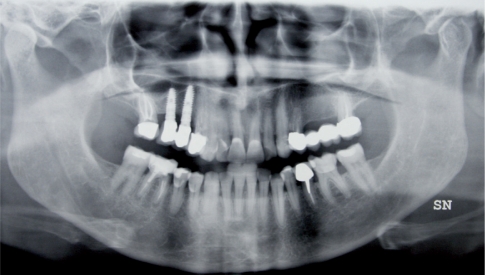
A 55 years old female patient suffering from a regular (every day), spontaneous pain due to mastication in the right region of the masseter, with muscular tension inducing cephalea in the right frontal region 2 or 3 times a week, was referred to our department. This pain started 4 years ago, when a prosthetic rehabilitation on implants was inserted in the right upper arch (Fig.1). The patient also suffered from frequent cervicalgia.
Figure 1.
Orthopantomography with the implant for prosthetic rehabilitation at the level of 15 and 16 visualized.
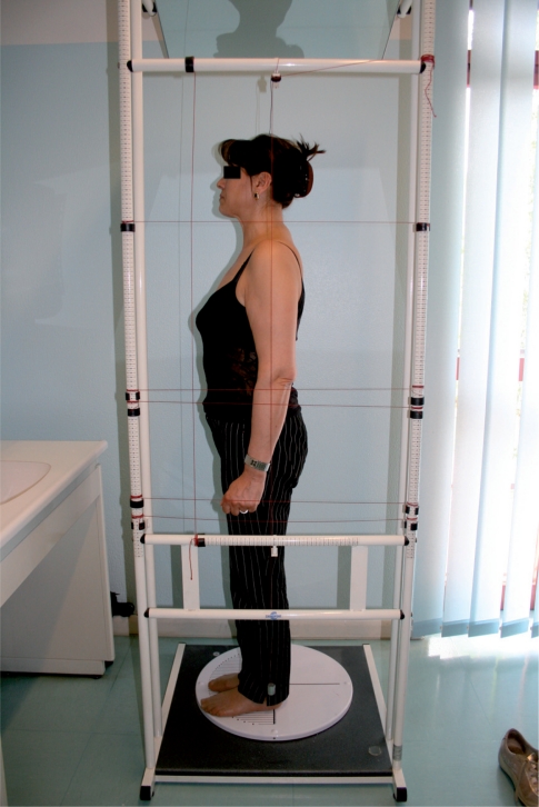
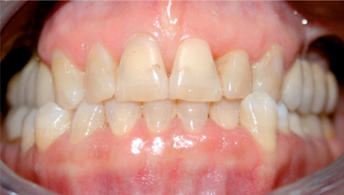
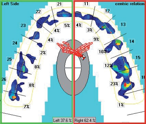
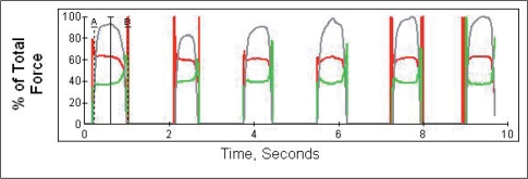
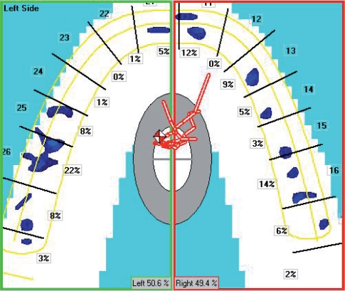
We started with the gnathological postural check-up, which includes: static postural analysis via scoliometer (Fig.2) and podoscope, evaluation of the skeletal and dental relations (Fig. 3), showing the occlusal parameters with the presence of crosses and dental inversion. We then made 3D and 2D (Fig. 4) occlusion images with the T-Scan (T-Scan III,Tec-Scan, Boston Usa, Dl Medica, Milan, Italy). These showed the occlusal loads to be distributed somewhat asymmetrically (60% on the right and 40% on the left) with particular overloading at the right posterior section, as shown also on the force/time diagram (Fig. 5). The dynamical evaluation of the occlusal load distribution demonstrated significant premature occlusal contact at the level of 14 in a temporal sequence, with an occlusal time of 0.876 seconds more than the physiological average. The computerized occlusal analysis was carried out detecting the centric contact points and the contact between the patient’s dental arches was checked 3 or 4 times in order to check the repeatability of the test (16, 17, 29).
Figure 2.
Static, postural evaluations on scoliometer, with slight postural asymmetry.
Figure 3.
Intra-oral picture, right section with cross of 23/33.
Figure 4.
2D display of the asymmetry of the occlusal loads on T-Scan III, right section with 60% of the load on the right and 40% on the left.
Figure 5.
Asymmetry of occlusal loads.
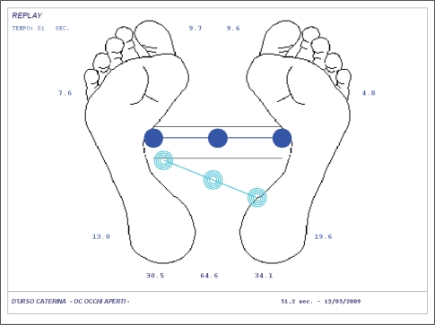
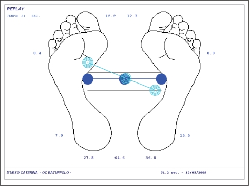
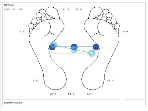
Using the Force Platform Correkta (Dl Medica, Milan Italy) in the physiologic rest position, we observed that the loads projected slightly towards the back with torsion. This was emphasized in centric position (Fig. 6) as if occlusion worsened postural balance. Inserting cotton rolls in the oral cavity seemed to improve the postural position (Fig. 7).
Figure 6.
Posturometry on force platform with accentuation of the projection of the loads towards the back and torsion in centric occlusion.
Figure 7.
Posturometry on force platform with improvement of the postural position inserting cotton rolls in the oral cavity.
As according to force platform protocol, the postural stabilometric analysis was carried out with eyes opened and closed, in rest position, in centric occlusion and with cotton rolls ensuring subjects are in orthostatic state, with the patient’s arms by her sides in a relaxed fashion, with feet slightly apart forming a 30° angle and heels 2 cm apart. (18)
The patient was advised to have a Michigan stabilization splint inserted in the upper arch, to be worn for 6 months, 16 hours a day, and to do some postural and physiotherapeutic exercises to improve cervical mobility and postural balance.
The stabilization splint was balanced using the T-Scan III, the insertion of the splint resulted in a better occlusal load distribution, with occlusal balance between the two arches and a better COF (centre of force) position (Fig. 8). These results can also be observed from the force/time diagram (Fig. 9). Force platform checks showed better postural balance after the insertion of the splint in the oral cavity, with the load brought forward and better torsion (Fig. 10).
Figure 8.
Best positioning of the occlusal centre of force (COF, little red and white dot) with the splint inserted in the oral cavity and a better distribution of the occlusal loads between the right and left hemiarches.
Figure 9.
On the force-time diagram the red and green lines coincide, as a result of arranging the loads symmetrically between the two hemiarches after the insertion of the splint.
Figure 10.
Improving the postural balance measured on force platform Correction with the splint inserted in the oral cavity.
After 4 months of wearing the splint, the masseter pain had significantly lessened, the cervical pain had stopped and the frequency of the bouts of cephalea had reduced.
The same clinical and instrumental improvements were observed stable at 6, 9, and 12 months.
Discussion and conclusions
Our experience in treating dysfunctional patients shows that a clinical and instrumental approach is best suited to diagnose and treat patients who often present postural and occlusal problems. These instruments are able to quantify and measure the obtained gnathological postural balance. However the instrumental analysis cannot replace, but merely supports the essential clinical analysis carried out according to international standards (RDC/TMD) in diagnostic literature (18, 24, 28, 29).
References
- 1.Ciancaglini R. Posture, occlusion and general health. Proceedings of the Research Forum. Milan, International Meeting in Clinical gnathology; 1997. [Google Scholar]
- 2.Michelotti A, Manzo P, Farella M, Martina R. Occlusione e postura :quali le evidenze di correlazione ? Minerva Stomatologica. 1999;48:525–34. [PubMed] [Google Scholar]
- 3.Ciancaglini R, Cerri C, Saggini R, Bellomo RG, Ridi R, Piscella V, Di Pancrazio L, Di Paolo C, Leonardi R, Greco M, Heir G. Posture and occlusion :hypothesis of correlation. J Stomat Occ Med. 2009;1:87–96. [Google Scholar]
- 4.Sakaguchi K, Metha N, Abdallah E, Forgione A. Examination of relationship between mandibular position and body posture. Cranio. 2007 Oct;25(4):237–49. doi: 10.1179/crn.2007.037. [DOI] [PubMed] [Google Scholar]
- 5.Bergamini M, Pierleoni F, Gizdulich A, Bergamini C. Dental occlusion and body posture : a surface EMG study. Cranio. 2008 Jan;26(1):25–32. doi: 10.1179/crn.2008.041. [DOI] [PubMed] [Google Scholar]
- 6.Tecco S, Salini V, Calvisi V, Colucci C, Orso CA, Festa F, D’Attilio M. Effects of anterior cruciate ligament (ACL)injury on postural control and muscle activity of head, neck and trunk muscles. J Oral Rehabilit. 2006 Aug;33(8):576–87. doi: 10.1111/j.1365-2842.2005.01592.x. [DOI] [PubMed] [Google Scholar]
- 7.Sforza C, Tartaglia GM, Solimene U, Morgun V, Kaspransky RR, Ferrario VF. Occlusion, sternocleidomastoid muscle activity and body sway:a pilot study in male astronauts. Cranio. 2006 Jan;24(1):43–9. doi: 10.1179/crn.2006.008. [DOI] [PubMed] [Google Scholar]
- 8.Chapman RJ. Principles of occlusion for implant prosthesis: guidelines for position, timing, Force of occlusal contacts. Quintessence International. 1989 Mar;20(7):473–480. [PubMed] [Google Scholar]
- 9.Bernasconi G, De Rysky C, Forniciti T. Computerized occlusal verification in venture frames. Dent Cadmos. 1990 Mar 15;58(4):72–4. [PubMed] [Google Scholar]
- 10.Makofosky HW, Sexton TR, Diamond DZ, Sexton MT. The effect of head posture on muscle contact position using the TScan system of occlusal analysis. Cranio. 1991 Oct;9(4):316–321. doi: 10.1080/08869634.1991.11678378. [DOI] [PubMed] [Google Scholar]
- 11.Lazic V, Todoritivic A, Zivkovic S, Marinovic Z. Computerized occlusal analysis in bruxism. Srp Arth Celok. 2006 Jan-Feb;134(1–2):22–9. doi: 10.2298/sarh0602022l. Serbian. [DOI] [PubMed] [Google Scholar]
- 12.Karey J, Craig M, Kerstein R, Radke J. Determining a relationship between applied occlusal load and articulating paper mark area. The Open Dent J. 2007;2007;1(1–7) doi: 10.2174/1874210600701010001. [DOI] [PMC free article] [PubMed] [Google Scholar]
- 13.Kerstein RB, Wright NR. Electromyographic and computer analyses of patiens suffering from chronic myofascial pain-dysfunction syndrome : before and after treatment with immediate complete anterior guidance development. J Prosthet Dent. 1991 Nov;66(5):677–86. doi: 10.1016/0022-3913(91)90453-4. Review. [DOI] [PubMed] [Google Scholar]
- 14.Tokumura K, Yamashita A. Study on occlusion analysis by means of T-Scan System. Its accuracy for measurement. Nihon Hotetsu Shika Gakkai Zasshi. 1989 Oct;33(5):1037–43. doi: 10.2186/jjps.33.1037. Japanese. [DOI] [PubMed] [Google Scholar]
- 15.Mizui M, Nabeshima F, Tosa J, Tanaka M, Kawazoe T. Quantitative analysis of occlusal balance in intercuspal position using the T-Scan system. Int J Prosthodont. 1994 Jan-Feb;:62–71. [PubMed] [Google Scholar]
- 16.Kerstein RB. Combining technologies: a computerized occlusal analysis system synchronized with a computerized electromyography system. Cranio. 2004 Apr;22(2):96–109. doi: 10.1179/crn.2004.013. [DOI] [PubMed] [Google Scholar]
- 17.Stevens CJ. Computerized occlusal implant management with the T-Scan II System :a case report. Dent Today. 2006 Feb;25(2):88–91. [PubMed] [Google Scholar]
- 18.Baldini A, Cioffi D, Rinaldi A. Gnatho-postural approach in Italian air force pilots: stabilometric evaluations. Annali di Stomatologia. 2009;LVIII(4):102–106. [Google Scholar]
- 19.Gomes de Oliveira S, Seraidarian PI, Landre J, Jr, Oliveira DD, Cavalcanti BN. Tooth displacement due to occlusal : a three-dimensional finite element study. J Oral Rehabililit. 2006 Dec;33(12):874–880. doi: 10.1111/j.1365-2842.2006.01670.x. [DOI] [PubMed] [Google Scholar]
- 20.Garg AK. Analyzing dental occlusion for implants : Tekscan’s, T-Scan III. Dent Implantol Update. 2007 Sep;18(9):65–70. [PubMed] [Google Scholar]
- 21.Saracoglu A, Ozpinar B. In vivo and in vitro evaluation of occlusal indicator sensitivity. J Prosthet Dent. 2002 Nov;88(5):522–6. doi: 10.1067/mpr.2002.129064. [DOI] [PubMed] [Google Scholar]
- 22.Patyk A, Lotzmann U, Scherer C, Kobes LW. Comparative analytic occlusal study of clinical use of TScan systems. ZWR. 1989 Sep;98(9):752–5. German. [PubMed] [Google Scholar]
- 23.Kalachev YS, Michailov TA, Iordanov PI. Study of occlusal articulation relationship with the help of T-Scan apparatus. Folia Med (Plovdiv) 2001;43(1–2):88–91. [PubMed] [Google Scholar]
- 24.Dworkin SF, LeResche L. Researarch diagnostic criteria for temporomandibular disorders:review, criteria, examinations and specifications, critique. J. Craniomandib Disord. 1992;6:301–355. [PubMed] [Google Scholar]
- 25.Baldini A, Beraldi A, Longoni S. La valutazione computerizzata dell’occlusione nelle riabilitazioni implanto-protesiche. Implantologia. Quintessenza Edizioni. 2008;3:71–77. [Google Scholar]
- 26.Baldini A. Importanza del T-Scan III nella valutazione dell’occlusione. Doctor Os. 2008;(4):124. Febbraio. XIX. 2008. [Google Scholar]
- 27.Baldini A. L’analisi computerizzata dell’occlusione in ortodonzia. Atti del XIX Congresso Internazionale Sido. 2008 Novembre;(20–22):134. Firenze. [Google Scholar]
- 28.Baldini A, Beraldi A, Nanussi A. Importanza clinica della valutazione computerizzata dell’occlusione. Dental Cadmos. 2009 Aprile;77(4):47–54. [Google Scholar]
- 29.Baldini A, Beraldi A, Longoni S. The importance of computerized occlusal analysis in the diagnosis and treatment of dysfunctional patients. Annales of Stomatology. 2009;LVI–II(4):117–124. [Google Scholar]


