Abstract
Psychiatric disorders, including autism spectrum disorders and schizophrenia, are extremely heritable complex genetic neurodevelopmental disorders. It is now possible to directly reprogram fibroblasts from psychiatric patients into human induced pluripotent stem cells (hiPSCs) and subsequently differentiate these disorder-specific hiPSCs into neurons. This means that researchers can generate nearly limitless quantities of live human neurons with genetic backgrounds that are known to result in psychiatric disorders, without knowing which genes are interacting to produce the disease state in each patient. With these new human-cell-based models, scientists can investigate the precise cell types that are affected in these disorders and elucidate the cellular and molecular defects that contribute to disease initiation and progression. Here, we present a short review of experiments using hiPSCs and other sophisticated in vitro approaches to study the pathways underlying psychiatric disorders.
Introduction
Psychiatric disorders are common yet debilitating conditions. Two of these disorders, autism spectrum disorders (ASDs) and schizophrenia (SCZD), combine to affect nearly 2% of the global population. ASDs are complex neurodevelopmental disorders characterized by severe impairments in social interactions, communication and behavior (Kanner, 1943; American Psychiatric Association, 2000). SCZD is characterized by three severe classes of symptoms: positive (hallucinations and delusions), negative (inability to speak, express emotion or find pleasure) and cognitive (deficits in attention, memory and planning) (American Psychiatric Association, 2000; Carpenter and Buchanan, 1994); it often leads to premature death from poverty, homelessness, substance abuse and poor health maintenance (Brown et al., 2000).
Of these two psychiatric disorders, twin studies estimate the heritability of ASDs to be greatest, as high as 90% (Ritvo et al., 1985), whereas estimates of the heritability of SCZD range from 70–85% (Tsuang et al., 2001; Sullivan et al., 2003). Genome-wide association scans for ASDs and SCZD suggest that common genetic loci influence susceptibility to many psychiatric disorders (Lewis et al., 2003; Fanous et al., 2007). It is now believed that many rare but penetrant mutations contribute to ASDs and SCZD, and that hundreds of common variants with mild effects are shared by SCZD and bipolar disorder (O’Donovan et al., 2008; Walsh et al., 2008; Moskvina et al., 2009).
It is generally very difficult to obtain live neurons from patients affected with ASDs or SCZD to study. In addition, because most psychiatric disorders are polygenic, few relevant animal models have been developed. Although transgenic mice demonstrate some abnormalities in behavior in specific experimental conditions (Clapcote et al., 2007; Hikida et al., 2007; Barros et al., 2009), it is difficult to determine how well these mouse models recapitulate the human condition because it is difficult to assess complex phenotypes such as hallucinations and delusions in mice. To date, human studies of these psychiatric disorders have relied primarily on brain imaging of patients, postmortem studies of brains and genetic studies of patient lymphocytes. Many notable insights have been made through these methods, but the molecular and cellular defects that contribute to disease initiation and progression in neurons have been difficult to elucidate.
Although they are poorly understood at the molecular and cellular levels, psychiatric disorders are now believed to be developmental disorders. Therefore, human-cell-based models representing genetic backgrounds known to develop these neurological diseases might be the best method to identify the neuronal defects associated with them. In this Commentary, we review recent studies that use sophisticated human-cell-based models to study the pathways underlying psychiatric disorders. We limit our discussion to approaches that focus on neuronal cell types, although non-neuronal cell-based approaches have also provided useful information (Box 1).
Box 1. Examples of existing cell-based models of psychiatric disease.
There is a long history of non-neuronal cell-based studies comparing patients and controls, particularly in the study of SCZD. The best established of these are studies of fibroblasts, lymphocytes and olfactory neural stem cells. Non-neuronal cells typically express lower levels of neuronal genes than do mature neurons, so it remains uncertain to what extent findings from these studies generate insights into the mechanism of disease.
Fibroblasts: skin fibroblasts have been used as cellular models of SCZD (Mahadik and Mukherjee, 1996). Although some comparisons between control and SCZD fibroblasts do not show robust patient-versus-control differences (Matigian et al., 2008), others find small differences (Matigian et al., 2010).
Lymphocytes: a small number of microarray analysis studies using immortalized lymphocytes derived from patients with SCZD have been reported; however, whether immortalized lymphocytes are a relevant alternative to neuronal tissue remains controversial (Matigian et al., 2010). Lymphocyte studies have identified aberrant expression of DISC1, serotonin transporter (5-HTT), dysbindin-1 and neuregulin-1 (NRG-1) (Hranilovic et al., 2004; Maeda et al., 2006; Yamamori et al., 2011) in samples from patients with SCZD.
Olfactory neural stem cells: Matigian et al. compared patient-derived olfactory neural stem cells from nine patients with SCZD and 14 controls, and identified SCZD-associated differences in gene expression and protein levels in neurodevelopmental pathways, including cell migration and axon guidance (Matigian et al., 2010).
Reprogramming patient fibroblasts to induced pluripotent stem cells
In 2006, Takahashi and Yamanaka identified four factors (Oct3/4, Klf4, Sox2 and Myc) whose transient expression was sufficient to directly reprogram mouse adult somatic cells into an induced pluripotent stem cell (iPSC) state (Takahashi and Yamanaka, 2006). One year later, groups led by Shinya Yamanaka and James Thomson (Takahashi et al., 2007; Yu et al., 2007) demonstrated for the first time that it was also possible to reprogram human fibroblasts.
iPSCs can be generated from either mouse or human skin cells and are nearly identical to embryonic stem (ES) cells with respect to gene expression, DNA methylation and chromatin modifications. Both mouse iPSCs (miPSCs) and human iPSCs (hiPSCs) are believed to be capable of differentiating into every cell type found in the adult (Maherali et al., 2007; Meissner et al., 2007; Takahashi et al., 2007; Wernig et al., 2007; Yu et al., 2007). Because hiPSCs can be derived from adult humans, after the development of disease, hiPSCs represent a potentially limitless source of human cells with which to study disease, even without knowing which genes are interacting to produce the disease state in an individual patient.
It is important to note that genetic and epigenetic mutations can and do occur during the reprogramming process. Sometimes hiPSC lines contain abnormal karyotypes after reprogramming (Mayshar et al., 2010), and large copy number variations (CNVs) have also been associated with both the reprogramming and differentiation of hiPSCs (Laurent et al., 2011). However, significantly more CNVs are present in early-passage hiPSCs than in higher-passage hiPSCs, fibroblasts or human ES cells; apparently, most novel CNVs generated during early stages of the reprogramming process are subsequently lost from the hiPSC population (Hussein et al., 2011). Additionally, among the 22 hiPSC lines studied, an average of five protein-coding point mutations were identified (Gore et al., 2011). Furthermore, hiPSCs share megabase-scale differentially methylated regions proximal to centromeres and telomeres that display incomplete reprogramming (Lister et al., 2011). Importantly, whereas hiPSCs do acquire genetic modifications in addition to epigenetic modifications, and although extensive genetic screening should become a standard procedure to ensure hiPSC safety before clinical use, it is extremely unlikely that de novo mutations affect disease-specific hiPSC lines in a meaningfully different way than control hiPSC lines. Although spontaneous mutations can occur during the reprogramming process, random mutations should not interfere with the ability to draw meaningful conclusions from hiPSC-based studies of psychiatric disorders when studies include comparisons of neurons generated from multiple hiPSC lines for each control and diseased patient.
Differentiating iPSCs to neurons
Work with ES cells first demonstrated methods to efficiently differentiate pluripotent stem cells into neurons (Tropepe et al., 2001). During embryonic development, embryonic neuroepithelia are believed to be first specified to a forebrain fate and are then subsequently transformed with caudalizing signals to midbrain/hindbrain and spinal cord fates in a gradient manner (Stern, 2001). Similarly, through the addition of various morphogens in vitro, ES cells and iPSCs can be directed to differentiate into regional identities including forebrain (Watanabe et al., 2005), midbrain/hindbrain (Kawasaki et al., 2000; Perrier et al., 2004) and spinal cord (Wichterle et al., 2002; Li et al., 2005). Owing to an excellent characterization of the transcription factors used to specify fate in embryonic mice, the best-characterized hiPSC differentiation protocol is generally accepted to be one for motor neurons (Di Giorgio et al., 2008; Dimos et al., 2008), although a highly efficient method for generating midbrain dopaminergic neurons was recently published (Chambers et al., 2009). Much of the developmental potential that is present in vivo seems to be maintained in vitro.
The choice of specific subtypes of neurons to be compared using hiPSC-derived neurons warrants careful consideration because the brain regions and cell types affected differ between diseases. Magnetic resonance imaging (MRI) studies have observed structural differences in the brains of many patients affected by psychiatric disorders compared with those of the normal population. For example, patients with SCZD often have decreased brain volume, particularly in the hippocampus, thalamus and cortex (Vita et al., 2006; Ellison-Wright et al., 2008), whereas patients with ASDs show transient increases in brain volume from ages 2 to 4 years, notably in the cortex, cerebellum and amygdala, coinciding with the onset of symptoms (Courchesne et al., 2007). Notably, although these MRI studies provide information regarding the brain regions that seem to be affected by these disorders, they do not reveal the cell types implicated in disease, but some insights have been provided through pharmacological studies. With respect to SCZD, for example, dopamine receptor antagonists, and perhaps also glutamate receptor (GLUR2/3) agonists, alleviate psychosis (Patil et al., 2007). Conversely, when administered to healthy human subjects, dopamine agonists (such as amphetamine) and glutamate antagonists (such as ketamine) induce hallucinations and cognitive deficits similar to those associated with SCZD (Krystal et al., 1994). Pharmacological studies of ASDs have been less successful in identifying abnormalities in the activity of a specific neurotransmitter.
Although the specific cell subtypes that should ideally be examined to provide insight into these diseases remain uncertain, it is generally believed that cortical neurons are a relevant cell type for the study of many psychiatric disorders. Rigorous validation of the neurons that are ultimately generated from hiPSCs is crucial for establishing the maturity and identity of the neuronal cultures that are generated in vitro. Electrophysiological characterization should verify that hiPSC neurons have membrane potentials, undergo induced action potentials and show evidence of spontaneous synaptic activity. Specific markers should be used to confirm neuronal identity. For example, mature dopaminergic neurons express tyrosine hydroxylase (TH) and aromatic L-amino acid decarboxylase (AADC) (Ang, 2006), whereas mature cortical neurons express CTIP2, a transcriptional repressor that is required for the maturation of cortical axons, and OTX1, a transcription factor that is highly expressed in layer 5 cortical neurons (Frantz et al., 1994; Weimann et al., 1999; Arlotta et al., 2005).
It is important to note that researchers cannot generate artificial brain structures in vitro, such as specific brain regions implicated in neurological disease – for example, the prefrontal cortex for studies of ASD or the hippocampus for studies of SCZD. Furthermore, the neural populations generated through directed differentiation protocols are invariably extremely heterogeneous. Although the relative frequency of a specific neuronal cell type might be favored, differentiated populations generally contain several types of neurons, as well as astrocytes, oligodendrocytes, neural precursors and even non-neural cells.
Phenotypes of psychiatric disorders can be identified in hiPSC-derived neurons
The first hiPSC-based models of psychiatric disease have now been reported. This strategy permits study of the innate neuron-specific deficits in psychiatric disease in a manner that is not confounded by environmental factors that typically plague studies of SCZD, such as treatment history, drug and alcohol abuse or poverty. These reports have all been published within the last year; each supports our contention that hiPSC-derived neurons from patients with psychiatric disorders show significant aberrations compared with those of healthy controls.
hiPSCs differentiate robustly to forebrain neurons and, by co-culturing hiPSC forebrain neurons with HEK293T cells, Kim et al. showed that HEK293T cells expressing wild-type NLGN3 and NLGN4, but not those containing ASD-associated mutations in these genes, can induce presynaptic differentiation in hiPSC-derived neurons (Kim et al., 2011). Although this work used only hiPSCs derived from the cells of healthy controls, these findings did establish that these cells are a viable model for the study of synaptic differentiation and that they function under normal and disorder-associated conditions.
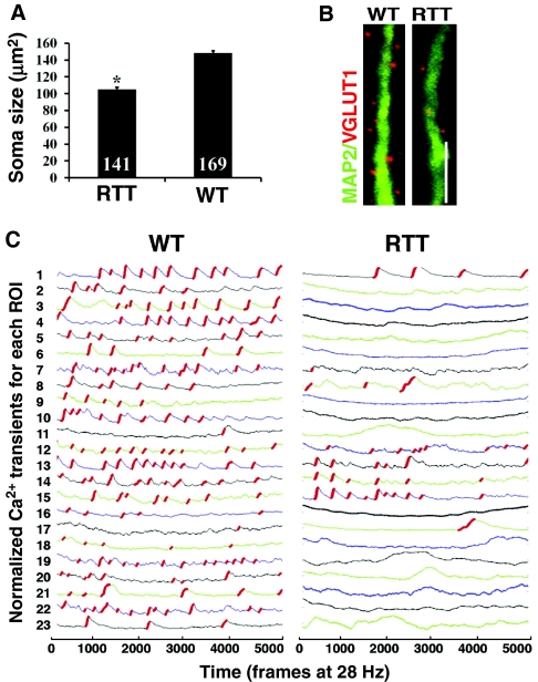
Using Rett syndrome (RTT) as an ASD genetic model, we generated hiPSCs from patients with RTT and found early alterations in developing hiPSC-derived neurons (Marchetto et al., 2010). Compared with controls, neurons derived from RTT hiPSCs had fewer synapses, reduced spine density, smaller soma size, altered Ca2+ signaling and electrophysiological defects (Fig. 1B,C). We also used RTT-hiPSC-derived neurons to test the capacity of drugs to rescue synaptic defects (Marchetto et al., 2010). Subsequently, a second group repeated the finding of reduced neuronal soma size in RTT-hiPSC-derived neurons (Cheung et al., 2011) (Fig. 1A). Together, these studies established that assays of neuronal size, synaptic structure and synaptic function can detect differences between control and disordered hiPSC-derived neurons in vitro, demonstrating that neurodevelopmental diseases can be modeled in this manner. Notably, RTT is usually associated with the complete loss of function of a single gene (MECP2) and shows rapid disease progression within the first years of life; the first studies of complex genetic forms of ASD have not yet been reported.
Fig. 1.
Cellular phenotypes of RTT. (A) hiPSC-derived neurons from RTT patients show decreased soma size. *P<0.0001, Student’s t-test. Adapted from Cheung et al. (Cheung et al., 2011), with permission. (B) RTT-hiPSC-derived neurons have reduced density of excitatory synapses along dendrites compared with neurons derived from healthy controls. The staining shows that hiPSC-derived neurons from RTT patients have fewer VGLUT1-positive glutamatergic synaptic puncta interspersed along MAP2-positive dendrites. (C) Tracking fluorescence intensity changes representing intracellular Ca2+ fluctuations provides evidence of reduced synaptic activity in RTT-hiPSC-derived neurons relative to controls. B and C are adapted from Marchetto et al. (Marchetto et al., 2010), with permission. ROI, region of interest; WT, wild type. Scale bar: 5 μm.
Mutations in DISC1 result in an extremely rare monogenic form of SCZD. Chiang et al. recently reported the generation of integration-free hiPSCs from SCZD patients with a DISC1 mutation (Chiang et al., 2011), but have not yet reported a characterization of neurons differentiated from these hiPSCs.
These first reports of hiPSCs generated from patients with psychiatric disease studied monogenetic disorders. Given that mouse models of RTT (Mecp2 null) and mutated DISC1 (dnDISC) exist, these early studies allow a direct comparison between in vitro hiPSC studies and in vivo animal experiments. It will be interesting to learn whether DISC1-hiPSC-derived neurons recapitulate the cellular phenotypes observed in dnDISC mice as faithfully as RTT-hiPSC-derived neurons recapitulated findings in Mecp2-null mice. With these important proof-of-principle validations of hiPSC models completed, future hiPSC-based studies must begin to take full advantage of the ability of hiPSCs to model complex genetic cases of psychiatric disease, in which interactions between as-yet-unidentified genes result in the disease state.
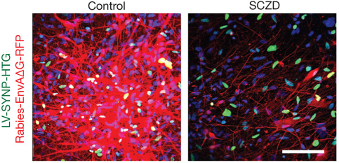
We recently reported the use of hiPSCs to model a complex genetic psychiatric disorder in which hiPSC-derived neurons from four patients with SCZD were compared with those from controls. SCZD-hiPSC-derived neurons had reduced neuronal connectivity, reduced outgrowths from soma, reduced PSD95 dendritic protein levels and altered gene expression profiles relative to controls (Fig. 2); defects in neuronal connectivity and gene expression were ameliorated following treatment with the antipsychotic drug loxapine (Brennand et al., 2011). Nearly 25% of genes that showed altered expression compared with controls had been previously implicated in SCZD, although the expression profiling data also suggested that a number of pathways not previously implicated in SCZD might contribute to the disorder, such as Notch signaling, cell adhesion and Slit-Robo-mediated axon guidance. We predict that, as future studies increase the number of SCZD-patient-derived neurons, core pathways of genes that contribute to this disorder will be identified.
Fig. 2.
Cellular phenotype of SCZD. Rabies virus transmission between neurons can be used to assay neuronal connectivity. SCZD-hiPSC-derived neurons show decreased transmission of a genetically engineered rabies virus designed specifically to indicate monosynaptic neuronal connectivity (Rabies-EnvAΔG-RFP). Adapted from Brennand et al. (Brennand et al., 2011), with permission. LV-SYNP-HTG, lentivirus expressing a fusion protein comprising histone 2B and green fluorescent protein, TVA and elements of the synapsin (SYN) promoter; used to label neurons for microscopic analysis. Scale bar: 80 μm.
hiPSC-based studies show great potential use to model complex genetic disorders for which the genes responsible for producing the disease state differ between individuals. We hope that hiPSCs will permit direct evaluations of several hypotheses that are not easy to address in psychiatric disease research, such as:
Does severity of clinical outcome predict magnitude of cellular phenotype?
Do genetic lesions correlate with neuronal gene expression differences?
Is clinical pharmacological response predictable by hiPSC neuronal drug response?
To discover the answers to these questions, substantially increased numbers of patient-specific hiPSC-derived neurons must be generated. Furthermore, hiPSC-derived neurons must be derived from better-characterized patient cohorts. By moving forward with cohorts of patients for whom clinical outcome, pharmacological response, MRI imaging and genotype data are available, hiPSC-based models of psychiatric disorders will enable direct correlations of clinical, cellular and molecular phenotypes in psychiatric disease.
Limitations of hiPSC-based modeling of psychiatric disease
The limitations of hiPSC-based approaches for studying psychiatric disease are mainly (1) neuron-to-neuron variability, (2) hiPSC-to-hiPSC variability and (3) patient-to-patient variability. Neuron-to-neuron variability encompasses differences between individual hiPSC-derived neurons from a single patient; it often reflects heterogeneity of neuronal subtypes within a neural population. Uniquely among the hiPSC reports discussed, Kim et al. attempted to direct the regional identity of their hiPSC neurons, and determined that treatment with a sonic hedgehog (SHH) agonist increased expression of the forebrain marker BF1 (also known as FOXG1), repressed the expression of the dorsal markers PAX6, EMX2 and GLI3, and increased the expression of the ventral markers SHH and NKX2.1 at the neural progenitor cell (NPC) stage, suggesting that some of their hiPSC neurons acquired an anterior ventral forebrain fate (Kim et al., 2011). Universally, hiPSC-based studies of psychiatric disorders to date have all been performed on mixed populations of neuronal subtypes, which are generally described as predominantly glutamatergic with a significant fraction of GABAnergic neurons and only rare dopaminergic neurons, as assessed by expression of VGLUT, GAD67 and TH, respectively. None of the reports described compares pure neuronal populations. The use of heterogeneous neuronal populations increases neuron-to-neuron variability in experimental assays, a limitation that could be overcome by studying fluorescence-activated cell sorting (FACS)-purified populations of neurons of a defined identity.
A second limitation of hiPSC studies is variability in hiPSC lines from a single patient. This variation might reflect differences in the integration of viruses used for gene delivery in reprogramming, variation in the extent of epigenetic reprogramming, spontaneous mutations resulting from reprogramming and expansion, differences in hiPSC culture technique, and even differences in the cell type of origin. Just as non-trivial differences in developmental potential exist among human ES cell lines and within subclones of individual ES cell lines cultured in different ways, substantial variability exists between the neural potency of individual hiPSC lines (Hu et al., 2010). For example, when the differentiation potential of 17 human ES cell lines was compared (Osafune et al., 2008), some lines exhibited a marked propensity to differentiate into specific lineages, in some cases showing greater than 100-fold differences in lineage-specific gene expression. This highlights the importance of comparing multiple hiPSC lines per patient.
A third major issue in hiPSC-based studies is the number of patients that are compared in each report and whether they are representative of the patient populations, particularly for complex diseases such as SCZD and ASDs. Moving forward, it is crucially important to recruit patients with well-defined clinical features as well as carefully matched healthy controls. Although we believe that effect sizes will be large enough to measure differences between controls and patients, it remains to be seen whether it will be feasible to carry out synaptic assays of hiPSC-derived neurons on a large scale.
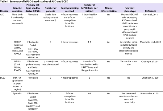
Because the number of neuronal differentiations, hiPSC lines and patients studied varies between published reports, we summarize the various methods in Table 1. Kim et al. included differentiations from one hiPSC line from each of two individuals in their models of synaptic disorders (Kim et al., 2011), whereas we generated RTT hiPSCs from four patients, and our synaptic assays compared one or two hiPSC lines from four control and three RTT patient lines (Marchetto et al., 2010). Cheung et al. generated three hiPSC lines from each of two female RTT patients, but analyzed hiPSC-derived neurons from only one RTT patient (Cheung et al., 2011). Of these three RTT hiPSC lines, random X-inactivation led to two MECP2-mutant lines and one isogenic control hiPSC line to which they were compared. In studies of SCZD, Chiang et al. generated at least two hiPSC lines from each of one control and two SCZD patients carrying DISC1 mutations, but did not perform any neuronal differentiations or synaptic assays (Chiang et al., 2011). Although we generated one to three hiPSC lines from each of five controls and four SCZD patients, we generally used two or three neural progenitor cell lines derived from one hiPSC line per individual in most neuronal assays (Brennand et al., 2011). To date, no studies of psychiatric disease have been able to compare synaptic maturation and/or function of neuronal differentiations from three hiPSC lines for each individual. Although these nested comparisons are extremely laborious, they will ultimately demonstrate the predominant source of variability in hiPSC experiments.
Table 1.
Summary of hiPSC-based studies of ASD and SCZD
To produce meaningful data, each hiPSC experiment should ideally compare multiple neuronal differentiations from multiple independent hiPSC lines from multiple patients. However, owing to issues of cost and time, particularly when characterizing synaptic maturation and function, it is not yet feasible to complete such large experiments. The development of more scalable assays will be essential in advancing this field. hiPSC-based studies will not replace MRI, postmortem and genetic studies of psychiatric disorders. Rather, we suggest that they are a new tool that will provide complementary approaches and insights for the study of a wide range of complex genetic disorders.
Direct reprogramming of fibroblasts to neurons
An alternative approach for generating patient-specific neurons to study complex psychiatric disorders has recently been reported. Starting from a pool of 19 candidate genes, researchers identified a combination of three factors – ASCL1 (also known as MASH1), BRN2 and MYT1L – that convert adult mouse fibroblasts into functional induced neurons (iNeurons) in vitro (Vierbuchen et al., 2010). The process is incredibly rapid, generating neurons that are capable of spontaneous action potentials and with functional synapses within 14 days. The conversion is also relatively efficient, occurring at an estimated rate of 1.8% to 7.7%. Direct reprogramming of human cells to iNeurons has also recently been demonstrated using several different combinations of NEUROD (Pang et al., 2011; Yoo et al., 2011) and/or the microRNAs miR-9 (Yoo et al., 2011) and/or miR-124 (Ambasudhan et al., 2011; Yoo et al., 2011). Additionally, expression of ASCL1 and two transcription factors that are crucial for dopaminergic differentiation, NURR1 and LMX1A, is sufficient to directly reprogram functional dopaminergic neurons from mouse and human fibroblasts (Caiazzo et al., 2011). The rapid experimental timeframe of iNeuron generation and the potential to reprogram to specific neuronal subtypes make this an appealing experimental strategy for in vitro models of neurological disease. However, a key limitation of iNeurons should be noted: unlike iPSCs, iNeurons do not have an inherent capacity for self-renewal. Therefore, large numbers of patient-derived fibroblasts (which have a finite capacity for replication) will probably be necessary to enable experimental analyses of iNeurons.
Two important additional issues must be considered when contemplating modeling psychiatric disorders in this manner. First, it remains to be determined whether bypassing neuronal differentiation and maturation will ‘shortcut’ the cellular phenotype of these neurodevelopmental disorders. For example, if ASDs ultimately result from abnormal synaptic maturation, it is possible that direct reprogramming would bypass the developmental window in which the ASD cellular phenotype can be observed in vitro. Second, if aberrant ASCL1, BRN2 or MYT1L activity were to contribute to psychiatric disorders, the persistent overexpression required for reprogramming might be sufficient to mask cellular phenotypes in vitro. Given that mutations disrupting MYT1L expression and binding have been linked to SCZD (Vrijenhoek et al., 2008; Riley et al., 2010), that BRN2 is known to regulate expression of KCNN3, a conductance calcium-activated potassium channel strongly implicated in SCZD (Sun et al., 2001), and that ASCL1 has been linked to Parkinson’s disease (Ide et al., 2005), it is not unreasonable to predict that overexpression of one or more of these key neuronal genes might affect the initiation or progression of a neurological disease.
A reasonable course of action to address this issue would be to determine whether the cellular and molecular phenotypes observed in RTT- and SCZD-hiPSC-derived neurons are recapitulated following the reprogramming of fibroblasts from the same patients directly to iNeurons. With this validation in place, we predict that many studies of psychiatric disorders using iNeurons will begin.
Conclusion
It is now possible to generate hiPSC-derived neurons from the fibroblasts of patients with psychiatric disorders. Defects in neuronal connectivity, synapse maturation and synaptic function have recently been reported in hiPSC-derived neurons from patients with RTT and SCZD. In addition, it might soon be possible to reprogram patient fibroblasts directly to iNeurons as an alternative in vitro model of psychiatric disorders. By combining these new cell-based human models with better-characterized cohorts of psychiatric patients, scientists will be able to more easily study the relationship between clinical, cellular and molecular phenotypes. We predict that future in vitro studies will help to elucidate not only the precise cell types affected in these disorders but also the cellular and molecular defects that contribute to the diseases.
Acknowledgments
K.J.B. is supported by a training grant from the California Institute for Regenerative Medicine. The Gage Laboratory is partially funded by CIRM Grant RL1-00649-1, The Lookout and Mathers Foundation, the Helmsley Foundation and Sanofi-Aventis.
Footnotes
COMPETING INTERESTS
The authors declare that they do not have any competing or financial interests.
REFERENCES
- Ambasudhan R., Talantova M., Coleman R., Yuan X., Zhu S., Lipton S. A., Ding S. (2011). Direct reprogramming of adult human fibroblasts to functional neurons under defined conditions. Cell Stem Cell 9, 113–118 [DOI] [PMC free article] [PubMed] [Google Scholar]
- Ang S. L. (2006). Transcriptional control of midbrain dopaminergic neuron development. Development 133, 3499–3506 [DOI] [PubMed] [Google Scholar]
- Arlotta P., Molyneaux B. J., Chen J., Inoue J., Kominami R., Macklis J. D. (2005). Neuronal subtype-specific genes that control corticospinal motor neuron development in vivo. Neuron 45, 207–221 [DOI] [PubMed] [Google Scholar]
- American Psychiatric Association (2000). Diagnostic and Statistical Manual of Mental Disorders, 4th edn – text revision (DSM-IV-TR). Washington, DC: American Psychiatric Association [Google Scholar]
- Barros C. S., Calabrese B., Chamero P., Roberts A. J., Korzus E., Lloyd K., Stowers L., Mayford M., Halpain S., Muller U. (2009). Impaired maturation of dendritic spines without disorganization of cortical cell layers in mice lacking NRG1/ErbB signaling in the central nervous system. Proc. Natl. Acad. Sci. USA 106, 4507–4512 [DOI] [PMC free article] [PubMed] [Google Scholar]
- Brennand K. J., Simone A., Jou J., Gelboin-Burkhart C., Tran N., Sangar S., Li Y., Mu Y., Chen G., Yu D., et al. (2011). Modelling schizophrenia using human induced pluripotent stem cells. Nature 473, 221–225 [DOI] [PMC free article] [PubMed] [Google Scholar]
- Brown S., Inskip H., Barraclough B. (2000). Causes of the excess mortality of schizophrenia. Br. J. Psychiatry 177, 212–217 [DOI] [PubMed] [Google Scholar]
- Caiazzo M., Dell’Anno M. T., Dvoretskova E., Lazarevic D., Taverna S., Leo D., Sotnikova T. D., Menegon A., Roncaglia P., Colciago G., et al. (2011). Direct generation of functional dopaminergic neurons from mouse and human fibroblasts. Nature 476, 224–227 [DOI] [PubMed] [Google Scholar]
- Carpenter W. T., Jr, Buchanan R. W. (1994). Schizophrenia. N. Engl. J. Med. 330, 681–690 [DOI] [PubMed] [Google Scholar]
- Chambers S. M., Fasano C. A., Papapetrou E. P., Tomishima M., Sadelain M., Studer L. (2009). Highly efficient neural conversion of human ES and iPS cells by dual inhibition of SMAD signaling. Nat. Biotechnol. 27, 275–280 [DOI] [PMC free article] [PubMed] [Google Scholar]
- Cheung A. Y., Horvath L. M., Grafodatskaya D., Pasceri P., Weksberg R., Hotta A., Carrel L., Ellis J. (2011). Isolation of MECP2-null Rett Syndrome patient hiPS cells and isogenic controls through X-chromosome inactivation. Hum. Mol. Genet. 20, 2103–2115 [DOI] [PMC free article] [PubMed] [Google Scholar]
- Chiang C. H., Su Y., Wen Z., Yoritomo N., Ross C. A., Margolis R. L., Song H., Ming G. L. (2011). Integration-free induced pluripotent stem cells derived from schizophrenia patients with a DISC1 mutation. Mol. Psychiatry 16, 358–360 [DOI] [PMC free article] [PubMed] [Google Scholar]
- Clapcote S. J., Lipina T. V., Millar J. K., Mackie S., Christie S., Ogawa F., Lerch J. P., Trimble K., Uchiyama M., Sakuraba Y., et al. (2007). Behavioral phenotypes of Disc1 missense mutations in mice. Neuron 54, 387–402 [DOI] [PubMed] [Google Scholar]
- Courchesne E., Pierce K., Schumann C. M., Redcay E., Buckwalter J. A., Kennedy D. P., Morgan J. (2007). Mapping early brain development in autism. Neuron 56, 399–413 [DOI] [PubMed] [Google Scholar]
- Di Giorgio F. P., Boulting G. L., Bobrowicz S., Eggan K. C. (2008). Human embryonic stem cell-derived motor neurons are sensitive to the toxic effect of glial cells carrying an ALS-causing mutation. Cell Stem Cell 3, 637–648 [DOI] [PubMed] [Google Scholar]
- Dimos J. T., Rodolfa K. T., Niakan K. K., Weisenthal L. M., Mitsumoto H., Chung W., Croft G. F., Saphier G., Leibel R., Goland R., et al. (2008). Induced pluripotent stem cells generated from patients with ALS can be differentiated into motor neurons. Science 321, 1218–1221 [DOI] [PubMed] [Google Scholar]
- Ellison-Wright I., Glahn D. C., Laird A. R., Thelen S. M., Bullmore E. (2008). The anatomy of first-episode and chronic schizophrenia: an anatomical likelihood estimation meta-analysis. Am. J. Psychiatry 165, 1015–1023 [DOI] [PMC free article] [PubMed] [Google Scholar]
- Fanous A. H., Neale M. C., Webb B. T., Straub R. E., Amdur R. L., O’Neill F. A., Walsh D., Riley B. P., Kendler K. S. (2007). A genome-wide scan for modifier loci in schizophrenia. Am. J. Med. Genet. B Neuropsychiatr. Genet. 144, 589–595 [DOI] [PubMed] [Google Scholar]
- Frantz G. D., Weimann J. M., Levin M. E., McConnell S. K. (1994). Otx1 and Otx2 define layers and regions in developing cerebral cortex and cerebellum. J. Neurosci. 14, 5725–5740 [DOI] [PMC free article] [PubMed] [Google Scholar]
- Gore A., Li Z., Fung H. L., Young J. E., Agarwal S., Antosiewicz-Bourget J., Canto I., Giorgetti A., Israel M. A., Kiskinis E., et al. (2011). Somatic coding mutations in human induced pluripotent stem cells. Nature 471, 63–67 [DOI] [PMC free article] [PubMed] [Google Scholar]
- Hikida T., Jaaro-Peled H., Seshadri S., Oishi K., Hookway C., Kong S., Wu D., Xue R., Andrade M., Tankou S., et al. (2007). Dominant-negative DISC1 transgenic mice display schizophrenia-associated phenotypes detected by measures translatable to humans. Proc. Natl. Acad. Sci. USA 104, 14501–14506 [DOI] [PMC free article] [PubMed] [Google Scholar]
- Hranilovic D., Stefulj J., Schwab S., Borrmann-Hassenbach M., Albus M., Jernej B., Wildenauer D. (2004). Serotonin transporter promoter and intron 2 polymorphisms: relationship between allelic variants and gene expression. Biol. Psychiatry 55, 1090–1094 [DOI] [PubMed] [Google Scholar]
- Hu B. Y., Weick J. P., Yu J., Ma L. X., Zhang X. Q., Thomson J. A., Zhang S. C. (2010). Neural differentiation of human induced pluripotent stem cells follows developmental principles but with variable potency. Proc. Natl. Acad. Sci. USA 107, 4335–4340 [DOI] [PMC free article] [PubMed] [Google Scholar]
- Hussein S. M., Batada N. N., Vuoristo S., Ching R. W., Autio R., Narva E., Ng S., Sourour M., Hamalainen R., Olsson C., et al. (2011). Copy number variation and selection during reprogramming to pluripotency. Nature 471, 58–62 [DOI] [PubMed] [Google Scholar]
- Ide M., Yamada K., Toyota T., Iwayama Y., Ishitsuka Y., Minabe Y., Nakamura K., Hattori N., Asada T., Mizuno Y., et al. (2005). Genetic association analyses of PHOX2B and ASCL1 in neuropsychiatric disorders: evidence for association of ASCL1 with Parkinson’s disease. Hum. Genet. 117, 520–527 [DOI] [PubMed] [Google Scholar]
- Kanner L. (1943). Autistic disturbances of affective contact. Nervous Child 2, 217–250 [PubMed] [Google Scholar]
- Kawasaki H., Mizuseki K., Nishikawa S., Kaneko S., Kuwana Y., Nakanishi S., Nishikawa S. I., Sasai Y. (2000). Induction of midbrain dopaminergic neurons from ES cells by stromal cell-derived inducing activity. Neuron 28, 31–40 [DOI] [PubMed] [Google Scholar]
- Kim J. E., O’Sullivan M. L., Sanchez C. A., Hwang M., Israel M. A., Brennand K., Deerinck T. J., Goldstein L. S., Gage F. H., Ellisman M. H., et al. (2011). Investigating synapse formation and function using human pluripotent stem cell-derived neurons. Proc. Natl. Acad. Sci. USA 108, 3005–3010 [DOI] [PMC free article] [PubMed] [Google Scholar]
- Krystal J. H., Karper L. P., Seibyl J. P., Freeman G. K., Delaney R., Bremner J. D., Heninger G. R., Bowers M. B., Jr, Charney D. S. (1994). Subanesthetic effects of the noncompetitive NMDA antagonist, ketamine, in humans. Psychotomimetic, perceptual, cognitive, and neuroendocrine responses. Arch. Gen. Psychiatry 51, 199–214 [DOI] [PubMed] [Google Scholar]
- Laurent L. C., Ulitsky I., Slavin I., Tran H., Schork A., Morey R., Lynch C., Harness J. V., Lee S., Barrero M. J., et al. (2011). Dynamic changes in the copy number of pluripotency and cell proliferation genes in human ESCs and iPSCs during reprogramming and time in culture. Cell Stem Cell 8, 106–118 [DOI] [PMC free article] [PubMed] [Google Scholar]
- Lewis C. M., Levinson D. F., Wise L. H., DeLisi L. E., Straub R. E., Hovatta I., Williams N. M., Schwab S. G., Pulver A. E., Faraone S. V., et al. (2003). Genome scan meta-analysis of schizophrenia and bipolar disorder, part II: schizophrenia. Am. J. Hum. Genet. 73, 34–48 [DOI] [PMC free article] [PubMed] [Google Scholar]
- Li X. J., Du Z. W., Zarnowska E. D., Pankratz M., Hansen L. O., Pearce R. A., Zhang S. C. (2005). Specification of motoneurons from human embryonic stem cells. Nat. Biotechnol. 23, 215–221 [DOI] [PubMed] [Google Scholar]
- Lister R., Pelizzola M., Kida Y. S., Hawkins R. D., Nery J. R., Hon G., Antosiewicz-Bourget J., O’Malley R., Castanon R., Klugman S., et al. (2011). Hotspots of aberrant epigenomic reprogramming in human induced pluripotent stem cells. Nature 471, 68–73 [DOI] [PMC free article] [PubMed] [Google Scholar]
- Maeda K., Nwulia E., Chang J., Balkissoon R., Ishizuka K., Chen H., Zandi P., McInnis M. G., Sawa A. (2006). Differential expression of disrupted-in-schizophrenia (DISC1) in bipolar disorder. Biol. Psychiatry 60, 929–935 [DOI] [PubMed] [Google Scholar]
- Mahadik S. P., Mukherjee S. (1996). Cultured skin fibroblasts as a cell model for investigating schizophrenia. J. Psychiatr. Res. 30, 421–439 [DOI] [PubMed] [Google Scholar]
- Maherali N., Sridharan R., Xie W., Utikal J., Eminli S., Arnold K., Stadtfeld M., Yachechko R., Tchieu J., Jaenisch R., et al. (2007). Directly reprogrammed fibroblasts show global epigenetic remodeling and widespread tissue contribution. Cell Stem Cell 1, 55–70 [DOI] [PubMed] [Google Scholar]
- Marchetto M. C., Carromeu C., Acab A., Yu D., Yeo G. W., Mu Y., Chen G., Gage F. H., Muotri A. R. (2010). A model for neural development and treatment of rett syndrome using human induced pluripotent stem cells. Cell 143, 527–539 [DOI] [PMC free article] [PubMed] [Google Scholar]
- Matigian N. A., McCurdy R. D., Feron F., Perry C., Smith H., Filippich C., McLean D., McGrath J., Mackay-Sim A., Mowry B., et al. (2008). Fibroblast and lymphoblast gene expression profiles in schizophrenia: are non-neural cells informative? PLoS ONE 3, e2412. [DOI] [PMC free article] [PubMed] [Google Scholar]
- Matigian N., Abrahamsen G., Sutharsan R., Cook A. L., Vitale A. M., Nouwens A., Bellette B., An J., Anderson M., Beckhouse A. G., et al. (2010). Disease-specific, neurosphere-derived cells as models for brain disorders. Dis. Model. Mech. 3, 785–798 [DOI] [PubMed] [Google Scholar]
- Mayshar Y., Ben-David U., Lavon N., Biancotti J. C., Yakir B., Clark A. T., Plath K., Lowry W. E., Benvenisty N. (2010). Identification and classification of chromosomal aberrations in human induced pluripotent stem cells. Cell Stem Cell 7, 521–531 [DOI] [PubMed] [Google Scholar]
- Meissner A., Wernig M., Jaenisch R. (2007). Direct reprogramming of genetically unmodified fibroblasts into pluripotent stem cells. Nat. Biotechnol. 25, 1177–1181 [DOI] [PubMed] [Google Scholar]
- Moskvina V., Craddock N., Holmans P., Nikolov I., Pahwa J. S., Green E., Owen M. J., O’Donovan M. C. (2009). Gene-wide analyses of genome-wide association data sets: evidence for multiple common risk alleles for schizophrenia and bipolar disorder and for overlap in genetic risk. Mol. Psychiatry 14, 252–260 [DOI] [PMC free article] [PubMed] [Google Scholar]
- O’Donovan M. C., Craddock N., Norton N., Williams H., Peirce T., Moskvina V., Nikolov I., Hamshere M., Carroll L., Georgieva L., et al. (2008). Identification of loci associated with schizophrenia by genome-wide association and follow-up. Nat. Genet. 40, 1053–1055 [DOI] [PubMed] [Google Scholar]
- Osafune K., Caron L., Borowiak M., Martinez R. J., Fitz-Gerald C. S., Sato Y., Cowan C. A., Chien K. R., Melton D. A. (2008). Marked differences in differentiation propensity among human embryonic stem cell lines. Nat. Biotechnol. 26, 313–315 [DOI] [PubMed] [Google Scholar]
- Pang Z., Yang N., Vierbuchen T., Ostermeier A., Fuentes D., Yang T., Citri A., Sebastiano V., Marro S., Südhof T., et al. (2011). Induction of human neuronal cells by defined transcription factors. Nature 476, 220–223 [DOI] [PMC free article] [PubMed] [Google Scholar]
- Patil S. T., Zhang L., Martenyi F., Lowe S. L., Jackson K. A., Andreev B. V., Avedisova A. S., Bardenstein L. M., Gurovich I. Y., Morozova M. A., et al. (2007). Activation of mGlu2/3 receptors as a new approach to treat schizophrenia: a randomized Phase 2 clinical trial. Nat. Med. 13, 1102–1107 [DOI] [PubMed] [Google Scholar]
- Perrier A. L., Tabar V., Barberi T., Rubio M. E., Bruses J., Topf N., Harrison N. L., Studer L. (2004). Derivation of midbrain dopamine neurons from human embryonic stem cells. Proc. Natl. Acad. Sci. USA 101, 12543–12548 [DOI] [PMC free article] [PubMed] [Google Scholar]
- Riley B., Thiselton D., Maher B. S., Bigdeli T., Wormley B., McMichael G. O., Fanous A. H., Vladimirov V., O’Neill F. A., Walsh D., et al. (2010). Replication of association between schizophrenia and ZNF804A in the Irish Case-Control Study of Schizophrenia sample. Mol. Psychiatry 15, 29–37 [DOI] [PMC free article] [PubMed] [Google Scholar]
- Ritvo E. R., Freeman B. J., Mason-Brothers A., Mo A., Ritvo A. M. (1985). Concordance for the syndrome of autism in 40 pairs of afflicted twins. Am. J. Psychiatry 142, 74–77 [DOI] [PubMed] [Google Scholar]
- Stern C. D. (2001). Initial patterning of the central nervous system: how many organizers? Nat. Rev. Neurosci. 2, 92–98 [DOI] [PubMed] [Google Scholar]
- Sullivan P. F., Kendler K. S., Neale M. C. (2003). Schizophrenia as a complex trait: evidence from a meta-analysis of twin studies. Arch. Gen. Psychiatry 60, 1187–1192 [DOI] [PubMed] [Google Scholar]
- Sun G., Tomita H., Shakkottai V. G., Gargus J. J. (2001). Genomic organization and promoter analysis of human KCNN3 gene. J. Hum. Genet. 46, 463–470 [DOI] [PubMed] [Google Scholar]
- Takahashi K., Yamanaka S. (2006). Induction of pluripotent stem cells from mouse embryonic and adult fibroblast cultures by defined factors. Cell 126, 663–676 [DOI] [PubMed] [Google Scholar]
- Takahashi K., Tanabe K., Ohnuki M., Narita M., Ichisaka T., Tomoda K., Yamanaka S. (2007). Induction of pluripotent stem cells from adult human fibroblasts by defined factors. Cell 131, 861–872 [DOI] [PubMed] [Google Scholar]
- Tropepe V., Hitoshi S., Sirard C., Mak T. W., Rossant J., van der Kooy D. (2001). Direct neural fate specification from embryonic stem cells: a primitive mammalian neural stem cell stage acquired through a default mechanism. Neuron 30, 65–78 [DOI] [PubMed] [Google Scholar]
- Tsuang M. T., Stone W. S., Faraone S. V. (2001). Genes, environment and schizophrenia. Br. J. Psychiatry 40 Suppl., s18–s24 [DOI] [PubMed] [Google Scholar]
- Vierbuchen T., Ostermeier A., Pang Z. P., Kokubu Y., Sudhof T. C., Wernig M. (2010). Direct conversion of fibroblasts to functional neurons by defined factors. Nature 463, 1035–1041 [DOI] [PMC free article] [PubMed] [Google Scholar]
- Vita A., De Peri L., Silenzi C., Dieci M. (2006). Brain morphology in first-episode schizophrenia: a meta-analysis of quantitative magnetic resonance imaging studies. Schizophr. Res. 82, 75–88 [DOI] [PubMed] [Google Scholar]
- Vrijenhoek T., Buizer-Voskamp J. E., van der Stelt I., Strengman E., Sabatti C., Geurts van Kessel A., Brunner H. G., Ophoff R. A., Veltman J. A. (2008). Recurrent CNVs disrupt three candidate genes in schizophrenia patients. Am. J. Hum. Genet. 83, 504–510 [DOI] [PMC free article] [PubMed] [Google Scholar]
- Walsh T., McClellan J. M., McCarthy S. E., Addington A. M., Pierce S. B., Cooper G. M., Nord A. S., Kusenda M., Malhotra D., Bhandari A., et al. (2008). Rare structural variants disrupt multiple genes in neurodevelopmental pathways in schizophrenia. Science 320, 539–543 [DOI] [PubMed] [Google Scholar]
- Watanabe K., Kamiya D., Nishiyama A., Katayama T., Nozaki S., Kawasaki H., Watanabe Y., Mizuseki K., Sasai Y. (2005). Directed differentiation of telencephalic precursors from embryonic stem cells. Nat. Neurosci. 8, 288–296 [DOI] [PubMed] [Google Scholar]
- Weimann J. M., Zhang Y. A., Levin M. E., Devine W. P., Brulet P., McConnell S. K. (1999). Cortical neurons require Otx1 for the refinement of exuberant axonal projections to subcortical targets. Neuron 24, 819–831 [DOI] [PubMed] [Google Scholar]
- Wernig M., Meissner A., Foreman R., Brambrink T., Ku M., Hochedlinger K., Bernstein B. E., Jaenisch R. (2007). In vitro reprogramming of fibroblasts into a pluripotent ES-cell-like state. Nature 448, 318–324 [DOI] [PubMed] [Google Scholar]
- Wichterle H., Lieberam I., Porter J. A., Jessell T. M. (2002). Directed differentiation of embryonic stem cells into motor neurons. Cell 110, 385–397 [DOI] [PubMed] [Google Scholar]
- Yamamori H., Hashimoto R., Verrall L., Yasuda Y., Ohi K., Fukumoto M., Umeda-Yano S., Ito A., Takeda M. (2011). Dysbindin-1 and NRG-1 gene expression in immortalized lymphocytes from patients with schizophrenia. J. Hum. Genet. 56, 478–483 [DOI] [PubMed] [Google Scholar]
- Yoo A. S., Sun A. X., Li L., Shcheglovitov A., Portmann T., Li Y., Lee-Messer C., Dolmetsch R. E., Tsien R. W., Crabtree G. R. (2011). MicroRNA-mediated conversion of human fibroblasts to neurons. Nature 476, 228–231 [DOI] [PMC free article] [PubMed] [Google Scholar]
- Yu J., Vodyanik M. A., Smuga-Otto K., Antosiewicz-Bourget J., Frane J. L., Tian S., Nie J., Jonsdottir G. A., Ruotti V., Stewart R., et al. (2007). Induced pluripotent stem cell lines derived from human somatic cells. Science 318, 1917–1920 [DOI] [PubMed] [Google Scholar]


