Abstract
Viruses are serious threats to human and animal health. Vaccines can prevent viral diseases, but few antiviral treatments are available to control evolving infections. Among new antiviral therapies, RNA interference (RNAi) has been the focus of intensive research. However, along with the development of efficient RNAi-based therapeutics comes the risk of emergence of resistant viruses. In this study, we challenged the in vitro propensity of a morbillivirus (peste des petits ruminants virus), a stable RNA virus, to escape the inhibition conferred by single or multiple small interfering RNAs (siRNAs) against conserved regions of the N gene. Except with the combination of three different siRNAs, the virus systematically escaped RNAi after 3 to 20 consecutive passages. The genetic modifications involved consisted of single or multiple point nucleotide mutations and a deletion of a stretch of six nucleotides, illustrating that this virus has an unusual genomic malleability.
INTRODUCTION
Since the discovery of antibiotics by Fleming in 1929 (20) and their generalized use against bacterial infections, viruses have become the major threats to human and animal health. A very limited number of antiviral treatments are available to control viral infections. In the past 20 years, several major pandemics of emerging or reemerging diseases occurred, such as severe acute respiratory syndrome (SARS) in humans (4, 51), H5N1 influenza in humans and poultry (6), and foot-and-mouth disease (35) and bluetongue (16) in ruminants. Though vaccines can prevent viral diseases, only antiviral drugs offer a therapeutic solution when the infection is already present. Today, one of the main challenges for virologists is to develop effective treatments. Current drugs are restricted by many factors, such as toxicity, complexity, cost, and the capacity of viruses to acquire resistance (10). Among new antiviral therapies explored in the past 10 years, RNA interference (RNAi) has been the focus of intensive research because it is a natural biological process in eukaryotic cells that can be diverted to the control of virus replication (19). Small interfering RNAs (siRNAs) act by knocking down the expression of a gene shortly after its transcription. This downregulation of posttranslational gene expression results from an enzymatic degradation of mRNA that takes place in the cell cytoplasm close to the nuclear pores (19, 43). The capacity of synthetic siRNAs to inhibit viral production was first established in vitro by introducing siRNAs into the cytoplasm of cultured cells (15). Subsequently, success with siRNAs against various viruses at the laboratory level led to the hope that these molecules could revolutionize antiviral therapy in humans and animals (18, 33, 38, 40, 45). However, the development of efficient RNAi-based therapeutics faces substantial challenges. Two of the most important issues are (i) the development of efficacious in vivo delivery systems (11, 52, 58) and (ii) the risk of emergence of resistant viruses (9, 57). Resistance can be acquired by genetic variation in viruses (mutation, deletion, recombination, and reassortment) because the activity of siRNAs is tightly sequence dependent. Thus, a single nucleotide mutation can entirely abrogate the antiviral effect. RNA viruses have a high propensity to modify their genomes and acquire resistance to siRNAs because of the high error rates of viral RNA-dependent RNA polymerase (42). To overcome this problem, current approaches are based on the use of a mixture of synthetic siRNAs against different conserved genome targets (7, 50).
Morbilliviruses include several pathogens of humans (measles virus [MV]) and terrestrial and marine mammals (canine distemper virus [CDV], rinderpest virus [RPV], peste des petits ruminants virus [PPRV], phocid distemper virus [PDV], and dolphin and porpoise morbilliviruses) (2). Vaccines are available for most of these diseases, but they are not generalized, and every year thousands of humans and small ruminants die. The genomes of morbilliviruses have good RNA sequence conservation and are more stable than those of other RNA viruses; they have an estimated mutation rate of 6.2 × 10⁻⁴ substitutions/site/year. Consequently, the morbillivirus clades diverged from each other possibly in the 11th to 12th centuries for the divergence between RPV and MV and in the first century for the divergence between PPRV and RPV/MV (21, 46). Among the six genes of these viruses, the most translated is the N gene, encoding the nucleoprotein, which plays a pivotal role in viral nucleic acid and protein synthesis, virus replication, and genome encapsidation (1). Considering the relative stability of the morbillivirus genome and the fact that three active siRNAs were previously identified in the most conserved regions of the N genes of morbilliviruses (36, 49), we hypothesized that the capacity of the virus to escape siRNA would be limited. To test this hypothesis, sequential passages of one model morbillivirus, PPRV, were carried out under variable concentrations of single or combined siRNAs against the three positions. The results show that the genome plasticity of morbilliviruses under pressure of siRNA activity can exceed the virus diversity observed in nature, thus illustrating that the control of relatively stable viruses by siRNA may not be as easy as initially foreseen.
MATERIALS AND METHODS
Cells, viruses, and siRNAs.
Vero cells were purchased from the European Collection of Cell Cultures, France (ECACC 84113001) and maintained in Eagle's minimum essential medium (EMEM) (Invitrogen, Fischer Scientific, France) supplemented with 10% fetal bovine serum (FBS) (Dutcher, France) and 2 mM l-glutamine (Invitrogen, Fischer Scientific, France). They were grown at 37°C with 5% CO2 in a humidified incubator.
Virus stock of the live-attenuated vaccine strain PPRV Nigeria 75/1 (GenBank accession number X74443) (13) was prepared by collecting the infected cell supernatant when cytopathic effect (CPE) affected about 80% of the cells. The cells were freeze-thawed 3 times and stored in aliquots at −80°C. Virus titers were estimated by the method of Reed and Muench (44) and expressed in 50% tissue culture infective doses (TCID50)/ml.
All siRNAs were synthesized and purified (≥80% purity) by Applied Biosystems (Courtaboeuf, France). Three siRNAs targeting conserved regions of the essential gene encoding the viral nucleoprotein (N) of PPRV were previously shown to prevent at least 80% of PPRV replication in vitro (36, 49): siNPPRV1 (sense, 5′GGAUCAACUGGUUUGAGAAtt3′; mRNA positions 480 to 498), siNPPRV6 (sense, 5′GGCGGUUCAUGGUAUCUCUtt3′; mRNA positions 741 to 759), and siNPPRV7 (sense, 5′GCAUUAGGCCUUCACGAGUtt3′; mRNA positions 899 to 917). Two additional siRNAs were derived from two escape mutants with modifications in the target sequence of siNPPRV1 (siNPPRV1mut1 [sense, 5′GGAUCGGCUGGUUUGAGAAtt3′] and siNPPRV1mut4 [sense, 5′GGAUCAGCUGGUUUGAGAAtt3′). An irrelevant siRNA was used as control (siMPPRV11; sense, 5′GGUAUUUUACAACAACACUtt3′) (in all sequences, lowercase letters represent the 2-nucleotide 3′ unpaired over hang).
siRNA transfection.
After 48 h of growth, Vero cells were trypsinized again and plated at 5 × 10⁴ cells per well in 48-well plates. Twenty-four hours later, different final concentrations of siRNAs (100, 33.3, 11.1, 3.7, and 0 nM) were complexed with 1 μg per well of Lipofectamine 2000 (Invitrogen, Fisher Scientific, Illkirch, France) in 50 μl of serum-free Opti-MEM I (Invitrogen, Fischer Scientific, France). After 20 min of incubation at room temperature, the siRNA-Lipofectamine 2000 complexes were added to wells containing 100 μl of serum-free MEM. Plates were incubated for 4 to 6 h at 37°C in 5% CO2. Afterwards, the cell supernatant was completed with 350 μl of MEM supplemented with 5% FBS and 2 mM l-glutamine. Twenty-four hours after transfection, cells were infected with 10-fold serial dilutions of the virus. Virus titrations were done in parallel to determine the exact multiplicity of infection (MOI) added to each well. Four days later, the siRNA silencing effect was evaluated by scoring the CPE on an inverted microscope. The scoring scale was as follows: −, CPE of 0%; +, CPE of between 1 and 25%; ++, CPE of between 26 and 50%; +++, CPE of between 51 and 75%; ++++, CPE of between 76 and 99%; and +++++, CPE of 100%. At the end of the transfection test, virus was harvested from the wells containing at least 25% CPE, pooled, freeze-thawed 3 times, and stored at −80°C until use in the next transfection test and detection of mutants. The suitability of CPE to detect RNA interference and the development of RNAi resistance was initially checked by comparison with an immunofluorescence test for virus detection. In brief, Vero cells were trypsinized and plated in 24-well plates. Twenty-four hours later, cells were transfected as described above. Twenty-four hours after transfection, cells were infected with 10-fold serial dilutions of the virus. Four days later, Vero cells were fixed with 3% paraformaldehyde. A tetramethyl rhodamine isocyanate (TRITC)-coupled monoclonal antibody (MAb) specific for the N gene of PPRV was appropriately diluted and added to each well. The plate was incubated for 30 min at 4°C and washed three times. All dilutions and washings were carried out using a phosphate-buffered saline (PBS) solution. The presence of the virus was then detected by microscopic observation of intracellular fluorescence. In addition, all mutants observed in the siNPPRV1 target were purified and their titers were determined for comparison with the parental virus titers.
Detection of mutant emergence and sequencing of the mutants.
To detect the presence of mutants in the infected cell cultures, total RNA was extracted with a Nucleospin RNA virus kit (Macherey Nalgen, Hoerdt, France) and cDNA was synthesized with a first-strand cDNA synthesis kit (GE Healthcare, Buckinghamshire, United Kingdom). The regions encompassing the targets of the three siRNAs were amplified with the primer pair NP63 forward (5′ACCGGCGTGATGATCAGCAT3′) and NP4 reverse (5′CCTCCTCCTGGTCCTCCAGAATCT3′). The amplified material was then sent to GATC Biotech (Konstanz, Germany) for sequencing. The mutations were discerned by the presence of a double peak in the chromatograms (one of them representing the original nucleotide and the other one the mutated nucleotide). According to the sequencing kit manufacturer, the detection limit for a minority population is 15 to 30%. The mutation was retained when the peak of the mutated nucleotide surpassed the original one.
To further analyze the relative distributions of the mutant and parental virus populations over the successive passages in cell cultures, a high-resolution-melting real-time PCR (PCR-HRM) was developed. This test discriminates parental from mutated viruses in a mixture and thus provides a kinetic determination of the relative proportions of the two viruses in the mixture. The target region of siNPPRV1 was amplified using oligonucleotides si1(77pb) forward (5′GGACCCTCGAGTGGAAGTAA3′) and si1(77pb) reverse (5′GCATCTTGCACCTCTATGTC3′) and a LightCycler 480 high-resolution-melting kit (Roche Applied Science, Mannheim, Germany) according to the supplier's recommendations (1× Master Mix, 3 mM MgCl2, 200 mM each primer). The melting curve of the amplified fragment was analyzed using LightCycler 480 Genotyping software in the “gene scanning” mode. A standard curve with different ratios of the original nonmutated and mutated population was used as reference for the quantification of the mutant and wild virus populations in each successive transfection test. In addition, all mutants observed in the siNPPRV1 target were plaque purified and titrated in comparison with the parental virus titer.
Verification of the role of mutation/deletion in the acquisition of RNAi resistance.
To confirm the role of mutation and deletion in the acquisition of RNAi resistance, a luciferase reporter system was designed, consisting of a plasmid in which the target sequence of siNPPRV1 was placed upstream from the coding sequence of the firefly luciferase-2 gene (siRNA-NPPR1-Firefly_Luciferase-2) in an expression plasmid vector (pCI-neo mammalian expression vector; Promega, Charbonnieres Les Bains, France). From that plasmid, the different mutations or deletion were generated by site-directed mutagenesis (QuikChange II XL site-directed mutagenesis kit; Stratagene) according to the supplier's recommendations. The plasmid siRNA-NPPR1-Firefly_Luciferase-2 was amplified with primers containing the desired mutations or deletion: siNPPRV1-mut1 (forward, 5′CGCCCTTGGATCGGCTGGTTTGAG3′; reverse, 5′CTCAAACCAGCCGATCCAAGGGCG3′), siNPPRV1-mut3 (forward, 5′CGCCCTTGGATAAACTGGTTTGAG3′; reverse, 5′CTCAAACCAGTTTATCCAAGGGCG3′), siNPPRV1-mut4 (forward 5′CGCCCTTGGATCAGCTGGTTTGAG3′; reverse, 5′CTCAAACCAGCTGATCCAAGGGCG3′), and siNPPRV1-del (forward, 5′CGAGAACGCCCTTAGAAAAACTGGTTTGAG3′; reverse, 5′CTCAAACCAGTTTTTCTAAGGGCGTTCTCG3′). The amplified products were treated with DpnI endonuclease, transformed into XL10-Gold ultracompetent cells (Stratagene) according to the instructions of the manufacturer, and cultivated on LB agar medium (Invitrogen, Fischer Scientific, France) under 1% ampicillin selection. The plasmids produced were purified with the PureLink Quick Plasmid Miniprep kit (Invitrogen, Fischer Scientific, France) and sequenced for confirmation of the desired mutations or deletion.
The expression capacities of these constructions and the capacities of different siRNAs to silence these constructions were assessed in vitro by cotransfection of Vero cells with 100 ng of each plasmid and 10 ng of each siRNA complexed with 0.25 μl/well of Lipofectamine 2000 (Fisher Scientifique, Illkirch, France). These tests were done in 96-well plates with 2 × 10⁴ Vero cells/well, according to the procedure described above. The in vitro luciferase activity was measured at 48 h posttransfection by bioimagery using an IVIS-Lumina II (Caliper Life Sciences, Villepinte, France) 10 min after the addition 150 μg/μl of d-luciferin potassium salt (Caliper Life Sciences, Villepinte, France). Irrelevant plasmids, siRNAs, and untransfected Vero cells were used as controls to determine the background bioluminescence signal. The bioluminescence signals obtained in 3-min periods were measured as total photon flux normalized for exposure time and surface area and expressed in photons/s/cm2/steradian (p/s/cm2/sr). Photons were quantified using Living Image software version 4.0 (Caliper Life Sciences, Villepinte, France).
Multiple alignments of the N genes of morbilliviruses and sequence analysis.
The nucleotide sequences of the N genes of PPRV, RPV, MV, and CDV were obtained from public databases or generated in our laboratory and aligned using Seaview version 4.2.11 (22, 23). The conservation of the positions targeted by the three siRNAs in the morbillivirus N gene was reassessed by multiple alignments of nucleic acid and amino acid sequences. The two regions of 19 nucleotides flanking the three siRNA targets were included in the alignment for the next analyses. The modifications observed in the targets of siRNAs N1, N6, and N7 were qualified by using a scoring system at all positions: (i) for nucleotide transition versus transversion, 1 = no change, 2 = transition, 3 = transversion, and 4 = transversion never observed in a morbillivirus; (ii) for changes in nucleotide position in the codon, 1 = no change, 2 = change in position 3, 3 = change in position 1, and 4 = change in position 2; (iii) for changes in relative codon usage preference, 1 = no change, 2 = no change in the amino acid and better codon usage preference, 3 = no change in the amino acid and worse codon usage preference, and 4 = change in the amino acid; and (iv) for changes of the amino acid, 1 = no change, 2 = synonymous change, 3 = nonsynonymous change, and 4 = nonsynonymous change never observed in a morbillivirus
The scores were then used in a statistical analysis to characterize the sequence events generated in this study (see below). In parallel, multiple alignments of the complete nucleotide sequences of morbillivirus N genes were used in phylogenetic reconstruction by a Bayesian approach (29). The general time-reversible model of correction with five gamma categories was used as proposed by Treefinder (March 2011 version; distributed by the author at www.treefinder.de) (32). The resulting trees and their corresponding multiple alignments were thus used in a dN/dS analysis implemented by Codeml in the PAML suite (59). In brief, the ratio of nonsynonymous to synonymous nucleotide substitutions is calculated over the whole N gene sequence. A dN/dS ratio of <1, 1, or >1 indicates negative, neutral, or positive selection, respectively. A molecular clock analysis on all PPRV N gene sequences available was also carried out using the BEAST software and a strict molecular clock model (14).
Statistical analyses.
The results of the tests with the firefly luciferase reporter system were compared with those of the nonparametric Wilcoxon test (55) and were adjusted by use of the Holm test (27). To assess the selection pressure effect of the three siRNAs on the N gene, the general framework of factorial methods was used (41). More specifically, multiple-factor analysis (MFA) as described by Escofier and Pagès (17) was used to assess the relationships between variables separated into the 4 scoring groups described in previous section for PPRV strains generated in this study and other wild-type viruses. By generating common factors for both variables and groups of variables, MFA accounts for the heterogeneity of groups of variables in terms of biological meaning and allows the identification of the main variables and groups of variables that differentiate PPRV strains with a special mutation pattern from those with the mutation patterns naturally observed. For this purpose, maps of the factorial planes were used to identify PPRV strains. To assess whether the mutation patterns of wild and siRNA-induced PPRV strains differed, a permutation test was carried out. The mean MFA scores of wild and siRNA-induced PPRV strains were calculated, and the Euclidean distance between the two resulting virus class centroids was computed. The null hypothesis was that this distance was observed by chance. To test this, the same distance was computed after a random permutation of virus class membership (either wild or siRNA-induced PPRV), and 9,999 iterations of this process were achieved. The initial value was appended to the 9,999 values obtained by the permutation process. The proportion of distances exceeding the initial value gave the P value of the test.
A survival analysis (28) was used to test any difference in the timely occurrence of the different modifications observed in the three siRNA targets according to the siRNA treatment. A mutation of a given nucleotide was considered a failure, and the number of transfection passages when this failure occurred gave the failure time. A Kaplan-Meier analysis (34) was used to graphically present the results. This graphic represents the “survival” curve of the original Nigeria 75/1 strain, which is the probability that this strain is unmodified and still sensitive to the siRNA treatment over 20 consecutive passages. For instance, when no modification occurs after 20 passages, the survival probability remains 1 over the 20 passages. To estimate the effect of each siRNA on the probability of nucleotide mutation in the targeted area of the N PPRV gene (19 nucleotides), we used a semiparametric Cox model of proportional mutation hazards (8). This model provided estimates of the relative risk (RR) of mutation. The mutation risk observed when using siNPPRV1 was selected as a baseline for the comparisons.
RESULTS
siRNAs targeting conserved regions of the N gene: reassessment of dose-effect responses.
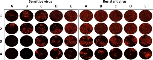
After infection of Vero cells, PPRV grew relatively fast, inducing a cytopathic effect (CPE) that was visible at 3 days postinfection and achieving its maximum titer in 4 to 5 days. The titers usually ranged from 104.5 to 106 TCID50/ml. In the presence of one or a combination of two or three siRNAs tested at a final concentration of 3.7 to 100 nM, the virus replication was inhibited as evidenced by a clear reduction of CPE development, confirmed by the reduction of virus-specific fluorescence (Fig. 1). This confirmed previous work showing an inhibition of CPE development and a reduction of the virus titers by 4 to 5 log10 units (36, 49). In contrast, the emergence and development of a mutant virus led to the development of CPE and immunofluorescence to the same extent as for the ones induced by the parental strain not treated with siRNAs (Fig. 1). In addition, when the five mutants generated in this study by the application of siNPPRV1 were plaque purified, they achieved titers of at least 103.7 and up to 105.1 TCID50/ml. In particular, the deletion mutant had a titer of 104.9 TCID50/ml. The titers of the mutants were in the range of their parental virus titer, illustrating that the mutated viruses did not have a significant reduction of their replication efficacy in vitro.
Fig 1.
Immunofluorescence detection of PPRV in Vero cells transfected with siNPPRV1. The cells were first transfected with 100, 33.3, 11.1, 3.7, and 0 nM siNPPRV1 (columns A to E, respectively). The day after, they were infected with Nigeria 75/1 vaccine virus (left panel) or with one of its escape mutants (si1mut1, right panel) at an MOI of 0.1 (row 1) and 10-fold serially diluted (rows 2 to 4). The presence of the virus was evidenced after 4 days of cell culture by a direct immunofluorescence test using a monoclonal antibody against the N protein coupled to TRITC. The left panel shows the dose/effect response with an RNAi-sensitive virus, while the right panel shows the results with a resistant virus.
PPRV easily escapes siRNA.
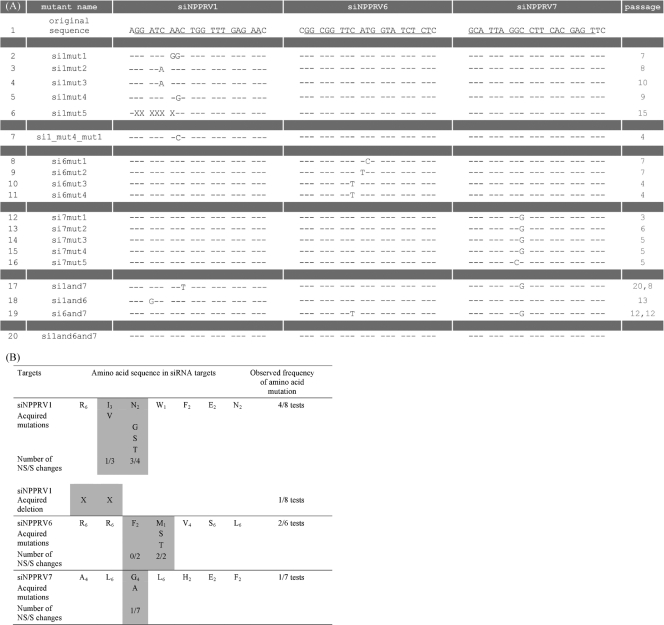
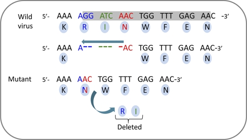
To examine the ability of PPRV to mutate and acquire resistance to siRNAs alone or in combination, consecutive transfections using different siRNA concentrations were carried out in vitro. For all single or combinations of siRNAs, a relapse of CPE inhibition was observed before the 20th passage in cell culture, except for the combination of the three siRNAs. By resequencing the presumed escape mutants, we observed either single or multiple point nucleotide mutations (synonymous or not) and a deletion of a stretch of six nucleotides in the siRNA targets (Fig. 2A). No other modifications in the PPRV N gene were evidenced. Mutations in the three target regions were concentrated at positions 3 to 7, 8 to 10, and 8 and 9 for siNPPRV1, -6, and -7, respectively. Consequently, a limited number of amino acid positions were modified. However, the frequency of amino acid substitutions at these positions was high, with one-half, one-third, and one-seventh of the mutants for siNPPRV1, -6, and -7, respectively (Fig. 2B). However, this frequency was in inverse proportion to the number of possible codons for the corresponding amino acid, thus showing a preference for synonymous substitutions when there is such an option. As anticipated, the deletion fulfilled the so-called “rule of six” observed in the genomes of paramyxoviruses, in which a multiple of six nucleotides in genome length is required for optimized replication (26, 47). On the other hand, a surprise lay in the frameshift introduced into the coding sequence. However, through an original relinkage, this shift resulted in the loss of only two amino acids (Fig. 3).
Fig 2.
Mutations and deletion resulting from siRNA selection in vitro. (A) siRNAs used in this study: siNPPRV1 (lines 2 to 6), siNPPRV1mut4 (line 7), siNPPRV6 (lines 8 to 11), siNPPRV7 (lines 12 to 16), siNPPRV1 and 7 (line 17), siNPPRV1 and -6 (line 18), siNPPRV6 and -7 (line 19), and siNPPRV1, -6, and -7 (line 20). Line 1 shows the original nucleotide sequence of PPRV vaccine strain Nigeria 75/1 (underlined letters show the exact siRNA targets). No modification was found after 20 consecutive transfections in vitro when three siRNAs were combined (line 20). The column “passage” indicates the number of consecutive transfections when a modification was first detected. (B) Amino acid mutations in the siRNA-targeted regions are shown below the original sequences. Subscripts in the original sequences indicate the number of possible codons for the corresponding amino acid. The gray areas indicate the amino acids where all modifications occurred (synonymous or nonsynonymous mutations and deletion).
Fig 3.
Deletion observed after 15 consecutive transfections. A deletion of six nucleotides in the siNPPRV1 target was observed. The deletion was not in the open reading frame (ORF) of the coding sequence but with one nucleotide shift. At the end, the shift resulted in the loss of only two amino acids (R and I); the rest of the protein remained unchanged.
The number of passages required for the detection of RNA interference resistance by single or double nucleic acid mutations ranged from 3 to 10. The deletion was obtained after 15 passages. Interestingly, with an association of two siRNAs, escape mutants were obtained only after 12 to 20 passages, demonstrating an additive inhibitory effect of the association. The combination of three siRNAs prevented the virus from acquiring complete resistance against the RNA interference in at least 20 consecutive passages. Modified siRNAs targeting two escape mutants (siNPPRV1mut1 and siNPPRV1mut4) also showed a strong interference with the mutant virus replication (data not shown). The possibility of a reversion or a next generation of escape mutants was tested by using siNPPRV1mut4 against the corresponding mutant. A new mutant emerged rapidly through another single nonsynonymous mutation, C/G, at the same position (Fig. 2A, test si1_mut_test4).
Development kinetics of the escape mutant within the parental virus populations over successive transfections in vitro.
A high-resolution-melting real-time PCR (PCR-HRM) was developed to analyze the relative proportions of the mutant and parental virus populations over the successive passages in cell cultures. This test was performed with the passages of si1mut3. Resistance to RNAi was first observed by CPE relapse at passage 8 out of 20 and later was confirmed by conventional Sanger resequencing at passage 10. With the PCR-HRM, the first change in the melting curve was detected at passage 5, which corresponds to the earliest passage where the resistant virus population could be traced back. The proportion of the mutant in the population then rapidly and gradually increased over the next passages. Up to passage 4, 100% of the virus population was composed of the parental virus, but the percentages fell to 95, 90, 75, 60, 55, 30, and 10% at passages 5 and 6, 7 and 8, 9, 10, 11, 12, and 13 to 15, respectively. At passage 19, the parental virus was not detectable.
Modifications in the N gene are directly responsible for RNAi escape.
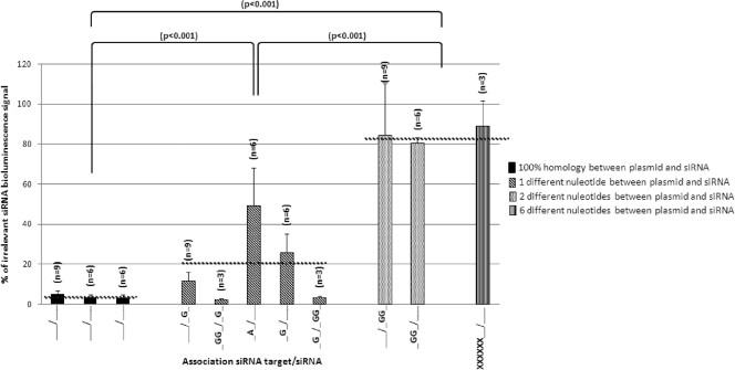
The resistance to RNAi may result from nucleotide modifications in the target sequence or from off-target alterations that may change the structure of the mRNA and hamper the accessibility of the target sequences to siRNA. To determine whether the mutations or the deletion observed in the siNPPRV1 target was directly responsible for RNAi escape, a cross-check luciferase reporter system was developed. The original or mutated target sequences were placed in the 5′ end of the coding sequence of the luciferase gene. The original and mutated siRNAs were then tested against both constructions. All mutations and the deletion obtained with siNPPRV1 were confirmed as being directly responsible for the RNAi escape (Fig. 4). These results confirm that siRNA off-target effects can be excluded. Interestingly, the deletion or double mutations showed the highest level of resistance against the original siRNA.
Fig 4.
Activities of different siRNAs on different targets in vitro. Vero cells were transfected with a reporter plasmid containing the firefly luciferase mRNA sequence and, upstream, the original or modified sequences of siNPPRV1 (siRNA-NPPRV1, siNPPRV1-mut1, siNPPRV1-mut3, or siNPPRV1-mut4 followed by firefly luciferase-2). They were cotransfected with different siRNAs (siNPPRV1, siNPPRV1mut1, and siNPPRV1mut4). The difference in sequence between target and siRNA is represented on the x axis (target sequence of siNPPRV1/sequence of siRNA). The signal level (y axis) was calculated in comparison with the signal obtained for the transfection with each plasmid and the irrelevant siMPPRV10. The number of repetitions (n) for each transfection is given above the histogram bars. The error bars represent the standard deviations of the repetitions. A significant difference was observed among the group with 100% homology between target and siRNA, the group with one nucleotide difference, and the group with two nucleotide differences or the deletion (P < 0.001). Horizontal broken lines represent the average of each of these groups.
siRNAs induce an unusual genetic variability in morbilliviruses.
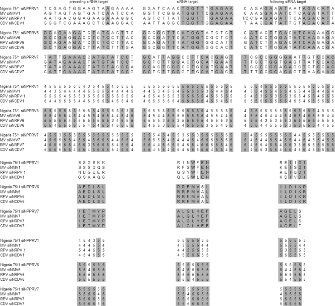
The conservation of the positions targeted by the three siRNAs in the morbillivirus N gene was reassessed by multiple alignments of nucleic acid and amino acid sequences retrieved in public databases and generated in our laboratory for PPRV. To compare the levels of sequence identity, the 19 nucleotides preceding and following the siRNA targets were also considered. In the 343 morbillivirus sequences analyzed, 52.5%, 49.1%, and 81.7% showed nucleic acid identity in the regions targeted by siRNAs N1, N6, and N7, respectively. These numbers are to be compared with 32.6%, 60.6%, and 73.1% identity in the left-flanking regions and 87.9%, 50.9%, and 63.7% in the right-flanking regions. For amino acid sequences, the identities were 76.1%, 96.5%, and 99.7% for the targeted regions, 57.4%, 99.4%, and 99.7% for the left-flanking regions, and 98.1%, 98.5%, and 99.7% for the right-flanking regions (Fig. 5).
Fig 5.
Conservation of target and flanking regions of the three siRNAs. The upper parts show the nucleotide conservation between vaccine strain Nigeria 75/1 and the consensus sequence of other morbilliviruses (vaccine strain Nigeria 75/1 was used by consensus for the PPRV sequences). The rest of the figure illustrates the nucleotide and amino acid conservation among the sequences of 25 peste des petits ruminants viruses (PPRV), 239 measles viruses (MV), 13 rinderpest viruses (RPV), and 66 canine distemper viruses (CDV). Numbers represent the level of identity within each morbillivirus group: 1, 0 to 25%; 2, 26 to 50%; 3, 51 to 75%; 4, 76 to 99%; and 5, 100%.
Within the 25 PPRV sequences, nucleotide/amino acid conservation was 100/100%, 96/100%, and 64/100% for siNPPRV1, siNPPRV6, and siNPPRV7, respectively. In comparison, the conservation in the flanking regions ranged from 60 to 88% and 52 to 100% for the nucleotide and amino acid sequences, respectively.
Six out of eight amino acid changes induced by siRNA were never encountered in the sequences of wild-type morbilliviruses. However, the positions (1 to 3) are not completely conserved within this genus (Fig. 5). The conservation of amino acids in the siRNA targets that may result from structural or functional constraints was further evaluated by a dN/dS analysis. The dN/dS analysis provides a quantification of the evolutionary pressures on proteins through the ratio of substitution rates at nonsynonymous and synonymous sites. The overall dN/dS ratio of the PPRV N gene was 0.144 ± 0.189 (minimum, 0.072; maximum, 1.429), with ratios of 0.089 ± 0.015, 0.095 ± 0.013, and 0.096 ± 0.013 at the siNPPRV1, -6, and -7 positions, respectively. These low dN/dS values confirm the existence of a negative selection pressure on the N gene and particularly in the three siRNA targets. For MV, the corresponding values were 0.200 ± 0.273 (minimum, 0.061; maximum, 1.493), 0.250 ± 0.309, 0.116 ± 0.097, and 0.091 ± 0.023, which were highly similar to those for PPRV. In contrast, when the escape PPRV mutants were included in the dN/dS analysis, only one value became higher than 1 (dN/dS = 1.302 ± 0.423) at position N145 in the siNPPRV1 region. This is the consequence of the accumulation of N/G145, N/S145, and N/T145 mutations in si1mut1, si1mut4, and si1_mut4_mut1 (Fig. 2). However, none of the 13 positions (including N145) found with a dN/dS ratio of >1 in the entire N protein were statistically considered positively selected (data not shown).
Using a molecular clock analysis after a Bayesian phylogenetic reconstruction on multiple alignments of sequences of PPRV N genes retrieved from the GenBank database (http://www.ncbi.nlm.nih.gov/GenBank/index.html) and others generated by our group, the predicted number of mutations per site per year was found to be 9.649 × 10⁻⁴. The numbers of mutations per site per passage under siRNA pressure were estimated to be 69.79 × 10⁻⁴, 103.38 × 10⁻⁴, 115.79 × 10⁻⁴, 18.44 × 10⁻⁴, and <8.77 × 10⁻⁴ for siNPPRV1, siNPPRV6, siNPPRV7, any combination of two siRNAs, and the association of the three siRNAs, respectively. In other words, the molecular evolution rates under the pressure of a single siRNA were 7 to 12 times higher than the natural molecular evolution rate of PPRV, and those of a combination of two siRNAs were 2 times higher.
This plasticity was further characterized by a multiple-factor analysis (41) of siRNA effects on sequence variations in the target regions in comparison with their flanking regions. The scoring system described in Materials and Methods was applied to any deviation from the nucleotide and amino acid sequences of the original strain Nigeria 75/1 (13). The results of multiple-factor analysis revealed a high level of divergence after consecutive siRNA treatments between the parental (wild) and the siRNA-induced PPRV strains (Fig. 6) (permutation test, P ≤ 10−4).
Fig 6.
Grouping of wild and siRNA-induced mutant PPRV strains according to their nucleotide mutation features. Coordinates of each PPRV strain were computed using a multiple-factor analysis. Each strain is represented by a dot on the factorial plane (axis 2, axis 3). Each group centroid is represented by a letter, M for siRNA-induced mutant PPRV strains and W for wild PPRV strains. Group membership is represented by a line segment joining each PPRV strain to its group centroid.
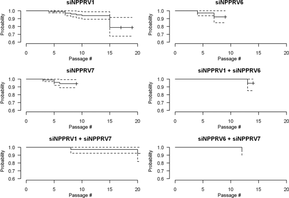
The results of the Kaplan-Meier analysis (34) are shown in Fig. 7. No mutation was observed over 20 passages when the 3 siRNAs were combined, which means that the probability of keeping the original vaccine strain Nigeria 75/1 unchanged and still sensitive to siRNA treatment (survival of the original strain) remained equal to 1 up to the 20th passage (not shown in Fig. 7). The probability of survival of strain Nigeria 75/1 started to decline after passage 7 when any of the 3 siRNAs were combined in pairs (last three graphs). In contrast, mutations appeared more rapidly, and consequently the survival probability decreased faster (between the 3rd and 10th passages), when a single siRNA was applied (first three graphs). The results of the Cox model (8) showed that, compared with siNPRV1, the mutation hazard was higher for siNPPRV6 (RR = 2.5) and siNPPRV7 (RR = 2.1), but not significantly different from that for siNPPRV1 (P = 0.11 and P = 0.13). Also, the RR was <1 for any two- or three-siRNA combination, but the difference was significant only for the association of siNPPRV6 and siNPPRV7 (RR = 0.2, P = 7 × 10⁻⁵).
Fig 7.
Survival (no-mutation) probability for Nigeria 75/1 under siRNA treatment. Curves were computed using a Kaplan-Meier survival analysis. The number of cell passages when the first mutation was observed was used as the failure time. Survival probability is shown by a solid line. Its 95% confidence interval is represented by dashed lines.
DISCUSSION
The discovery of RNAi has provided powerful tools for biological research and drug discovery. RNAi has also expanded therapeutic strategies, with the potential to treat a wide range of different diseases, including viral infections. Many viruses have been successfully targeted by RNAi (24, 25, 49). Moreover, RNAi-based therapies are currently advancing from basic research to clinical trials. In fact, several RNAi-based antiviral drugs are currently being tested in clinical trials (5, 25). One of the main advantages of RNAi is its high specificity for the targeted sequences. In contrast, RNAi is highly prone to sequence divergence and is thus poorly flexible, in relation to the capacity of viruses to evolve rapidly. Indeed, viral pathogens can quickly and readily escape inhibition conferred by siRNAs (3, 9, 37). Pioneer studies evidenced the high specificity of RNAi by evaluating the activity of an siRNA with one or several mismatches relative to the target RNA sequence (30, 56). These studies showed that a single nucleotide mismatch may be sufficient to reduce the silencing effect. The emergence of viral escape mutants is a major challenge for the clinical application of RNAi. Clearly, an important strategy in designing siRNAs to limit the risk of escape mutants would be to target conserved viral sequences with a strong negative selection pressure.
In this context, this work intended to explore the propensity of morbilliviruses to escape RNA interference targeting three conserved regions of the viral genome. siRNAs directed to these regions were previously shown to strongly repress the replication of measles, rinderpest, and peste des petits ruminants viruses (36, 49). They all target region III, the most conserved part of the N gene (85 to 90% nucleotide identity within all morbilliviruses, compared to 75 to 83%, 40%, and 17 to 30% identity for regions I, II, and IV, respectively) (12). The high conservation of the third region suggests a negative selection pressure possibly linked to structural or functional constraints in the virus replication cycle. Nonetheless, this study showed that the generation of escape mutants with mutations in this region was unexpectedly easy. Alterations in the N gene ranged from a single or two nucleotide mutations (with or without amino acid substitution) to a rather large deletion of six nucleotides. In other studies, many viruses have been found to develop variants capable of escaping from RNAi by nucleotide substitutions or by partial or complete deletion of the target sequence (9, 54). A single-nucleotide mismatch between the siRNA and the morbillivirus target sequence was shown here to alter the level of silencing, in agreement with the results of Das et al. (9) and Sabariegos et al. (48) on human immunodeficiency virus type 1 (HIV-1), in which one nucleotide mismatch between the siRNA and its target sequence provided partial resistance to RNAi. In contrast, von Eije et al. (53) recently analyzed more than 500 HIV-1 escape mutants, and when essential and highly conserved sequences were targeted, deletion was never observed. Interestingly, the deletion observed in the N gene of PPRV still satisfies the so-called “rule of six,” by which the genome of a morbillivirus must be a multiple of 6 for efficient replication (26, 47), and although it is not in the frame of the coding sequence, it results only in a shorter protein. This is a striking demonstration of the extraordinary plasticity of morbilliviruses, whereas the PPR morbillivirus could simply escape RNAi by a single mutation. Accordingly, the level of resistance to RNAi was correlated to the extent of modifications in the target sequence. The loss of RNAi activity was more pronounced, with the deletion of six nucleotides and dual mutations compared to single mutations, suggesting in the former case a better adaptive response to bypass RNA interference. Although we postulated that there was a strong structural or functional constraint on the conserved regions selected in this study, when a second siRNA (siNPPRV1mut4) was applied to a first generation of viral escape mutants, there was no reversion to the original virus. On the contrary, a novel mutation was rapidly acquired, which conferred new RNAi resistance. This is an additional illustration of the high level of malleability of the morbillivirus genome.
All synonymous or nonsynonymous mutations were at positions 3 to 10 in the siRNA target and affected amino acid positions 2 to 4, regarding nonsynonymous changes. The deletion encompassed the same region. This indicates either that the critical residues for the activity of our siRNAs are restricted to the first half of the target sequence or that the second half of the sequence cannot be modified without deleterious consequences for virus viability. A large proportion of mutants (8/18) had a modification in their N protein. The majority of these modifications (6/8) were never observed in nature in other morbilliviruses. Such protein malleability was unexpected since the dN/dS analysis of the N genes of PPRV and MV rendered ratios lower than 0.150, indicating a general negative selection pressure on this protein. The dN/dS ratios in the siRNA target regions were even lower than that (0.095), which supports the notion that our siRNAs were well designed for conserved regions of the N coding sequence. Surprisingly, in the siPPRV1 target, a dN/dS ratio of >1 was obtained with the siRNA treatment at position N145. It is unclear how a preference for nonsynonymous substitutions can be driven by pressure applied by siRNA on the mRNA. This may be the consequence of a combination of constraints, including (i) the critical residues that must be mutated to escape RNA interference, (ii) the number of alternative codons available for synonymous substitutions, and (iii) the tolerance of the protein for amino acid substitutions. Since nucleotide mutations in escape mutants were concentrated in positions encoding amino acids with limited genetic code degeneracy, it is not surprising that almost half of our mutants had a modified N protein. When looking at the genetic divergence achieved with the siRNA selection pressure, the mutation rates per site and passage were 7 to 12 times higher than the mutation rate per site and year in wild-type morbilliviruses when a single siRNA was applied and 2 times higher when a combination of two siRNAs was applied. In contrast, the combination of three siRNAs reduced the evolution rate observed in vitro to the corresponding value observed in nature. Such a genomic plasticity in morbilliviruses was unexpected, since they are relatively stable viruses. Here, PPRV has a nucleotide substitution/site/year rate of 9.64 × 10⁻⁴, which is slightly higher than that for measles virus (6.2 × 10⁻⁴) but still lower than those for other RNA viruses such as enteroviruses and retroviruses (34 × 10−4 and 25 × 10−4, respectively) (31) In addition to the large genomic malleability peculiar to morbilliviruses, these considerations underline that inappropriate siRNA concentrations for therapeutic use in the field could accelerate the evolution and possibly the emergence of strains with modified properties such as a more virulent phenotype. To reduce the risk of escape mutants, it is therefore recommended to use appropriate concentrations of a combination of at least two siRNAs targeting different regions within the same gene or in different genes. In this study, the emergence of escape mutants was delayed by 6 to 13 passages when two siRNAs were simultaneously delivered in the cell culture, and the association of three siRNAs prevented any escape over 20 passages in cell culture. A similar result was obtained by Kusov et al. (39), who efficiently prevented resistant HIV-1 variants by using siRNAs targeting various sites in the HIV-1 nonstructural genes. In such a therapeutic scenario, all siRNAs will be potent viral inhibitors, and the cumulative pressure may be enough to block the emergence of escape mutants.
This study provides insight into the capacity of morbilliviruses to force adaptive responses to escape siRNA targeting well-conserved regions of their genomes. This capacity went up to, for the first time, a significant deletion in an essential viral gene, resulting in the production of a shorter protein. These results constitute a critical obstacle to the use of RNAi as an antiviral therapy. On the other hand, the emergence of escape mutants was prevented after 20 passages in cell culture in the presence of three combined siRNAs, which could be an acceptable strategy to address the risk of treatment failure with siRNA.
ACKNOWLEDGMENTS
This work was supported by Centre de Coopération International en Recherche Agronomique pour le Développement and Languedoc-Roussillon Région. This study was also supported by the European Union Network of Excellence for Epizootic Disease Diagnosis and Control (EPIZONE, no. FOOD-CT-2006-016236) and by CIRAD (ATP “Emergences”).
We thank Anne Xuèreb, Philippe Gauthier, and Maxime Galan for help and advice on real-time PCR-HRM.
Footnotes
Published ahead of print 9 November 2011
REFERENCES
- 1. Banyard AC, Grant RJ, Romero CH, Barrett T. 2008. Sequence of the nucleocapsid gene and genome and antigenome promoters for an isolate of porpoise morbillivirus. Virus Res. 132:213–219 [DOI] [PubMed] [Google Scholar]
- 2. Barrett TC, Pastoret PP, Taylor WP. 2006. Rinderpest and peste des petits ruminants: virus plagues of large and small ruminants, Elsevier Academic Press, Amsterdam, Netherlands [Google Scholar]
- 3. Boden D, Pusch O, Lee F, Tucker L, Ramratnam B. 2003. Human immunodeficiency virus type 1 escape from RNA interference. J. Virol. 77:11531–11535 [DOI] [PMC free article] [PubMed] [Google Scholar]
- 4. Borgundvaag B, et al. 2004. SARS outbreak in the greater Toronto area: the emergency department experience. CMAJ 171:1342–1344 [DOI] [PMC free article] [PubMed] [Google Scholar]
- 5. Castanotto D, Rossi JJ. 2009. The promises and pitfalls of RNA-interference-based therapeutics. Nature 457:426–433 [DOI] [PMC free article] [PubMed] [Google Scholar]
- 6. Claas EC, et al. 1998. Human influenza A H5N1 virus related to a highly pathogenic avian influenza virus. Lancet 351:472–477 (Erratum, Lancet 351:1292.) [DOI] [PubMed] [Google Scholar]
- 7. Colbère-Garapin F, Blondel B, Saulnier A, Pelletier I, Labadie K. 2005. Silencing viruses by RNA interference. Microbes Infect. 7:767–775 [DOI] [PMC free article] [PubMed] [Google Scholar]
- 8. Cox DR, Oakes D. 1984. Analysis of survival data. Chapman & Hall/CRC Press, London, United Kingdom [Google Scholar]
- 9. Das AT, et al. 2004. Human immunodeficiency virus type 1 escapes from RNA interference-mediated inhibition. J. Virol. 78:2601–2605 [DOI] [PMC free article] [PubMed] [Google Scholar]
- 10. Dave RS, Pomerantz RJ. 2003. RNA interference: on the road to an to alternate therapeutic strategy. Rev. Med. Virol. 13:373–385 [DOI] [PubMed] [Google Scholar]
- 11. De Fougerolles AR. 2008. Delivery vehicles for small interfering RNA in vivo. Hum. Gene Ther. 19:125–132 [DOI] [PubMed] [Google Scholar]
- 12. Diallo A, Barrett T, Barbron M, Meyer G, Lefevre PC. 1994. Cloning of the nucleocapsid protein gene of peste-des-petits-ruminants virus—relationship to other morbilliviruses. J. Gen. Virol. 75:233–237 [DOI] [PubMed] [Google Scholar]
- 13. Diallo A, Taylor WP, Lefèvre PC, Provost A. 1989. Attenuation of a strain of rinderpest virus: potential homologous live vaccine. Rev. Elev. Med. Vet. Pays. Trop. 42:311–319 [PubMed] [Google Scholar]
- 14. Drummond AJ, Rambaut A. 2007. BEAST: Bayesian evolutionary analysis by sampling trees. BMC Evol. Biol. 7:214. [DOI] [PMC free article] [PubMed] [Google Scholar]
- 15. Elbashir SM, et al. 2001. Duplexes of 21-nucleotide RNAs mediate RNA interference in cultured mammalian cells. Nature 411:494–498 [DOI] [PubMed] [Google Scholar]
- 16. Elbers ARW, et al. 2008. Field observation during the Bluetongue serotype 8 epidemic in 2006. I. Detection of first outbreaks and clinical signs in sheep and cattle in Belgium, France and The Netherlands. Prev. Vet. Med. 87:21–30 [DOI] [PubMed] [Google Scholar]
- 17. Escofier B, Pagès J. 1994. Multiple factor analysis (AFMULT package). Comput. Stat. Data Anal. 18:121–140 [Google Scholar]
- 18. Ferreira HL, Spilki FR, Servan de Almeida R, Santos MMAB, Arns CW. 2007. Inhibition of avian metapneumovirus (AMPV) replication by RNA interference targeting nucleoprotein gene (N) in cultured cells. Antiviral Res. 74:77–81 [DOI] [PubMed] [Google Scholar]
- 19. Fire A, et al. 1998. Potent and specific genetic interference by double-stranded RNA in Caenorhabditis elegans. Nature 391:806. [DOI] [PubMed] [Google Scholar]
- 20. Fleming A. 1929. On the antibacterial action of cultures of Penicillium with special reference to their use in situation of H. influenzae. Br. J. Exp. Pathol. 10:226 [Google Scholar]
- 21. Furuse Y, Suzuki A, Oshitani H. 2010. Origin of measles virus: divergence from rinderpest virus between the 11th and 12th centuries. Virol. J. 7:52. [DOI] [PMC free article] [PubMed] [Google Scholar]
- 22. Galtier N, Gouy M, Gautier C. 1996. SEAVIEW and PHYLO_WIN: two graphic tools for sequence alignment and molecular phylogeny. Comput. Appl. Biosci. 12:543–548 [DOI] [PubMed] [Google Scholar]
- 23. Gouy M, Guindon S, Gascuel O. 2010. SeaView version 4: a multiplatform graphical user interface for sequence alignment and phylogenetic tree building. Mol. Biol. Evol. 27:221–224 [DOI] [PubMed] [Google Scholar]
- 24. Haasnoot J, Berkhout B. 2006. RNA interference: its use as antiviral therapy. Handb. Exp. Pharmacol. 173:117–150 [DOI] [PMC free article] [PubMed] [Google Scholar]
- 25. Haasnoot J, Westerhout EM, Berkhout B. 2007. RNA interference against viruses: strike and counterstrike. Nat. Biotechnol. 25:1435–1443 [DOI] [PMC free article] [PubMed] [Google Scholar]
- 26. Hausmann S, Jacques JP, Kolakofsky D. 1996. Paramyxovirus RNA editing and the requirement for hexamer genome length. RNA 2:1033–1045 [PMC free article] [PubMed] [Google Scholar]
- 27. Holm S. 1979. A simple sequentially rejective multiple test procedure. Scand. J. Stat. 6:65–70 [Google Scholar]
- 28. Hosmer DW, Lemeshow S. 1999. Applied survival analysis: regression modeling of time to event data, John Wiley, New York, NY [Google Scholar]
- 29. Huelsenbeck JP, Ronquist F. 2001. MRBAYES: Bayesian inference of phylogenetic trees. Bioinformatics 17:754–755 [DOI] [PubMed] [Google Scholar]
- 30. Jacque JM, Triques K, Stevenson M. 2002. Modulation of HIV-1 replication by RNA interference. Nature 418:435–438 [DOI] [PMC free article] [PubMed] [Google Scholar]
- 31. Jenkins GM, Rambaut A, Pybus OG, Holmes EC. 2002. Rates of molecular evolution in RNA viruses: a quantitative phylogenetic analysis. J. Mol. Evol. 54:156–165 [DOI] [PubMed] [Google Scholar]
- 32. Jobb G, von Haeseler A, Strimmer K. 2004. TREEFINDER: a powerful graphical analysis environment for molecular phylogenetics. BMC Evol. Biol. 4:18. [DOI] [PMC free article] [PubMed] [Google Scholar] [Retracted]
- 33. Kanda T, Kusov Y, Yokosuka O, Gauss-Müller V. 2004. Interference of hepatitis A virus replication by small interfering RNAs. Biochem. Biophys. Res. Commun. 318:341–345 [DOI] [PubMed] [Google Scholar]
- 34. Kaplan EL, Meier P. 1958. Non parametric estimation from incomplete observations. J. Am. Stat. Assoc. 53:457–481 [Google Scholar]
- 35. Keeling MJ, et al. 2001. Dynamics of the 2001 UK foot and mouth epidemic: stochastic dispersal in a heterogeneous landscape. Science 294:813. [DOI] [PubMed] [Google Scholar]
- 36. Keita D, Servan de Almeida R, Libeau G, Albina E. 2008. Identification and mapping of a region on the mRNA of morbillivirus nucleoprotein susceptible to RNA interference. Antiviral Res. 80:158–167 [DOI] [PubMed] [Google Scholar]
- 37. Konishi M, et al. 2006. siRNA-resistance in treated HCV replicon cells is correlated with the development of specific HCV mutations. J. Viral Hepat. 13:756–761 [DOI] [PubMed] [Google Scholar]
- 38. Krönke J, et al. 2004. Alternative approaches for efficient inhibition of hepatitis C virus RNA replication by small interfering RNAs. J. Virol. 78:3436–3446 [DOI] [PMC free article] [PubMed] [Google Scholar]
- 39. Kusov Y, Kanda T, Palmenberg A, Sgro JY, Gauss-Müller V. 2006. Silencing of hepatitis A virus infection by small interfering RNAs. J. Virol. 80:5599–610 [DOI] [PMC free article] [PubMed] [Google Scholar]
- 40. Liu YP, Haasnoot J, ter Brake O, Berkhout B, Konstantinova P. 2008. Inhibition of HIV-1 by multiple siRNAs expressed from a single microRNA polycistron. Nucleic Acids Res. 36:2811–2824 [DOI] [PMC free article] [PubMed] [Google Scholar]
- 41. Manly BFJ. 2006. Randomization, bootstrap and Monte Carlo methods in biology, Chapman & Hall/CRC Press, Laramie, WY [Google Scholar]
- 42. Nichol ST, Arikawa J, Kawaoka Y. 2000. Emerging viral diseases. Proc. Natl. Acad. Sci. U. S. A. 97:12411–12412 [DOI] [PMC free article] [PubMed] [Google Scholar]
- 43. Plasterk RA. 2002. RNA silencing: the genome's immune system. Science 296:1263–1265 [DOI] [PubMed] [Google Scholar]
- 44. Reed LJ, Muench H. 1938. A simple method of estimating fifty percent endpoints. Am. J. Hyg. 27:493–497 [Google Scholar]
- 45. Reuter T, Weissbrich B, Schneider-Schaulies S, Schneider-Schaulies J. 2006. RNA interference with measles virus N, P, and L mRNAs efficiently prevents and with matrix protein mRNA enhances viral transcription. J. Virol. 80:5951–5957 [DOI] [PMC free article] [PubMed] [Google Scholar]
- 46. Rima BK, Wishaupt RGA, Welsh MJ, Earle JAP. 1995. The evolution of morbilliviruses: a comparison of nucleocapsid gene sequences including a porpoise morbillivirus. Vet. Microbiol. 44:127–134 [DOI] [PubMed] [Google Scholar]
- 47. Roux L. 2005. Dans le genome des Paramyxovirinae, les promoteurs et leurs activités sont façonnés par la “régle de six.” Virologie 9:19–34 [DOI] [PubMed] [Google Scholar]
- 48. Sabariegos R, Giménez-Barcons M, Tàpia N, Clotet B, Martínez MA. 2006. Sequence homology required by human immunodeficiency virus type 1 to escape from short interfering RNAs. J. Virol. 80:571–577 [DOI] [PMC free article] [PubMed] [Google Scholar]
- 49. Servan de Almeida R, Keita D, Libeau G, Albina E. 2007. Control of ruminant morbillivirus replication by small interfering RNA. J. Gen. Virol. 88:2307–2311 [DOI] [PubMed] [Google Scholar]
- 50. Shin D, Lee H, Kim SI, Yoon Y, Kim M. 2009. Optimization of linear double-stranded RNA for the production of multiple siRNAs targeting hepatitis C virus. RNA 15:989–910 [DOI] [PMC free article] [PubMed] [Google Scholar]
- 51. Silverman A, Simor A, Loutfy MR. 2004. Toronto emergency medical services and SARS. Emerg. Infect. Dis. 10:1688–1689 [DOI] [PMC free article] [PubMed] [Google Scholar]
- 52. Takeshita F, Hokaiwado N, Honma K, Banas A, Ochiya T. 2009. Local and systemic delivery of siRNAs for oligonucleotide therapy. Methods Mol. Biol. 487:83–92 [DOI] [PubMed] [Google Scholar]
- 53. von Eije KJ, ter Brake O, Berkhout B. 2008. Human immunodeficiency virus type 1 escape is restricted when conserved genome sequences are targeted by RNA interference. J. Virol. 82:2895–2903 [DOI] [PMC free article] [PubMed] [Google Scholar]
- 54. Westerhout EM, Ooms M, Vink M, Das AT, Berkhout B. 2005. HIV-1 can escape from RNA interference by evolving an alternative structure in its RNA genome. Nucleic Acids Res. 33:796–804 [DOI] [PMC free article] [PubMed] [Google Scholar]
- 55. Wilcoxon F. 1945. Individual comparisons by ranking methods. Biometrics 1:80–83 [Google Scholar]
- 56. Wilson JA, Richardson CD. 2005. Hepatitis C virus replicons escape RNA interference induced by a short interfering RNA directed against the NS5b coding region. J. Virol. 79:7050–7058 [DOI] [PMC free article] [PubMed] [Google Scholar]
- 57. Wu HL, et al. 2005. RNA interference-mediated control of hepatitis B virus and emergence of resistant mutant. Gastroenterology 128:708–716 [DOI] [PMC free article] [PubMed] [Google Scholar]
- 58. Xie FY, Woodle MC, Lu PY. 2006. Harnessing in vivo siRNA delivery for drug discovery and therapeutic development. Drug Discov. Today 11:67–73 [DOI] [PMC free article] [PubMed] [Google Scholar]
- 59. Yang Z. 2007. PAML 4: a program package for phylogenetic analysis by maximum likelihood. Mol. Biol. Evol. 24:1586–1591 [DOI] [PubMed] [Google Scholar]


