Abstract
Cancer metastasis to the thyroid is rare. Primary sites that have been reported to metastasize to the thyroid gland include breast and kidney. There are very few case reports describing metastasis of a lung primary cancer to the thyroid gland. We describe a case of a never-smoker patient with metastatic adenocarcinoma of the lung and a malignant thyroid mass presenting simultaneously. The thyroid biopsy could not distinguish whether it was a metastasis or incidentally found thyroid primary carcinoma. Based on EGFR mutational status it was determined that he has a primary lung cancer and thyroid metastasis. He was started on erlotinib and has had a marked response in both the lung and the thyroid masses. The presence of mutations within the EGFR gene can help distinguish lung adenocarcinoma from other carcinomas like thyroid cancer. This is the first published case of primary lung adenocarcinoma with metastasis to thyroid and with EGFR mutation.
Keywords: lung cancer, thyroid cancer, non-small cell lung cancer, lung adenocarcinoma, thyroid adenocarcinoma, thyroid nodule, EGFR, L858R EGFR mutation, Erlotinib
Introduction
Cancer metastasis to the thyroid is rare. Primary sites that have been reported to metastasize to the thyroid gland include breast and kidney. There are very few case reports describing metastasis of a lung primary cancer to the thyroid gland. We report a case of a patient who presented with two large masses, lung mass and thyroid mass at the same time. Biopsy of both showed adenocarcinoma. Pathology and IHC was not able to differentiate whether this was two separate malignancies or one primary with metastasis. The EGFR gene mutation was the key for the final diagnosis. This is the first reported case of lung cancer with L858R EGFR mutation positivity and metastasis to the thyroid gland.
Case report
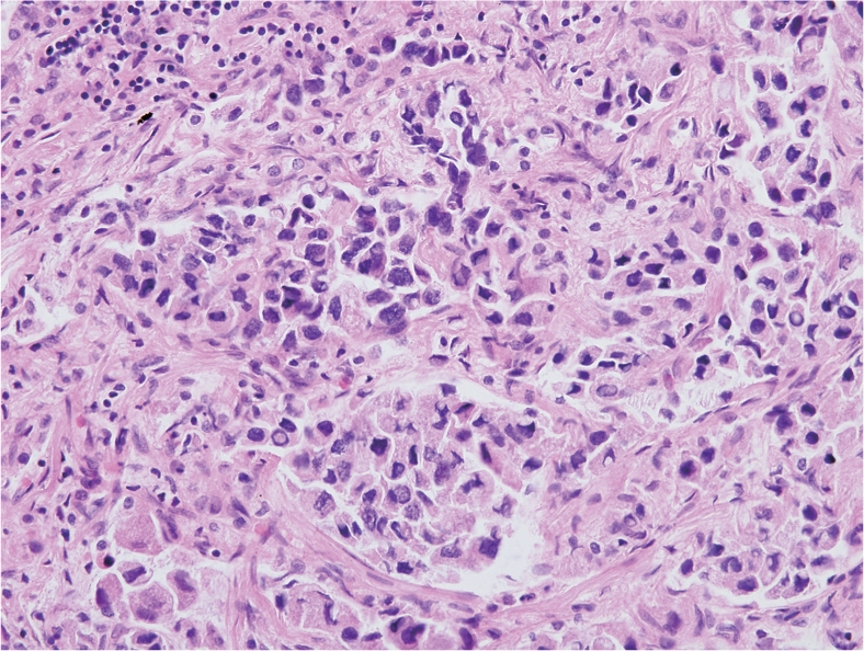
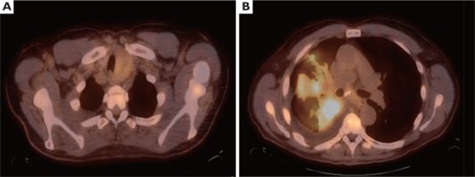
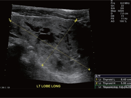
A 68-year-old male never smoker presented with progressive fatigue, shortness of breath, cough and weight loss over several months. Chest X-ray revealed a right-sided pneumonia, which was not resolving with appropriate antibiotics. Subsequent body PET/ CT scan revealed FDG-avid left thyroid mass measuring 3.8x5.2 cm with SUV of 3, along with a right hilar mass measuring 8.4x6.2 cm intensely FDG-avid SUV of 8.9; with diffuse mediastinal lymphadenopathy and a small right pleural effusion (Figure 1). Thyroid ultrasound (Figure 2) with fine needle biopsy revealed malignancy, which was CK7 and TTF-1 positive (Figure 3). CT guided lung biopsy revealed adenocarcinoma of the lung.
Figure 1. PET/ CT scan with FDG-avid left thyroid mass measring 3.8 x 5.2 cm with SUV of 3 (A) and right hilar mass measuring 8.4 x 6.2 cm intensely FDG-avid with SUV of 8.9 (B).
Figure 2. Ultrasound of the thyroid showing large mass replacing the left lobe composed of both cystic and solid components.
Figure 3. Clusters of loosely cohesive carcinoma cells with hyperchromatic nuclei showing occasional intranuclear cytoplasmic inclusions are present in alveolar air spaces.

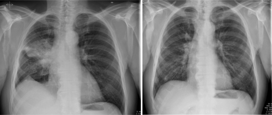
It was initially difficult to determine whether this represented two separate malignancies, lung cancer with metastasis to thyroid, or a separate thyroid cancer. The tumor specimen from the lung demonstrated L858R EGFR mutation in Exon 21, confirming that this was all primary adenocarcinoma of the lung and not thyroid cancer. He was started on Erlotinib. He had an excellent clinical and radiographic response to the treatment after just 4 weeks, with reduction in his symptoms and radiographic improvement. After 8 months of follow up he continues to be in remission. His originally described palpable thyroid mass measuring 4x5 cm is no longer palpable; and his most recent CXR showed no evidence of disease (Figure 4).
Figure 4. Chest X-ray (AP) before starting therapy and 8 months later while on treatment with erlotinib showing remarkable and durable response to therapy.
Discussion
Lung cancer is the second most common malignancy and the leading cause of cancer death in both men and women. Adenocarcinoma accounts for 40% of non-small cell lung cancers (1). Most never smokers have adenocarcinoma histology (2). Common sites of metastasis include brain, bones, adrenal glands, contralateral lung and liver.
Metastasis to the thyroid gland is rare and is found mainly in autopsy cases (3). The most common malignancies, which are known to metastasize to the thyroid, include the breast, lung and kidney (4). With the advent of PET scan, incidental primary tumors of thyroid can be observed. Metastasis to the thyroid accounts for 2%–3% of all clinical cases with malignant tumors of the thyroid (4).
Accurate diagnosis of a thyroid lesion as a primary or metastatic neoplasm is a significant challenge; primary and secondary lesions do not show specific features on imaging studies, such as computed tomography or ultrasound. Pathological diagnosis remains the gold standard for diagnosis. In our case, Immunohistochemistry was positive for pankeratin, CK7 and TTF-1. CK7 and TTF-1 do not differentiate between lung and thyroid carcinoma.
The epidermal growth factor receptor (EGFR) is a transmembrane glycoprotein with an intrinsic tyrosine kinase (TK) activity. Erlotinib selectively binds to the ATP-binding site of the (EGFR)-TK, which inhibits receptor tyrosine kinases, and is utilized for patients with advanced NSCLC (5). Activating mutation of exon 19 or exon 21 is observed primarily in never smokers. Patients harboring EGFR mutations are likely to respond to EGFR inhibitors like erlotinib (6),(7).
We describe a case of a never-smoker patient with metastatic adenocarcinoma of the lung and a malignant thyroid mass presenting simultaneously. The thyroid biopsy could not distinguish whether it was a metastasis or incidentally found thyroid primary carcinoma. Based on EGFR mutational status it was determined that he has a primary lung cancer and thyroid metastasis. He was started on erlotinib and has had a marked response in both the lung and the thyroid masses. The presence of mutations within the EGFR gene can help distinguish lung adenocarcinoma from other carcinomas like thyroid cancer. This is the first published case of primary lung adenocarcinoma with metastasis to thyroid and with EGFR mutation.
accounts for 2%–3% of all clinical cases with malignant tumors of the thyroid (4).
Accurate diagnosis of a thyroid lesion as a primary or metastatic neoplasm is a significant challenge; primary and secondary lesions do not show specific features on imaging studies, such as computed tomography or ultrasound. Pathological diagnosis remains the gold standard for diagnosis. In our case, Immunohistochemistry was positive for pankeratin, CK7 and TTF-1. CK7 and TTF-1 do not differentiate between lung and thyroid carcinoma.
The epidermal growth factor receptor (EGFR) is a transmembrane glycoprotein with an intrinsic tyrosine kinase (TK) activity. Erlotinib selectively binds to the ATP-binding site of the (EGFR)-TK, which inhibits receptor tyrosine kinases, and is utilized for patients with advanced NSCLC (5). Activating mutation of exon 19 or exon 21 is observed primarily in never smokers. Patients harboring EGFR mutations are likely to respond to EGFR inhibitors like erlotinib (6),(7).
We describe a case of a never-smoker patient with metastatic adenocarcinoma of the lung and a malignant thyroid mass presenting simultaneously. The thyroid biopsy could not distinguish whether it was a metastasis or incidentally found thyroid primary carcinoma. Based on EGFR mutational status it was determined that he has a primary lung cancer and thyroid metastasis. He was started on erlotinib and has had a marked response in both the lung and the thyroid masses. The presence of mutations within the EGFR gene can help distinguish lung adenocarcinoma from other carcinomas like thyroid cancer. This is the first published case of primary lung adenocarcinoma with metastasis to thyroid and with EGFR mutation.
Footnotes
No potential conflict of interest.
References
- 1.Travis WD. Pathology of lung cancer. Clin Chest Med. 2002;23:65–81, viii. doi: 10.1016/s0272-5231(03)00061-3. [DOI] [PubMed] [Google Scholar]
- 2.Subramanian J, Govindan R. Lung cancer in never smokers: a review. J Clin Oncol. 2007;25:561–70. doi: 10.1200/JCO.2006.06.8015. [DOI] [PubMed] [Google Scholar]
- 3.Lam KY, Lo CY. Metastatic tumors of the thyroid gland: a study of 79 cases in Chinese patients. Arch Pathol Lab Med. 1998;122:37–41. [PubMed] [Google Scholar]
- 4.Nakhjavani MK, Gharib H, Goellner JR, van Heerden JA. Metastasis to the thyroid gland. A report of 43 cases. Cancer. 1997;79:574–8. doi: 10.1002/(sici)1097-0142(19970201)79:3<574::aid-cncr21>3.0.co;2-#. [DOI] [PubMed] [Google Scholar]
- 5.Siegel-Lakhai WS, Beijnen JH, Schellens JH. Current knowledge and future directions of the selective epidermal growth factor receptor inhibitors erlotinib (Tarceva) and gefitinib (Iressa) Oncologist. 2005;10:579–89. doi: 10.1634/theoncologist.10-8-579. [DOI] [PubMed] [Google Scholar]
- 6.Pao W, Miller V, Zakowski M, Doherty J, Politi K, Sarkaria I, et al. EGF receptor gene mutations are common in lung cancers from “never smokers” and are associated with sensitivity of tumors to gefitinib and erlotinib. Proc Natl Acad Sci U S A. 2004;101:13306–11. doi: 10.1073/pnas.0405220101. [DOI] [PMC free article] [PubMed] [Google Scholar]
- 7.Lynch TJ, Bell DW, Sordella R, Gurubhagavatula S, Okimoto RA, Brannigan BW, et al. Activating mutations in the epidermal growth factor receptor underlying responsiveness of non-small-cell lung cancer to gefitinib. N Engl J Med. 2004;350:2129–39. doi: 10.1056/NEJMoa040938. [DOI] [PubMed] [Google Scholar]


