Abstract
Osteoarthritis, characterized by the breakdown of articular cartilage in synovial joints, has long been viewed as the result of “wear and tear”1. Although low-grade inflammation is detected in osteoarthritis, its role is unclear2–4. Here we identify a central role for the inflammatory complement system in the pathogenesis of osteoarthritis. Through proteomic and transcriptomic analyses of synovial fluids and membranes from individuals with osteoarthritis, we find that expression and activation of complement is abnormally high in human osteoarthritic joints. Using mice genetically deficient in C5, C6, or CD59a, we show that complement, and specifically the membrane attack complex (MAC)-mediated arm of complement, is critical to the development of arthritis in three different mouse models of osteoarthritis. Pharmacological modulation of complement in wild-type mice confirmed the results obtained with genetically deficient mice. Expression of inflammatory and degradative molecules was lower in chondrocytes from destabilized joints of C5-deficient mice than C5-sufficient mice, and MAC induced production of these molecules in cultured chondrocytes. Furthermore, MAC co-localized with matrix metalloprotease (MMP)-13 and with activated extracellular signal-regulated kinase (ERK) around chondrocytes in human osteoarthritic cartilage. Our findings indicate that dysregulation of complement in synovial joints plays a critical role in the pathogenesis of osteoarthritis.
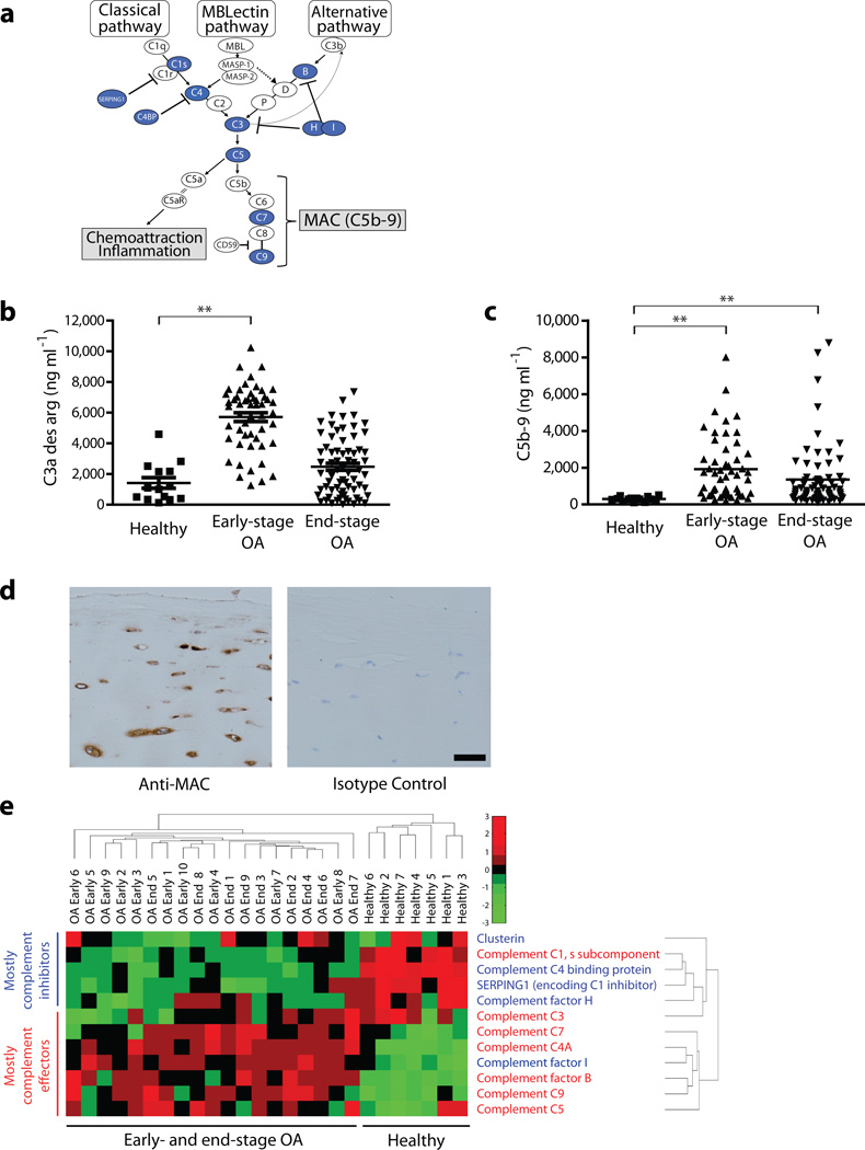
The pathogenesis of osteoarthritis is unclear, and there are currently no treatments that prevent the development of osteoarthritis. Seeking to gain insight into osteoarthritis, we used mass spectrometry to identify proteins aberrantly expressed in synovial fluid—the fluid that bathes the synovial joints—of individuals with osteoarthritis. We discovered that proteins of the complement system are differentially expressed in osteoarthritic compared to healthy synovial fluids (Supplementary Table 1). Using less-sensitive proteomic techniques, we previously detected ten of these twelve differentially expressed complement proteins in osteoarthritic synovial fluids5. The complement system consists of three distinct pathways that converge at the formation of the C3 and C5 convertases, enzymes that mediate activation of the C5a anaphylatoxin and formation of MAC (comprising the complement effectors C5b-9) (Fig. 1a)6. Components of the classical (C1s and C4A) and alternative (factor B) pathways, the central components C3 and C5, and the C5, C7, and C9 components of MAC were all aberrantly expressed in synovial fluids from individuals with osteoarthritis (Fig. 1a and Supplementary Table 1).
Figure 1.
Complement components are aberrantly expressed and activated in human osteoarthritic joints. (a) Schematic of the complement cascade; blue-filled circles denote the complement effectors and inhibitors identified as aberrantly expressed in osteoarthritic synovial fluid. (b,c) ELISA quantification of (b) C3a des arg and of (c) the soluble form of MAC (complement effectors C5b-9) in synovial fluids from healthy individuals (n = 14) and from individuals with early-stage osteoarthritis (n = 52) or end-stage osteoarthritis (n = 69). **P ≤ 0.01 by one-way ANOVA and Dunnett’s post-hoc test. (d) Immunohistochemical staining of MAC in cartilage from individuals with end-stage osteoarthritis. Isotype-matched antibodies were used as negative controls. Staining is representative of that seen in samples from 4 different individuals with osteoarthritis. Scale bar, 100 µm. (e) Cluster analysis of gene-expression profiles in microarray datasets from synovial membranes from healthy individuals (downloaded from the NCBI Gene Expression Omnibus) and from individuals with early-stage or end-stage osteoarthritis (experimentally determined). Analysis was limited to the set of genes encoding the complement-related proteins differentially expressed in RA compared to healthy synovial fluid (Supplementary Table 1). The scale bar represents fold change in gene expression compared to the reference control. Complement effectors are shown in red text, and complement inhibitors in blue text.
Validating our proteomic results, ELISA analysis showed that levels of C3a (Fig. 1b) and C5b-9 (Fig. 1c) are significantly higher in synovial fluids from individuals with early-stage osteoarthritis than synovial fluids from healthy individuals. Thus, complement activation occurs in synovial joints early in the course of osteoarthritis and persists, albeit at a lower level, during the late phases of osteoarthritis (Fig. 1c). Moreover, immunohistochemical analysis revealed the presence of MAC in synovium (data not shown) and around chondrocytes in cartilage (Fig. 1d) from individuals with end-stage osteoarthritis, consistent with previous findings7–9.
To determine whether the synovium is a source of complement components, we analyzed the expression of genes encoding complement-related proteins (those identified in synovial fluid; Supplementary Table 1) in synovial membranes from individuals with osteoarthritis and from healthy individuals. Analysis by unsupervised hierarchical clustering revealed two major clusters: one containing all the expression profiles from individuals with osteoarthritis (both early- and end-stage), and one containing all the profiles from healthy individuals (Fig. 1e and Supplementary Fig. 1). Interestingly, expression of transcripts encoding the complement effectors C7, C4A, factor B, C9, and C5 was markedly higher, and expression of transcripts encoding the complement inhibitors clusterin, factor H, C4-binding protein, and C1 inhibitor was markedly lower, in osteoarthritic compared to healthy synovial membranes. Our results suggest that the synovial membrane may take on a pathogenic role in osteoarthritis by contributing to excessive complement activation.
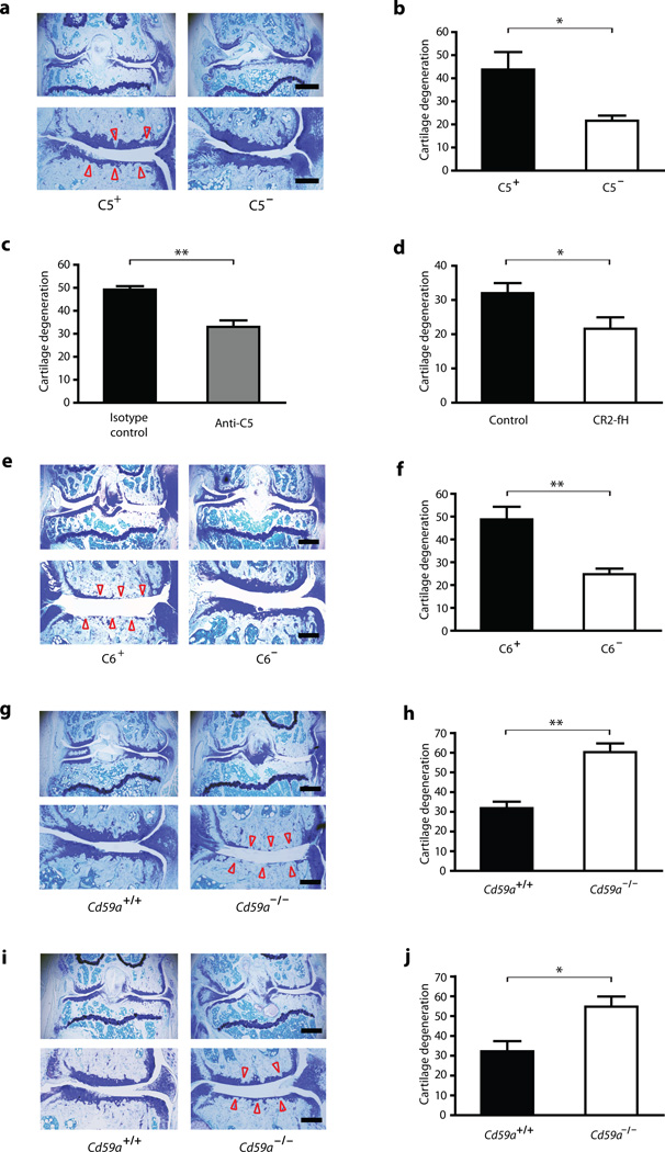
To investigate the role of complement in the pathogenesis of osteoarthritis, we used a mouse model of osteoarthritis induced by medial meniscectomy10. In humans, tearing of the meniscus often requires meniscectomy, which is a risk factor for knee osteoarthritis11. Because the C5 effector lies at the nexus of the complement cascade (Fig. 1a), we surgically induced osteoarthritis in C5-deficient (C5−)12 and C5-sufficient (C5+) mice. Sixteen weeks after surgery, C5− mice exhibited substantially less cartilage loss, osteophyte formation, and synovitis than did C5+ mice (Fig. 2a,b and Supplementary Fig. 2d). By contrast, osteoarthritis in this model was not affected by genetic deficiency in C3 (data not shown). That C3−/− mice were not protected against osteoarthritis can be explained by the observation that compensatory mechanisms operate in C3−/− mice: coagulation factors compensate for the lack of C3, allowing C5 activation to proceed even in the absence of C313. Corroborating our findings in the C5− congenic mouse strain, treatment with a neutralizing monoclonal antibody to C514 attenuated osteoarthritis in wild-type mice (Fig. 2c). We also tested the effect on osteoarthritis of CR2-fH—a fusion protein that inhibits activation of C3 and C515. Administration of CR2-fH attenuated the development of osteoarthritis in wild-type mice (Fig. 2d).
Figure 2.
The complement cascade, acting through its MAC effector arm, is crucial for the development of osteoarthritis in three different mouse models. (a,e,g,i) Toluidine-blue-stained sections of the medial region of mouse stifle joints. (a) Representative cartilage degeneration in C5+ and C5− mice subjected to medial meniscectomy. (b) Quantification of cartilage degeneration in (a) (n = 5 mice per group). (c) Quantification of cartilage degeneration in wild-type mice subjected to medial meniscectomy and then treated i.p. with 750 µg of either the C5-specific monoclonal antibody BB5.1 or an isotype-control antibody (n = 5 mice per group). (d) Quantification of cartilage degeneration in wild-type mice subjected to medial meniscectomy and then treated i.v. with 250 µg of CR2-fH or with PBS (n = 5 mice per group). (e) Representative cartilage degeneration in C6+ and C6− mice subjected to medial meniscectomy. (f) Quantification of cartilage degeneration in (e) (n = 13 mice per group). (g) Representative cartilage degeneration in Cd59a+/+ and Cd59a−/− mice subjected to medial meniscectomy. (h) Quantification of cartilage degeneration in (g) (n = 10 mice per group). (i) Representative cartilage degeneration in Cd59a+/+ and Cd59a−/− mice subjected to DMM. (j) Quantification of cartilage degeneration in (i) (n = 5 mice per group). Arrowheads indicate areas of cartilage degeneration. Scale bars: low-magnification (uppermost) images, 500 µm; higher-magnification (lower) images, 200 µm. Bar-chart data are the mean + s.e.m. *P < 0.05, **P < 0.01, by t test.
We next determined whether the MAC-mediated effector arm of the complement cascade is important in osteoarthritis. We found that mice deficient in C6, an integral component of the MAC (see Fig. 1a), were protected against the development of osteoarthritis and synovitis induced by medial meniscectomy (Fig. 2e,f and Supplementary Fig. 2d). Conversely, mice deficient in CD59a, an inhibitor of MAC6 (see Fig. 1a), developed more severe osteoarthritis and synovitis than their wild-type littermates (Fig. 2g,h and Supplementary Fig. 2a,d).
Not only meniscectomy but also meniscal tearing per se can lead to the development of osteoarthritis in humans11. We therefore also examined the role of complement in the destabilization of the medial meniscus (DMM) model of osteoarthritis16–18. We found that deficiency in CD59a accentuated the osteoarthritic phenotype in mice subjected to DMM (Fig. 2i,j and Supplementary Fig. 2b–d). Deficiency in CD59a also accentuated the milder osteoarthritis that developed spontaneously in aged mice (Supplementary Fig. 3). These findings suggest that MAC-mediated complement activity plays a pathogenic role in osteoarthritis of disparate etiologies.
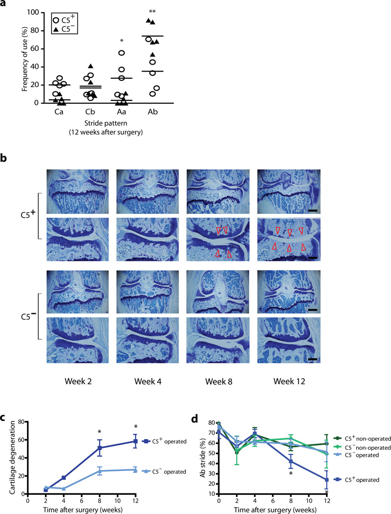
Resistance to the development of histological osteoarthritis in complement-deficient mice translated to functional benefit. Twelve weeks after medial meniscectomy, C5− mice maintained normal gait, whereas C5+ mice developed abnormal gait (Fig. 3a). Time-course studies revealed that neither C5+ nor C5− mice mice exhibited proteoglycan loss or cartilage degeneration two and four weeks after medial meniscectomy (Fig. 3b,c). This period of latency is similar to that observed in the DMM model16 and in humans who have undergone medial meniscectomy11. Eight and twelve weeks after surgery, however, C5+ mice exhibited significant proteoglycan and cartilage loss and synovitis, while C5− mice did not (Fig. 3b,c and Supplementary Fig. 4). The osteoarthritic phenotype was pronounced in these mice, most likely owing to their genetic background and age at the time of surgery, both factors that influence the severity of mouse osteoarthritis16.
Figure 3.
C5 deficiency protects against the progressive development of osteoarthritic joint pathology and gait dysfunction. (a) Gait analysis of C5+ and C5− mice 12 weeks after medial meniscectomy (n = 5 mice per group). C5− mice used the Ab stride pattern (the sequence of paw strides being right front—right hind—left front—left hind) with normal frequency, whereas C5+ mice used this pattern significantly less frequently. *P < 0.05, **P < 0.01 by t test. Results are representative of two independent experiments. (b) Histological analysis of articular cartilage at serial time points after medial meniscectomy. Representative toluidine-blue-stained sections of the medial region of stifle joints are presented; arrowheads show areas of cartilage degeneration. Scale bars: low-magnification (upper) images, 500 µm; high-magnification (lower) images, 200 µm. (c) Quantification of cartilage degeneration in C5+ and C5− mice subjected to medial meniscectomy (C5+ operated and C5− operated). *P ≤ 0.05 by t test comparing C5+ operated and C5− operated. Data are the mean ± s.e.m. (d) Gait analysis of C5+ operated and C5− operated mice, and of C5+ non-operated and C5− non-operated mice, at serial time points after medial meniscectomy. *P ≤ 0.05 by t test comparing C5+ operated mice and C5− operated mice. At week 8, n = 6 for C5+ operated, n = 6 for C5− operated; at week 12, n = 4 for C5+ operated, n = 4 for C5− operated; at all time points n ≥ 4 for C5+ non-operated and n = 3 for C5− non-operated.
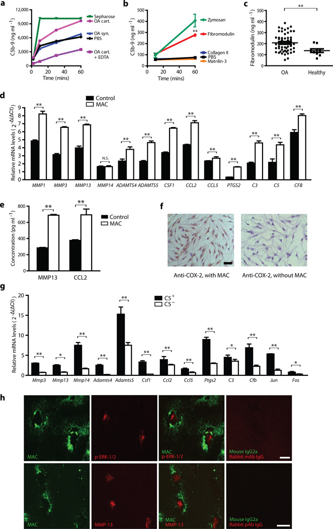
Products of dysregulated cartilage remodeling and repair may contribute to joint inflammation in osteoarthritis19–22. We examined the ability of osteoarthritic cartilage or specific components of the extracellular matrix (ECM) of cartilage to activate complement in vitro. Pulverized osteoarthritic cartilage induced the formation of C5b-9, as did the ECM components fibromodulin and aggrecan but not the ECM components type II collagen and matrilin-3 (Fig. 4a,b and Supplementary Fig. 5). Fibromodulin, which can bind C1q and activate the complement cascade23, was present at higher levels in osteoarthritic compared to healthy synovial fluid (Fig. 4c). Other cartilage ECM components, such as cartilage oligomeric matrix protein, are also detected at abnormally high levels in osteoarthritic synovial fluid and can activate complement19,20. Together, these results suggest that release or exposure cartilage ECM components may contribute to the pathophysiology of osteoarthritis by activating complement.
Figure 4.
Cartilage ECM components can induce MAC formation, and MAC induces chondrocyte expression of inflammatory and catabolic molecules. ELISA quantification of C5b-9 (soluble MAC) in (a) 67% human serum incubated with 20 µg ml−1 of pulverized human osteoarthritic cartilage (OA cart) or synovium (OA syn), or (b) 10% human serum incubated with 20 µg ml−1 of recombinant cartilage ECM components. Sepharose and zymosan are positive controls; PBS and EDTA negative controls. **P ≤ 0.01 by one-way ANOVA with Dunnett’s post-hoc test comparing each cartilage component with PBS. Data are the mean of triplicate values ± s.d. and representative of three independent experiments. (c) ELISA quantification of fibromodulin in synovial fluids from individuals with osteoarthritis (n = 50) and healthy individuals (n = 9). **P ≤ 0.01 by t test. (d–f) qPCR analysis of relative mRNA expression (d), ELISA analysis of protein expression (e), and immunocytochemical analysis of COX-2 expression (f) in human chondrocytes incubated with or without MAC for 72 hours. Scale bar, 50 µm. *P ≤ 0.05; **P ≤ 0.01 by t test. Data are the mean ± s.d. and representative of three independent experiments. (g) mRNA expression in chondrocytes from C5+ and C5− mice (n = 4 mice per group) subjected to DMM. Data are the mean + s.e.m. of triplicates and representative of results from 4 mice from 2 independent experiments. *P ≤ 0.05; **P ≤ 0.01 by fixed-effect ANOVA taking into account both destabilized and non-destabilized joints. (h) Immunofluorescent analysis of p-ERK1/2, MMP-13, and MAC co-localization in human osteoarthritic cartilage. Scale bar, 10 µm.
Our in situ (Fig. 1d) and in vivo (Fig. 2e–j and Supplementary Figs. 2,3) findings indicate that MAC is important in mediating complement-related cartilage damage in osteoarthritis. But how might MAC damage cartilage? Extensive deposition of MAC induces cell lysis and necrotic cell death, whereas sublytic MAC can activate signaling pathways that drive the expression of proinflammatory and catabolic molecules24. Because many of the MAC-encircled chondrocytes in osteoarthritic cartilage appear morphologically intact (Figs. 1d, 4h and Supplementary Fig. 6), we examined whether MAC induces the expression of proinflammatory and degradative enzymes in osteoarthritis. We first examined the expression of genes encoding such molecules in cultured chondrocytes coated with sublytic levels of MAC. Sublytic MAC increased the chondrocytes’ expression of multiple genes implicated in osteoarthritis: those encoding cartilage-degrading enzymes17,18,22 (MMPs, ADAMTSs); inflammatory cytokines25 (CCL2, CSF1, and CCL5); and cyclo-oxygenase 226 (Fig. 4d–f). Sublytic MAC also induced the expression of complement effectors (Fig. 4d); chondrocyte production of complement may thus synergize with complement derived from synovial membrane to amplify pathogenic complement signaling in osteoarthritis.
We next examined the effect of complement deficiency on the in vivo expression of these genes in destabilized mouse joints. Twenty weeks after DMM surgery, chondrocytes from the destabilized joints of C5-deficient mice, which are MAC deficient and protected against osteoarthritis (Figs. 2a,b and Fig. 3), expressed lower levels of these inflammatory and degradative mediators than did chondrocytes from the destabilized joints of C5-sufficient mice (Fig. 4g). mRNA levels of Jun and Fos, proinflammatory transcription factors whose expression is induced by MAC27,28, were also lower (Fig. 4g). Moreover, in human osteoarthritic cartilage, MAC co-localized with MMP-13 and with activated ERK-1/2 (Fig. 4h and Supplementary Fig. 6), a kinase that mediates resistance to MAC-mediated cell lysis29 and stimulates the expression of MMP-13 by inducing the expression of Fos30.
Here we show that the complement cascade is crucial to the pathogenesis of osteoarthritis. Cartilage ECM components released by or exposed in osteoarthritic cartilage may trigger the complement cascade. Additionally, dysregulation of gene expression in joint tissues may contribute to a local preponderance of complement effectors over inhibitors in osteoarthritis, permitting complement activation to proceed unchecked. Complement activation in turn results in the formation of MAC on chondrocytes, which either kills the cells or causes them to produce matrix-degrading enzymes, inflammatory mediators, and further complement effectors—all of which promote joint pathology.
Recent findings suggest that low-grade complement activation contributes to the development of other degenerative diseases, such as age-related macular degeneration31 and Alzheimer’s disease32. We propose that osteoarthritis can be added to this list of diseases. Our findings provide rationale for targeting the complement system as a disease-modifying therapy for osteoarthritis.
Supplementary Material
ACKNOWLEDGEMENTS
These studies were supported by a Rehabilitation Research and Development Merit Award from the Department of Veterans Affairs and US National Heart, Lung, and Blood Institute Proteomics Center contract N01 HV 28183 (W.H.R.); Northern California Chapter Arthritis Foundation Postdoctoral Fellowship Award (A.L.R.); New York Chapter Arthritis Foundation/Merck Osteoarthritis Research Fellowship Award, the Atlantic Philanthropies, American College of Rheumatology Research and Education Fund, and the Association of Specialty Professors (C.R.S.); and the Frankenthaler and Kohlberg Foundations (M.K.C).
Footnotes
Accession codes. Experimentally determined microarray data is available at GEO (accession code GSE32317).
AUTHOR CONTRIBUTIONS
A.L.R. and W.H.R. initiated the investigation of complement in osteoarthritis, and Q.W. conducted key studies. Q.W., A.L.R., D.M. Larsen, H.H.W., and W.H.R. conducted the studies of osteoarthritis in mouse models. A.E., A.L.R., and M.S. performed the gait analysis studies. Q.W. and H.H.W. performed the in vitro MAC deposition experiments. C.M.L. and J.J.S. performed the immunohistochemical analyses of cartilage. J.F.C., G.B., S.Y.R., L.P., S.R.G., R.G., and D.M. Lee conducted or contributed to the proteomic analysis of osteoarthritic synovial fluid. S.Y.R. and D.M. Lee performed the ELISA analysis of osteoarthritic and healthy synovial fluids. C.R.S. and M.K.C. performed the gene-expression analysis of synovium, and G.B., R.G., and D.M. Lee contributed to the analysis of these datasets. A.L.R., C.M.L., J.J.S., and I.H. performed the in vitro complement activation and stimulation assays using samples provided by S.B.G. V.M.H., J.M.T. and N.B. provided the C5-specific antibody and the CR2-fH fusion protein; T.W-C the C6− and Cd59a−/− mice. V.M.H., T.M.L., and D.M. Lee provided scientific input. T.M.L., A.L.R., and W.H.R. wrote the manuscript, and Q.W., C.R.S, T.W-C., S.R.G., M.K.C., V.M.H., and D.M. Lee edited the manuscript. All authors analyzed the data and approved the final manuscript.
COMPETING FINANCIAL INTERESTS
D.M. Lee is currently employed by Novartis Pharma, AG. D.M. Lee and R.G. own equity in Synostics, Inc. J.M.T. and M.V.H. are consultants for Alexion Pharmaceuticals, Inc.
REFERENCES
- 1.Felson DT. Clinical practice. Osteoarthritis of the knee. N Engl J Med. 2006;354:841–848. doi: 10.1056/NEJMcp051726. [DOI] [PubMed] [Google Scholar]
- 2.Goldring MB, Goldring SR. Osteoarthritis. J Cell Physiol. 2007;213:626–634. doi: 10.1002/jcp.21258. [DOI] [PubMed] [Google Scholar]
- 3.Pelletier JP, Martel-Pelletier J, Abramson SB. Osteoarthritis, an inflammatory disease: potential implication for the selection of new therapeutic targets. Arthritis Rheum. 2001;44:1237–1247. doi: 10.1002/1529-0131(200106)44:6<1237::AID-ART214>3.0.CO;2-F. [DOI] [PubMed] [Google Scholar]
- 4.Hill CL, et al. Synovitis detected on magnetic resonance imaging and its relation to pain and cartilage loss in knee osteoarthritis. Ann Rheum Dis. 2007;66:1599–1603. doi: 10.1136/ard.2006.067470. [DOI] [PMC free article] [PubMed] [Google Scholar]
- 5.Gobezie R, et al. High abundance synovial fluid proteome: distinct profiles in health and osteoarthritis. Arthritis Res Ther. 2007;9:R36. doi: 10.1186/ar2172. [DOI] [PMC free article] [PubMed] [Google Scholar]
- 6.Kemper C, Atkinson JP. T-cell regulation: with complements from innate immunity. Nature Reviews Immunology. 2007;7:9–18. doi: 10.1038/nri1994. [DOI] [PubMed] [Google Scholar]
- 7.Cooke TD, Bennett EL, Ohno O. The deposition of immunoglobulins and complement in osteoarthritic cartilage. Int Orthop. 1980;4:211–217. doi: 10.1007/BF00268158. [DOI] [PubMed] [Google Scholar]
- 8.Corvetta A, et al. Terminal complement complex in synovial tissue from patients affected by rheumatoid arthritis, osteoarthritis and acute joint trauma. Clin Exp Rheumatol. 1992;10:433–438. [PubMed] [Google Scholar]
- 9.Cooke TD. Significance of immune complex deposits in osteoarthritic cartilage. J Rheumatol. 1987;14:77–79. Spec No. [PubMed] [Google Scholar]
- 10.Kamekura S, et al. Osteoarthritis development in novel experimental mouse models induced by knee joint instability. Osteoarthritis Cartilage. 2005;13:632–641. doi: 10.1016/j.joca.2005.03.004. [DOI] [PubMed] [Google Scholar]
- 11.Englund M, Lohmander LS. Risk factors for symptomatic knee osteoarthritis fifteen to twenty-two years after meniscectomy. Arthritis Rheum. 2004;50:2811–2819. doi: 10.1002/art.20489. [DOI] [PubMed] [Google Scholar]
- 12.Gervais F, Stevenson M, Skamene E. Genetic control of resistance to Listeria monocytogenes: regulation of leukocyte inflammatory responses by the Hc locus. J Immunol. 1984;132:2078–2083. [PubMed] [Google Scholar]
- 13.Huber-Lang M, et al. Generation of C5a in the absence of C3: a new complement activation pathway. Nat Med. 2006;12:682–687. doi: 10.1038/nm1419. [DOI] [PubMed] [Google Scholar]
- 14.Banda NK, et al. Mechanisms of effects of complement inhibition in murine collagen-induced arthritis. Arthritis Rheum. 2002;46:3065–3075. doi: 10.1002/art.10591. [DOI] [PubMed] [Google Scholar]
- 15.Banda NK, et al. Targeted inhibition of the complement alternative pathway with complement receptor 2 and factor H attenuates collagen antibody-induced arthritis in mice. J Immunol. 2009;183:5928–5937. doi: 10.4049/jimmunol.0901826. [DOI] [PMC free article] [PubMed] [Google Scholar]
- 16.Glasson SS. In vivo osteoarthritis target validation utilizing genetically-modified mice. Curr Drug Targets. 2007;8:367–376. doi: 10.2174/138945007779940061. [DOI] [PubMed] [Google Scholar]
- 17.Glasson SS, et al. Deletion of active ADAMTS5 prevents cartilage degradation in a murine model of osteoarthritis. Nature. 2005;434:644–648. doi: 10.1038/nature03369. [DOI] [PubMed] [Google Scholar]
- 18.Stanton H, et al. ADAMTS5 is the major aggrecanase in mouse cartilage in vivo and in vitro. Nature. 2005;434:648–652. doi: 10.1038/nature03417. [DOI] [PubMed] [Google Scholar]
- 19.Happonen KE, et al. Regulation of complement by cartilage oligomeric matrix protein allows for a novel molecular diagnostic principle in rheumatoid arthritis. Arthritis Rheum. 2010;62:3574–3583. doi: 10.1002/art.27720. [DOI] [PMC free article] [PubMed] [Google Scholar]
- 20.Neidhart M, et al. Small fragments of cartilage oligomeric matrix protein in synovial fluid and serum as markers for cartilage degradation. Br J Rheumatol. 1997;36:1151–1160. doi: 10.1093/rheumatology/36.11.1151. [DOI] [PubMed] [Google Scholar]
- 21.Scanzello CR, Plaas A, Crow MK. Innate immune system activation in osteoarthritis: is osteoarthritis a chronic wound? Curr Opin Rheumatol. 2008;20:565–572. doi: 10.1097/BOR.0b013e32830aba34. [DOI] [PubMed] [Google Scholar]
- 22.Sofat N. Analysing the role of endogenous matrix molecules in the development of osteoarthritis. Int J Exp Pathol. 2009;90:463–479. doi: 10.1111/j.1365-2613.2009.00676.x. [DOI] [PMC free article] [PubMed] [Google Scholar]
- 23.Sjoberg A, Onnerfjord P, Morgelin M, Heinegard D, Blom AM. The extracellular matrix and inflammation: fibromodulin activates the classical pathway of complement by directly binding C1q. J Biol Chem. 2005;280:32301–32308. doi: 10.1074/jbc.M504828200. [DOI] [PubMed] [Google Scholar]
- 24.Bohana-Kashtan O, Ziporen L, Donin N, Kraus S, Fishelson Z. Cell signals transduced by complement. Mol Immunol. 2004;41:583–597. doi: 10.1016/j.molimm.2004.04.007. [DOI] [PubMed] [Google Scholar]
- 25.Scanzello CR, et al. Synovial inflammation in patients undergoing arthroscopic meniscectomy: molecular characterization and relationship to symptoms. Arthritis Rheum. 2011;63:391–400. doi: 10.1002/art.30137. [DOI] [PMC free article] [PubMed] [Google Scholar]
- 26.Attur M, et al. Prostaglandin E2 exerts catabolic effects in osteoarthritis cartilage: evidence for signaling via the EP4 receptor. J Immunol. 2008;181:5082–5088. doi: 10.4049/jimmunol.181.7.5082. [DOI] [PubMed] [Google Scholar]
- 27.Badea TD, et al. Sublytic terminal complement attack induces c-fos transcriptional activation in myotubes. J Neuroimmunol. 2003;142:58–66. doi: 10.1016/s0165-5728(03)00261-3. [DOI] [PubMed] [Google Scholar]
- 28.Rus HG, Niculescu F, Shin ML. Sublytic complement attack induces cell cycle in oligodendrocytes. J Immunol. 1996;156:4892–4900. [PubMed] [Google Scholar]
- 29.Kraus S, Seger R, Fishelson Z. Involvement of the ERK mitogen-activated protein kinase in cell resistance to complement-mediated lysis. Clin Exp Immunol. 2001;123:366–374. doi: 10.1046/j.1365-2249.2001.01477.x. [DOI] [PMC free article] [PubMed] [Google Scholar]
- 30.Litherland GJ, et al. Protein kinase C isoforms zeta and iota mediate collagenase expression and cartilage destruction via STAT3- and ERK-dependent c-fos induction. J Biol Chem. 2010;285:22414–22425. doi: 10.1074/jbc.M110.120121. [DOI] [PMC free article] [PubMed] [Google Scholar]
- 31.Daiger SP. Genetics. Was the Human Genome Project worth the effort? Science. 2005;308:362–364. doi: 10.1126/science.1111655. [DOI] [PMC free article] [PubMed] [Google Scholar]
- 32.Rogers J, et al. Complement activation by β-amyloid in Alzheimer disease. Proc. Natl. Acad. Sci. USA. 1992;89:10016–10020. doi: 10.1073/pnas.89.21.10016. [DOI] [PMC free article] [PubMed] [Google Scholar]
- 33.Huber R, et al. Identification of intra-group, inter-individual, and gene-specific variances in mRNA expression profiles in the rheumatoid arthritis synovial membrane. Arthritis Res Ther. 2008;10:R98. doi: 10.1186/ar2485. [DOI] [PMC free article] [PubMed] [Google Scholar]
- 34.Irizarry RA, et al. Summaries of Affymetrix GeneChip probe level data. Nucleic Acids Res. 2003;31:e15. doi: 10.1093/nar/gng015. [DOI] [PMC free article] [PubMed] [Google Scholar]
- 35.Lin AC, et al. Modulating hedgehog signaling can attenuate the severity of osteoarthritis. Nat Med. 2009;15:1421–1425. doi: 10.1038/nm.2055. [DOI] [PubMed] [Google Scholar]
- 36.Zhou W, et al. Predominant role for C5b-9 in renal ischemia/reperfusion injury. J Clin Invest. 2000;105:1363–1371. doi: 10.1172/JCI8621. [DOI] [PMC free article] [PubMed] [Google Scholar]
- 37.Holt DS, et al. Targeted deletion of the CD59 gene causes spontaneous intravascular hemolysis and hemoglobinuria. Blood. 2001;98:442–449. doi: 10.1182/blood.v98.2.442. [DOI] [PubMed] [Google Scholar]
- 38.Frei Y, Lambris JD, Stockinger B. Generation of a monoclonal antibody to mouse C5 application in an ELISA assay for detection of anti-C5 antibodies. Mol Cell Probes. 1987;1:141–149. doi: 10.1016/0890-8508(87)90022-3. [DOI] [PubMed] [Google Scholar]
- 39.Gabriel AF, Marcus MA, Honig WM, Walenkamp GH, Joosten EA. The CatWalk method: a detailed analysis of behavioral changes after acute inflammatory pain in the rat. J Neurosci Methods. 2007;163:9–16. doi: 10.1016/j.jneumeth.2007.02.003. [DOI] [PubMed] [Google Scholar]
- 40.Chen Y, et al. Terminal complement complex C5b-9-treated human monocyte-derived dendritic cells undergo maturation and induce Th1 polarization. Eur J Immunol. 2007;37:167–176. doi: 10.1002/eji.200636285. [DOI] [PubMed] [Google Scholar]
Associated Data
This section collects any data citations, data availability statements, or supplementary materials included in this article.


