Abstract
Many studies have reported spontaneous spinal epidural hematoma (SSEH). Although most cases are idiopathic, several are associated with thrombolytic therapy or anticoagulants. We report a case of SSEH coincident with acute myocardial infarction (AMI), which caused serious neurological deficits. A 56 year old man presented with chest pain accompanied with back and neck pain, which was regarded as an atypical symptom of AMI. He was treated with nitroglycerin, aspirin, low molecular weight heparin, and clopidogrel. A spinal magnetic resonance image taken after paraplegia developed 3 days after the initial symptoms revealed an epidural hematoma at the cervical and thoracolumbar spine. Despite emergent decompressive surgery, paraplegia has not improved 7 months after surgery. A SSEH should be considered when patients complain of abrupt, strong, and non-traumatic back and neck pain, particularly if they have no spinal pain history.
Keywords: Acute myocardial infarction; Hematoma, epidural, spinal; Paraplegia; Thrombolytic therapy; Anticoagulants
Introduction
A spontaneous spinal epidural hematoma (SSEH) is rare; however, it causes severe neurological deficits unless treated in a timely manner. In extremely rare cases, the use of thrombolytic therapy or anticoagulants in patients with acute myocardial infarction (AMI) cause SSEHs.1),2) We report a patient with an SSEH and an AMI, who received anticoagulants and developed a spinal cord injury.
Case
A 56 year old man, who complained of severe neck and back pain beginning as chest pain, visited the emergency room (ER). He had no medical history of pain, particularly originating from the spine. The chest pain occurred after an argument with another individual. He visited the ER immediately after the chest pain occurred. The laboratory findings showed elevated creatine kinase (CK), the muscle brain isoenzyme of CK, and troponin-I to 276 IU/L, 13.15 ng/mL, and 9.746 ng/mL, respectively. He was diagnosed with a non-ST-segment elevation myocardial infarction. He was treated with nitroglycerin, 250 mg aspirin following 200 mg per day, 60 mg enoxaparin every 12 hours, and 300 mg clopidogrel following 75 mg per day in the intensive care unit. Despite that the chest pain and laboratory findings improved, his back and neck pain progressed slowly with a visual analogue pain scale value of 8 or 9. He did not respond to pain killers, including opioids, and then abruptly developed paraplegia 3 days after the initial chest pain onset. He consulted the department of neurosurgery without a coronary angiographic evaluation.
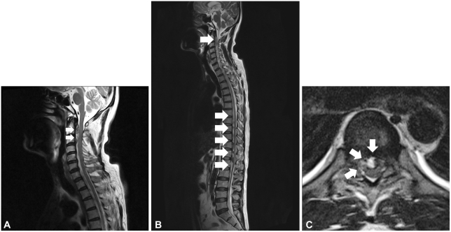
Spinal magnetic resonance images (MRI) obtained after the paraplegia showed a T1 low and T2 mixed heterogeneous signal intensity mass at the ventral epidural space from C2 to C4 and from T7 to L1, respectively. The mass compressed the cornus medullaris at the T10/L1 level and the spinal cord at the level of C2/3. Some high signals from the spinal cord were observed on T2 weighted images, indicating a spinal cord injury. Some of the T1 weighted images showed abnormally high signal intensity, indicating a multistage hemorrhage (Fig. 1). No abnormal coagulation findings were observed, including prothrombin time, activated partial thrombin time, bleeding time, and platelet count. He underwent an emergent operation to decompress the spinal cord.
Fig. 1.
T2 weighted sagittal cervical magnetic resonance images. A: lentiform heterogeneous mass (arrows) located in the ventral epidural space from C2 to 3 compressed the cervical cord. B: T2 weighted spinal sagittal MRI. Heterogeneous mass (arrows) from T7 to L1 compressed the spinal cord. Abnormally high signal intensity of the spinal cord at the thoracolumbar junction indicated a spinal cord injury. C: T2 weighted axial section at the thoracolumbar junction revealed a spinal hematoma (arrows) in front of the spinal cord. Some high-signal intensity hematomas indicate multistage hemorrhages.
A right-side hemilaminectomy was performed from T7 to L1 under general anesthesia in the prone position. A hematoma was located at the ventral epidural space and had compressed the dura. The hematoma was hard, with no spinal cord pulsation. No active bleeding and no abnormal vascular structure were observed. Vital signs were stable during surgery.
The patient recovered from general anesthesia with no complications, but his neurological symptoms did not change. He started 75 mg clopidogrel per day and 12.5 mg carvedlol per day 14 days after surgery A coronary angiographic evaluation was not performed, as he declined the treatment.
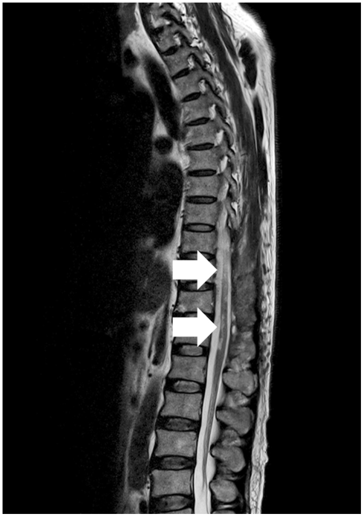
A follow-up MRI 2 months after surgery revealed several abnormal T2 weighted high-signal intensities and spinal cord atrophy, although no residual hematoma was found at the cervical and thoracolumbar areas (Fig. 2). No neurological recovery had occurred 7 months after the initial deficit.
Fig. 2.

T2 weighted whole-spine sagittal magnetic resonance image obtained 2 months after surgery. No residual hematoma was evident. However, multiple abnormal high signal intensities (arrows) indicated spinal cord atrophy after the cord injury.
Discussion
The clinical presentation of acute coronary syndrome (ACS) is generally chest or left arm pain, and a transient murmur, hypotension, or diaphoresis occasionally indicate ACS. ACS is associated with a severely impaired prognosis and requires prompt and efficient treatment. Chest pain may radiate to the arms, jaws, and back.3-5) In cases of atypical chest pain, dizziness, confusion, dyspnea, fatigue, perspiration, shortness of breath, weakness, atypical abdominal pain, or elbow pain may lead doctors to believe that the problem is not cardiac related, although acute chest pain is absent in 43% of ACS cases.6-9) In one report, solitary back pain without any significant clinical manifestation was regarded as an atypical symptom of AMI.10) Although a detailed pain history is essential, there is little possibility that axial back pain is associated with ACS.
A SSEH is relatively rare (0.1 patients/100,000) and includes <1% of spinal epidural-space occupied lesions.11) Some controversies exist about the origin of SSEH. Most researchers assert that it originates from the epidural venous system; however, an arterial source has been more persuasively proposed when considering the relatively acute progression of SSEH. Usually SSEH presents with sudden, severe neck or back pain that tends to progress, and neurological manifestations usually occur after the back pain.11) SSEH is related to organic vascular disease, hemodialysis, coagulation disorders, and anticoagulant therapy for stroke.12-16) In rare cases, thrombolytic therapy for coronary ischemia follows SSEH.1),17) Immediate surgical decompression of the neural structures is the treatment of choice before the neurological deficit progresses. Conservative management has been considered in cases without neurological deficit.11) Preoperative neurological state is correlated with postoperative outcome. The time interval from diagnosis to surgery and the progress of the clinical manifestations also have relationships with postoperative outcome; a shorter progressive interval usually predicts a poorer recovery. Progressive intervals <12 hours are correlated with a poor prognosis.11) Mortality is correlated with a high frequency of cervical or cervicothoracic hematomas. Patients with cardiovascular disease and those undergoing anticoagulant therapy also have a poor prognosis.18)
Patients with heart disease or circulation disorders including those of the cerebrovascular system are increasing in frequency. Although it is unknown how many patients take anticoagulants, antihypertensives, and undergo thrombolytic therapy, there are increased number of these patients compared to the past. SSEH associated with anticoagulants or thrombolytic therapy in patients with ACS has also possibly increased. In the present case, the neck and back pain were thought to be atypical symptoms of ACS initially, because the patient presented with typical chest pain and elevated cardiac enzymes.
Anticoagulant use may be related with SSEH progression. Although our patient had poor prognostic factors, such as cardiovascular disease, undergoing anticoagulant therapy, and required intensive care, a detailed history including the nature of the pain and rapid imaging tests may have provided a better prognosis. Newly developing or sustained back pain during ACS treatment should not be neglected and should not be regarded as an atypical symptom of ACS. We believe that an aggressive imaging study including a spinal MRI should be performed for patients with AMI who complain of persistent neck or back pain while undergoing thrombolytic or anticoagulant therapy.
We report a case of paraplegia due to SSEH, which was thought to be aggravated while the patient was undergoing anticoagulant therapy for ACS. SSEH is a rare disease; however, it can result in severe mortality or morbidity unless treated in a timely manner. The present case suggests that a cautious evaluation should be considered in a patient who complains of persistent neck and back pain and is undergoing anticoagulation therapy for AMI, which could be regarded as atypical symptoms of ACS.
Footnotes
The authors have no financial conflicts of interest.
References
- 1.Chan KC, Wu DJ, Ueng KC, et al. Spinal epidural hematoma following tissue plasminogen activator and heparinization for acute myocardial infarction. Jpn Heart J. 2002;43:417–421. doi: 10.1536/jhj.43.417. [DOI] [PubMed] [Google Scholar]
- 2.Subbiah M, Avadhani A, Shetty AP, Rajasekaran S. Acute spontaneous cervical epidural hematoma with neurological deficit after low-molecular-weight heparin therapy: role of conservative management. Spine J. 2010;10:e11–e15. doi: 10.1016/j.spinee.2010.04.011. [DOI] [PubMed] [Google Scholar]
- 3.Akhtar MM, Iqbal YH, Kaski JC. Managing unstable angina and non-ST elevation MI. Practitioner. 2010;254:25–30. [PubMed] [Google Scholar]
- 4.Kumar A, Cannon CP. Acute coronary syndromes: diagnosis and management, part I. Mayo Clin Proc. 2009;84:917–938. doi: 10.4065/84.10.917. [DOI] [PMC free article] [PubMed] [Google Scholar]
- 5.Riegel B, Hanlon AL, McKinley S, et al. Differences in mortality in acute coronary syndrome symptom clusters. Am Heart J. 2010;159:392–398. doi: 10.1016/j.ahj.2010.01.003. [DOI] [PMC free article] [PubMed] [Google Scholar]
- 6.Lee HO. Typical and atypical clinical signs and symptoms of myocardial infarction and delayed seeking of professional care among blacks. Am J Crit Care. 1997;6:7–13. [PubMed] [Google Scholar]
- 7.McSweeney JC, Cody M, O'Sullivan P, Elberson K, Moser DK, Garvin BJ. Women's early warning symptoms of acute myocardial infarction. Circulation. 2003;108:2619–2623. doi: 10.1161/01.CIR.0000097116.29625.7C. [DOI] [PubMed] [Google Scholar]
- 8.Lee H, Bahler R, Park OJ, Kim CJ, Lee HY, Kim YJ. Typical and atypical symptoms of myocardial infarction among African-Americans, whites, and Koreans. Crit Care Nurs Clin North Am. 2001;13:531–539. [PubMed] [Google Scholar]
- 9.Løvlien M, Schei B, Hole T. Women with myocardial infarction are less likely than men to experience chest symptoms. Scand Cardiovasc J. 2006;40:342–347. doi: 10.1080/14017430600913199. [DOI] [PubMed] [Google Scholar]
- 10.Atypical presentation of myocardial infarction in the outpatient setting, a lapse in clinical knowledge and inadequate data transfer constitute a potentially fatal error. Int J Qual Health Care. 2003;15:179. doi: 10.1093/intqhc/mzg027. doi: 10.1093/intqhc/mzg027. [DOI] [PubMed] [Google Scholar]
- 11.Liu Z, Jiao Q, Xu J, Wang X, Li S, You C. Spontaneous spinal epidural hematoma: analysis of 23 cases. Surg Neurol. 2008;69:253–260. doi: 10.1016/j.surneu.2007.02.019. [DOI] [PubMed] [Google Scholar]
- 12.Kitagawa RS, Mawad ME, Whitehead WE, Curry DJ, Luersen TG, Jea A. Paraspinal arteriovenous malformations in children. J Neurosurg Pediatr. 2009;3:425–428. doi: 10.3171/2009.2.PEDS08427. [DOI] [PubMed] [Google Scholar]
- 13.Sung JH, Hong JT, Son BC, Lee SW. Clopidogrel-induced spontaneous spinal epidural hematoma. J Korean Med Sci. 2007;22:577–579. doi: 10.3346/jkms.2007.22.3.577. [DOI] [PMC free article] [PubMed] [Google Scholar]
- 14.Dimou J, Jithoo R, Morokoff A. Spontaneous spinal epidural haematoma in a geriatric patient on aspirin. J Clin Neurosci. 2010;17:142–144. doi: 10.1016/j.jocn.2009.03.021. [DOI] [PubMed] [Google Scholar]
- 15.Deger SM, Emmez H, Bahadirli K, et al. A spontaneous spinal epidural hematoma in a hemodialysis patient: a rare entity. Intern Med. 2009;48:2115–2118. doi: 10.2169/internalmedicine.48.2335. [DOI] [PubMed] [Google Scholar]
- 16.Tagaya A, Miyajima Y, Sakamoto M, et al. Spontaneous spinal epidural hematoma in an infant with undiagnosed hemophilia A. Pediatr Int. 2010;52:296–298. doi: 10.1111/j.1442-200X.2010.03033.x. [DOI] [PubMed] [Google Scholar]
- 17.Cultrera F, Passanisi M, Giliberto O, Giuffrida M, Mancuso P, Ventura F. Spinal epidural hematoma following coronary thrombolysis: a case report. J Neurosurg Sci. 2004;48:43–47. [PubMed] [Google Scholar]
- 18.Groen RJ, van Alphen., HA Operative treatment of spontaneous spinal epidural hematomas: a study of the factors determining postoperative outcome. Neurosurgery. 1996;39:494–508. doi: 10.1097/00006123-199609000-00012. discussion 508-9. [DOI] [PubMed] [Google Scholar]


