Abstract
The synthesis and initial biological evaluation of 3-carboranyl thymidine analogues (3CTAs) that are (radio)halogenated at the closo-carborane cluster is described. Radiohalogenated 3CTAs have the potential to be used in the radiotherapy and imaging of cancer, as they may be selectively entrapped in tumor cells through monophosphorylation by human thymidine kinase 1 (hTK1). Two strategies for the synthesis of a 127I-labeled form of a specific 3CTA, previously designated as N5, are described: 1) direct iodination of N5 with iodine monochloride (ICl) and aluminum chloride (AlCl3) to obtain N5-127I and 2) initial monoiodination of o-carborane to 9-iodo-o-carborane followed by its functionalization to N5-127I. The former strategy produced N5-127I in low yields along with di-, tri-, and tetra-iodinated N5 as well as decomposition products, whereas the latter method produced only N5-127I in high yields. N5-127I was subjected to nucleophilic halogen- and isotope exchange reactions using Na79/81Br and Na125I, respectively, in the presence of Herrmann's catalyst to obtain N5-79/81Br and N5-125I, respectively. Two intermediate products formed using the second strategy, 1-(tert-butyldimethylsilyl)-9-iodo-o-carborane and 1-(tert-butyldimethylsilyl)-12-iodo-o-carborane, were subjected to x-ray diffraction studies to confirm that substitution at a single carbon atom of 9-iodo-o-carborane resulted in the formation of two structural isomers. To the best of our knowledge, this is the first report of halogen and isotope exchange reactions of B-halocarboranes that have been conjugated to a complex biomolecule. Human TK1 phosphorylation rates of N5, N5-127I, and N5-79/81Br ranged from 38.0 % to 29.6% relative to that of thymidine, the endogenous hTK1 substrate. The in vitro uptake of N5, N5-127I, and N5-79/81Br in L929 TK1 (+) cells was 2.0 ×, 1.8 ×, and 1.4 × greater than that in L929 TK1 (−) cells.
Keywords: 3-Carboranyl thymidine analogues (3CTAs), (radio)halogenated carboranes, (radio)halogenated 3CTAs, thymidine kinase, nucleophilic isotope exchange, selective monoiodination at the carborane cage, tetraiodination at the carborane cage, phosphoryl transfer assay, cancer therapy and imaging
INTRODUCTION
Radiopharmaceuticals containing carbon-bound radiohalogens play an important role in a variety of therapy and imaging applications.2 There is, however, a convincing body of evidence indicating that most radiopharmaceutical biomolecules (e.g. antibodies, carbohydrates, growth factors) that contain radiolabeled boron clusters are less susceptible to dehalogenation under physiological conditions than their counterparts containing conventional radiohalogen-carbon bonds.3 The vast majority of these radiopharmaceuticals possess negatively charged boron clusters, such as nido-carborane(1-) (C2B9H12−), closo-monocarborane(1-) (C1B11H12−), decaborate(2) (B10H102−) or dodecaborate (B12H122−).3 By analogy to electrophilic radiohalogenation of tyrosine residues in proteins,2 labeling of these negatively charged boron clusters mainly was carried out with electrophilic halogen species produced in situ by the action of Chloramine T or Iodogen.3 Nucleophilic labeling is another widely used strategy for the introduction of carbon-bound radiohalogens in biomolecules.2,4,5 Methods employing iron-, copper-, and palladium-catalyzed nucleophilic halogen exchange at simple boron cluster structures were developed6–11 but they have not as yet been applied to the radiohalogenation of more complex biologically active carborane-containing compounds. Nucleophilic halogen exchange can be carried out at hydrophobic neutral boron clusters, and thus could be a useful alternative in the radiohalogenation of low molecular weight boron cluster containing bioconjugates that may rely on hydrophobic properties for biological activity.12
Thymidine (dThd) analogues with a hydrophobic boron cluster (closo-carborane) attached through spacers to the N3 position (3-carboranyl thymidine analogues or 3CTAs) may be good examples for such compounds. Both boronated and non-boronated N3-substituted dThd analogues have attracted considerable attention in a variety of biomedical applications in recent years.13–21 3CTAs have been evaluated as boron delivery agents for Boron Neutron Capture Therapy (BNCT) of cancer.19,22 One of the lead 3CTAs that was identified is N5 (3, Figure 1, Scheme 1).14,23 This agent appears to selectively accumulate within cancer cells following monophosphorylation by human thymidine kinase 1 (hTK1), which is overexpressed in cancer cells. This form of selective entrapment of nucleoside analogues in tumor cells has been referred to as “Kinase Mediated Trapping” (KMT).13 Furthermore, 3CTAs did not appear to be substrates of nucleoside catabolizing enzymes such as 5′-nucleotidases (5′-NTs) and nucleoside phosphorylases, and preliminary data indicated that these compounds might pass cell membranes by passive diffusion.12,14,23 It is conceivable that cage-monoradioiodinated forms of 3 (e.g. N5-125I, [13a or 13b, Scheme 3]) could be used in the radiotherapy and imaging of cancer, as well as for BNCT related biodistribution/pharmacokinetic experiments. They may be superior to conventional radioiodinated nucleosides, such 5-[125I]iodo-2′-deoxyuridine.24–27 Under physiological conditions, the latter is cleaved within a few minutes by nucleoside phosphorylases to 5-[125I]iodouracil. This is followed by rapid dehalogenation, which can lead to the accumulation of significant quantities of radioiodine in the thyroid and the stomach.24–27
Figure 1.
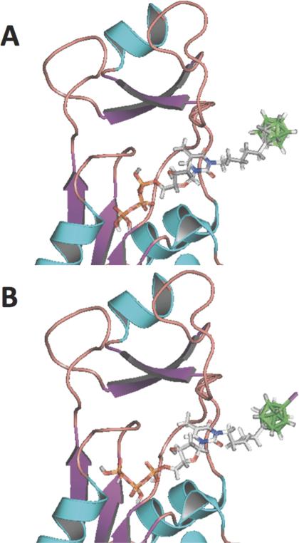
Docking poses of the 5'-triphosphate forms of 3 (A) and 4b (B) in a homology model of hTK1 (PDB ID # 1W4R) that was developed from the crystal structure of Thermotoga maritima Thymidine Kinase (TmTK, PDB ID # 2QPO).
Scheme 1.
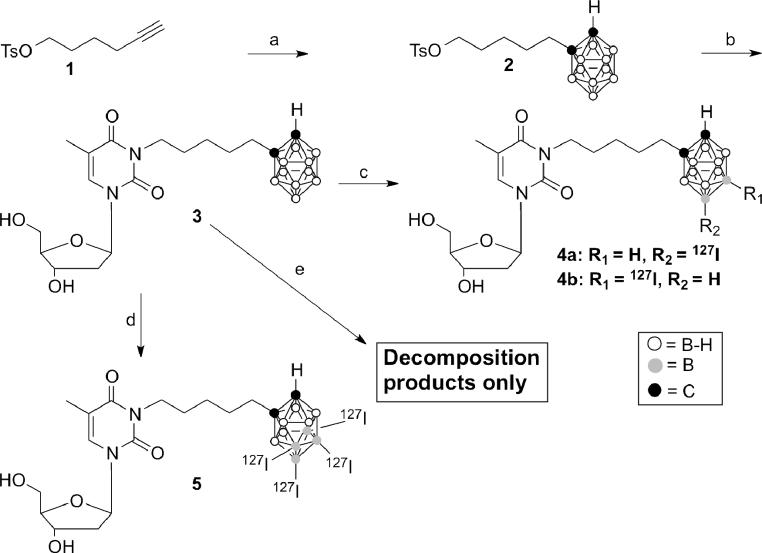
Reagents and conditions: a) B10H14, (bmim)+Cl−, toluene, 120°C, 10 min; b) dThd, K2CO3, DMF/acetone (1:1), 40°C, 24 h; c) ICl, AlCl3, DCM, 0°C, 2 h; d) ICl, AlCl3, DCM, 40°C, 48 h; e) ICl, DCM, rt, 1 h.
Scheme 3.
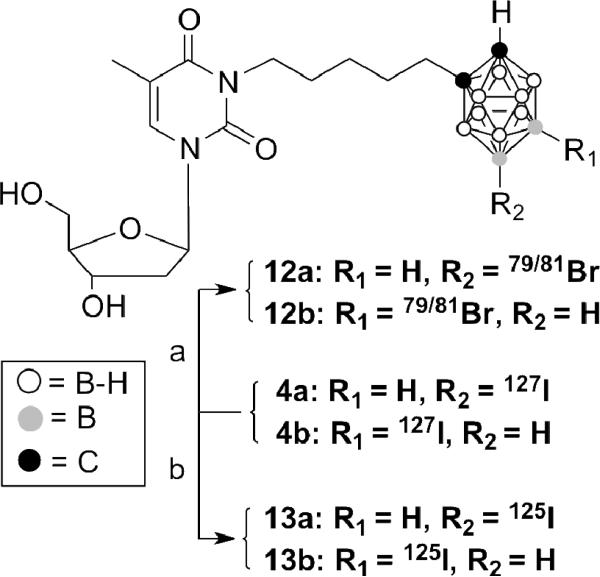
Reagents and conditions: a) Na79/81Br, Herrmann's catalyst, DMF, 110°C, 1h; b) Na125I, Herrmann's catalyst, DMF, 110°C, 1h
In this paper we describe strategies for the controlled introduction of a single iodine-127 atom to the closo-o-carborane cluster of 3, the halogen and the isotope exchange of iodine-127 to bromine-79/81 and iodine-125, respectively, via palladium catalyzed nucleophilic halogen/isotope exchange, and the initial biological evaluation of cage-halogenated forms of 3 in phosphoryl transfer assays with hTK1 and in cell uptake experiments with L929 TK1 (+)- and L929 TK1 (−) cells. To the best of our knowledge, this is the first description of this type of nucleophilic halogen/isotope exchange reaction at a neutral boron cluster in a complex biomolecule. The methodology that we have developed could be applicable for the synthesis of a wide range of low molecular weight bioconjugates containing neutral radioiodinated boron clusters for bioimaging and the treatment of cancer.
EXPERIMENTAL SECTION
NMR spectra were obtained on a Bruker Avance 400 at The Ohio State University College of Pharmacy (400 MHz for 1H, 100 MHz for 13C and 128 MHz for 11B). Chemical shifts (δ) are reported in ppm from internal deuterated chloroform, deuterated acetone or an external BF3•Et2O standard. Coupling constants are reported in Hz. High Resolution – Electron Spray ionization (HR-ESI) mass spectra were obtained at The Ohio State University Campus Chemical Instrumentation Center on a Micromass Q-TOF II- and a Micromass LCT spectrometer and at the University of Illinois Mass Spectrometry Laboratory (Urbana-Champaign, Illinois) on a Waters Q-TOF Ultima Tandem Quadrupole/Time-of-Flight spectrometer. Electron Impact (EI) mass spectra were obtained at the University of Illinois Mass Spectrometry Laboratory (Urbana-Champaign, Illinois) on a Micromass 70-VSE Double Focusing Sector spectrometer on TSSPro3.0 system. For all carborane-containing compounds, the mass of the most intensive peak of the isotopic pattern was reported for a 80% 11B to 20% 10B distribution. Measured patterns agreed with calculated patterns. Melting points were measured in sealed capillaries on a Thomas-Hoover capillary melting point apparatus and are uncorrected. Silica gel 60 (0.063–0.200 mm) from Merck was used for gravity column chromatography whereas silica gel 60 (0.015–0.049 mm) from EM science was used for flash column chromatography. Pre-coated glass-backed TLC plates with silica gel 60 F254 (0.25-mm layer thickness) from Dynamic Adsorbents (Norcross, GA) were used for TLC studies. For all non-radioactive materials, (semi-)preparative HPLC purification was performed either with a Gemini 5μ C18 column (21.20 mm × 250 mm, 5 μm particle size), supplied by Phenomenex Inc. CA, USA or with a Supelco Discovery® HS C18 column (10 mm × 250 mm, 10 μm particle size) supplied by Sigma Aldrich (MO, USA). A LiChrocart® 250–4 HPLC cartridge packed with LiChrospher® RP-18 stationary phase (4 mm × 250 mm, 5 μm particle size) supplied by EM Science, NJ, USA was used for analytical reversed-phase chromatography. For all radioactive materials, semi-preparative HPLC purification was performed with a Supelco Discovery® HS C18 column (10 mm × 250 mm, 10 μm particle size). A Beckman Ultrasphere® column (4.6 mm × 250 mm, 5 μm particle size) supplied by Beckmann Coulter Inc., CA, USA, was used for analytical reversed-phase chromatography. Further general experimental conditions are provided in the Supplementary Information.
5-(o-Carboran-1-yl)pentyl 4-methylbenzenesulfonate (2)28
6-Heptynyl 4-methylbenzenesulfonate (1)29 (1.0 g, 3.7 mmol), decaborane (240 mg, 2 mmol), and 1-butyl-3-methylimidazolium chloride (bmim+ Cl−) (300 mg, 1.72 mmol) were vigorously stirred in anhydrous toluene at 120 °C for 10 minutes. Subsequently, the reaction mixture was diluted with CH2Cl2 and filtered through silica gel. The filtrate was evaporated and the residue was purified by column chromatography using hexanes: EtOAc (2:1) to afford 2 (420 mg, 55%) as a white solid. 1HNMR (CDCl3) data were the same as those obtained previously for the same compound using a different procedure.28 The measured melting point was slightly lower than the one previously reported (105–106 °C vs. 109–110 °C).
3-(5-(o-Carboran-1-yl) pentyl) thymidine (3)28
To a solution of 2 (650 mg, 1.7 mmol) in DMF/acetone (1:1, 15 mL) were added thymidine (500 mg, 2.06 mmol) and potassium carbonate (700 mg, 5.07 mmol). The solution was stirred for 24 h at 50 °C. The reaction mixture was filtered and the filtrate was concentrated in vacuo. The residue was purified by column chromatography using CH2Cl2/CH3COCH3 (4:1) as the solvent system to give compound 3 (490 mg, 64%) as a wax-like solid. Spectroscopic data were consistent with those previously reported for the same compound.28
3-(5-(9-Iodo-o-carboran-1-yl)pentyl)thymidine and 3-(5-(12-iodo-o-carboran-1-yl)pentyl)thymidine (4a/4b, [N5-I]) [Strategy 1]
To a stirred solution of 3 28 (25 mg, 0.055 mmol) and AlCl3 (73 mg, 0.55 mmol) in anhydrous CH2Cl2, ICl (1M in CH2Cl2) was added dropwise (55 μL, 0.055 mmol) at 0°C. The reaction mixture was stirred for 2 h at at 0°C, quenched with a saturated solution of Na2S2O3, and extracted with CH2Cl2. The organic layer was separated and the aqueous layer washed 2 × with 10 mL CH2Cl2. The combined organic layers were washed with brine, dried over magnesium sulfate, and evaporated in vacuo. The product was crudely separated from decomposition products by column chromatography using CH2Cl2: MeOH (10:1) or, alternatively, PhMe:CH3CN (3:2) as the solvent system. Rf. 0.34 (CH2Cl2:MeOH, 10:1). Final purification, primarily by separation from di-iodinated product, was accomplished by preparative RP-18 HPLC (acetonitrile/water 60:40) to furnish 4a/4b (5.0 mg, 16%) as a white foam. 1HNMR (CD3COCD3) δ 0.6x2013;4.0 (m, 9H, BH), 1.32 (m, 4H, -CH2), 1.57 (m, 8H, CH2-CH2), 1.83 (s, 6H, -CH3), 2.25 x2013;2.39 (m, 8H, -CH2-Ccarborane and H-2′), 3.78 (d, 4H, H-5′, J = 2.7 Hz), 3.86 (m, 4H, -CH2-N), 3.93 (m, 2H, H-3′), 4.26 (m, 2H, -OH), 4.41 (d, 2H, H-4′, J = 3.2 Hz), 4.49 (s, 2H, -OH), 4.90 (s, 1H, H-Ccarborane), 5.10 (s, 1H, H-Ccarborane), 6.34 (t, 2H, H-1′, J = 6.8 Hz,), 7.83 (s, 2H, H-6 11B {1HNMR (CD3COCD3) δ. −17.79 (s, 1B, B12-I, B9-I), −16.15 (s, 1B, B9-I/B12-I), −11.04 (m, 12B, B3, B4, B5, B6, B7, B11), −7.19 (s, 4B, B8, B10), −3.96 (s, 1B, B12-H/ B9-H), −0.58 (s, 1B, B9-H/B12-H). 11B NMR (CD3COCD3) δ −17.93 (s, 1B), −16.26 (s, 1B), −11.17 (m, 12B), −7.33 (d, 4B, J = 156.2 Hz), −4.10 (d, 1B, J = 145.7 Hz), −0.70 (d, 1B, J = 153.5 Hz). HPLC retention time = 9.91 min (RP-18 analytical HPLC, 1 mL flow rate, solvent system: CH3CN:H2O, 60:40, isocratic elution). MS (HR-ESI) C17H33B10IN2o5 (M+ Na)+ calcd 603.2324, found 603.2344. N5-I2: HPLC retention time = 11.21 min (RP-18 analytical HPLC, 1 mL flow rate, solvent system: CH3CN:H2O, 60:40, isocratic elution). MS (HR-ESI), C17H32B10I2N2O5 (M + Na) + calcd 729.1259, found 729.1310.
3-(5-(8,9,10,12-Tetra-iodo-o-carboran-1-yl)pentyl]thymidine (5, N5-I4)
To a stirred solution 328 (100 mg, 0.22 mmol) and AlCl3 (440 mg, 3.3 mmol) in anhydrous CH2Cl2, 1 M ICl solution in CH2Cl2 (1.1 mL, 1.1 mmol) was added dropwise at 0°C. The reaction mixture was stirred for 2 h at 0° C and then refluxed for two days. The reaction mixture was quenched with a saturated solution of Na2S2O3 and extracted with CH2Cl2. The organic layer was separated and the aqueous layer was washed 2 × 10 mL of CH2Cl2. The combined organic layers were washed with brine, dried over anhydrous MgSO4, and evaporated in vacuo. The product was crudely separated from decomposition products by column chromatography using CH2Cl2: MeOH (10:1) or, alternatively, toluene:acetonitrile (3:2) as the solvent system. Rf. 0.34 (CH2Cl2: MeOH, 10:1). Final purification, primarily by separation from tri-iodinated product was accomplished by preparative RP-18 HPLC (CH3CN:H2O, 60:40, isocratic elution) to furnish 5 (27 mg, 13 %) of as a yellow solid. HPLC retention time = 13.81 min (RP-18 analytical HPLC, 1 mL flow rate, solvent system: CH3CN:H2O, 60:40, isocratic elution). 1H-NMR (CD3COCD3) δ.0.6–4.0 (m, 6H, BH), 1.33 (m, 2H, -CH2), 1.61 (m, 4H, -CH2-CH2), 1.83 (s, 3H, -CH3), 2.23 (dd, 2H, H-2′, J = 4.7Hz, J = 6.7 Hz,), 2.45 (m, 2H, -CH2-Ccarborane), 3.78 (m, 2H, H-′), 3.88 (m, 2H, -CH2-N), 3.94 (dd, 1H, H-3′, J = 3.1Hz, J = 6.1 Hz), 4.27 (s, 1H,-OH), 4.42 (s, 1H, -OH), 4.49 (s, 1H, H-4′), 5.72 (s, 1H, H-Ccarborane), 6.34 (t, 1H, H-1′, J = 6.8 Hz), 7.84 (s, 1H, H-6). 11B {1H} NMR (CD3COCD3) δ −17.8 (s, 4B, B8-I, B9-I, B10-I and B12-I), −12.1 (s, 1B), −10.2, −8.4 (m, 5B) 11B NMR (CD3COCD3) δ −17.9 (s, 4B, B8, B9, B10 and B12), −12.1 (d, 1B, J = 205.2 Hz), −10.1, −8.3 (m, 5B). MS (HR-ESI) C17H30B10I4N2O5 (M + H) + calcd 960.9371, found 960.9373. N5-I3: HPLC retention time = 13.60 min (RP-18 analytical HPLC, 1 mL flow rate, solvent system: CH3CN:H2O, 60:40, isocratic elution). MS (HR-ESI) C17H31B10I3N2O5 (M + Na)+ calcd 855.0226, found 855.0277.
9-Iodo-o-carborane (7)30,31
To a solution of o-carborane (6) (2.0 g, 14 mmol) in anhydrous CH2Cl2 and was added dropwise ICl (27.7 mmol, 27.7 mL of a 1M solution in CH2Cl2) at 25 °C. The reaction mixture was stirred for 5h at 40 °C, quenched with a saturated aqueous solution of Na2S2O3, and extracted with CH2Cl2. The organic layer was washed with brine, dried (MgSO4), and evaporated in vacuo. The residue was purified by column chromatography to afford 7 (2.7 g, 72%) as a off-white solid. Rf. 0.38 (hexanes: ethyl acetate, 10:4). 1H-NMR (CD3COCD3) δ 1.53–3.21 (m, 9H, BH), 4.74 (s, 1H, H-Ccarborane), 4.95 (s, 1H, HCcarborane).. 11B {1H} NMR (CD3COCD3) δ −16.5 (s, 1B, B9-I), −13.7 (s, 2B, B3, B6), −12.8 (s, 2B, B4,B5), −12.1 (s, 2B, B7, B11), −7.3 (s, 2B, B8, B10), −1.1 (s, 1B, B12). 11B NMR (CD3COCD3) δ −16.5 (s, 1B), −13.1 (m, 6B), −7.3 (d, 2B, J = 155.0 Hz), −1.2 (d, 1B, J = 151.2 Hz). MS (HR-EI) C2B10H9I (M) + calcd 270.0909, found 270.0912.
1-(tert-Butyldimethylsilyl)-9-iodo-o-carborane (8a) and 1-(tert-butyldimethylsilyl)-12-iodo-ocarborane (8b)
To a stirred solution of 7 (2.1 g, 7.8 mmol) in anhydrous THF at −78 °C was added dropwise a 2.5 M solution of n-BuLi in hexanes (3.26 mL, 8.15 mmol). The reaction mixture was allowed to stir for 30 min at room temperature, cooled to 0 °C and tert-butyldimethylsilyl chloride (1.29 g, 8.55 mmol) in 15 mL of anhydrous THF was added dropwise. The solution was refluxed overnight, carefully quenched with 10 mL of water, and extracted with 60 mL of diethyl ether. The layers were separated and the aqueous layer was extracted with 2 × 30 mL of diethyl ether. The combined organic layers were dried (MgSO4) and concentrated in vacuo. The crude residue was purified by column chromatography using pentane to afford 8a/8b as colorless oil (2.0 g, 66 %) Rf. 0.24–0.31. 1H-NMR (CD3COCD3) δ 0.30 (s, 6H, -Si(CH3)2), 0.33 (s, 6H, -Si(CH3)2), 1.04 (s, 9H, -C(CH3)3), 1.07 (s, 9H, -C(CH3)3), 1.47–2.23 (m,9H, BH), 4.52 (s, 1H, H-Ccarborane), 4.74 (s, 1H, H-Ccarborane). MS (HR-EI) C8H25B10ISi (M) + calcd 384.1774, found 384.1774.
A quantity of 100 mg of the mixture of 8a and 8b was separated by column chromatography using pentane as the solvent system to give 58 mg of 8a (Rf. 0.31, mp 105–106 °C) and 31 mg of 8b (Rf. 0.24, mp 125–126 °C). 8a: 1H-NMR (CD3COCD3) δ 0.33 (s, 6H, -Si(CH3)2), 1.09 (s, 9H, -C(CH3)3), 1.47−2.23 (m, 9H, BH), 4.77 (s, 1H, H-Ccarborane). 11B {1H} NMR (CD3COCD3) δ −15.0 (s, 1B, B9-I), −11.7 (m, 4B, B3, B4, B5, B6), −8.9 (s, 2B, B7, B11), −4.6 (s, 2B, B8, B10), 2.3 (s, 1B, B12). 11B NMR (CD3COCD3) δ −15.0 (s, 1B), −10.4 (m, 6B), −4.6 (d, 2B, J = 152.6Hz), 2.3 (d, 1B, J = 157.9Hz, B12. MS (HR-EI) C8H25B10ISi (M) + calcd 384.1774, found 384.1758. 8b: 1H-NMR (CD3COCD3) δ 0.31 (s, 6H, -Si (CH3)2), 1.04 (s, 9H, -C(CH3)3), 1.47−2.23 (m, 9H, BH), 4.55 (s, 1H, H-Ccarborane). 11B {1H} NMR (CD3COCD3) δ −14.4 (s, 1B, B12-I), −12.3 (m, 4B, B3, B4, B5, B6), −11.0 (m, 2B, B7, B11), −5.2 (s, 2B, B7, B11), −0.2 (s, 1B, B9). 11B NMR (CD3COCD3) δ −14.3 (s, 1B), −13.0 (s, 1B), −10.4 (m, 1B, J=76.7Hz, J = 168.5Hz), −5.2 (d, 1B, J = 154.3Hz), −0.3 (d, 1B, J = 152.1Hz). MS (HR-EI) C8H25B10ISi (M) + calcd 384.1774, found 384.1791.
1-(tert-Butyldimethylsilyl)-2-(5-(tert-butyldimethylsilyloxy)pentyl)-9-iodo-o-carborane and 1-(tert-butyl dimethylsilyl)-2-(5-(tert-butyldimethyl silyloxy)pentyl)-12-iodo-o-carborane (9a/9b)
To a stirred solution of a mixture of 8a and 8b (6.52 g, 17 mmol) in 35 mL of anhydrous THF at −78 °C was added dropwise a 2.5 M solution of n-BuLi in hexanes (8.48 mL, 21.2 mmol). The reaction mixture was stirred for 30 min at room temperature, then cooled to 0 °C, and tert-butyldimethylsilyl chloride (8.21 g, 22 mmol) dissolved in THF (20 mL) was added dropwise. The solution was stirred for 24 h, quenched with 20 mL of water, and extracted with 60 mL of diethyl ether. The separated aqueous layer was extracted 2 × 30 mL of diethyl ether, the combined organic layers dried (MgSO4), and concentrated in vacuo. The crude residue was purified by flash chromatography to afford 9a/9b (6.5 g, 66 %). Rf. 0.72 (pentane: diethyl ether, 30:1). 1H-NMR (CD3COCD3) δ 0.05 (s, 6H, -Si(CH3)2), 0.05 (s, 6H, -Si(CH3)2), 0.39 (s, 6H, -Si(CH3)2), 0.43 (s, 6H, -Si(CH3)2), 0.89 (s, 9H, -C(CH3)3), 0.90 (s, 9H, - C(CH3)3), 1.09 (s, 9H, -C(CH3)3), 1.12 (s, 9H, -C(CH3)3), 1.34 – 1.67 (m, 12H, CH2-CH2-CH2), 1.3–3.27 (m, 18H, BH), 2.22 (m, 2H, CH2-Ccarborane), 2.34 (m, 2H, CH2-Ccarborane), 3.63 (dd, 4H, -CH2-OTBDMS, J = 6.2 Hz, J = 13.2 Hz). 11B {1H} NMR (CD3COCD3) δ −15.9 (s, 1B, B12-I/B9-I), −14.2 (s, 1B, B9-I/B12-I), −9.0 (m, 12B, B3, B4, B5, B6, B7, B11), −5.3 (m, 4B, B8, B10), −2.5 (s, 1B, B12-H/B9-H), 2.0 (s, 1B, B9-H/B12-H). 11B NMR (CD3COCD3) δ −15.9 (s, 1B), −14.2 (s, 1B), −9.2 (m, 12B), −5.3 (m, 4B), −2.4 (d, 1B, J = 148.6Hz), 2.1 (d, 1B, J = 148.8Hz). MS (HR-EI) C19H49B10IOSi2 (M-56)− calcd 528.2744, found 528.2760.
5-(9-Iodo-o-carboran-1-yl)pentan-1-ol and 5-(12-iodo-o-carboran-1-yl)pentan-1-ol (10a/10b)
To a solution of 9a/9b (2.37g, 4.05 mmol) in 40 mL of THF was added dropwise at −78 °C a 1.0 M solution of tetrabutylammonium fluoride (TBAF) in THF (6.1 mL, 6.1 mmol). The reaction mixture was stirred at 4 °C for 30 min, acidified with 10% methanolic-HCl (20 mL), and then stirred for an additional 30 min at 4 °C. The reaction mixture was extracted with ethyl acetate (3 × 35 mL), the combined organic layers washed with water and then saturated aqueous NaHCO3, dried over anhydrous magnesium sulfate, and concentrated in vacuo. The residue was purified by column chromatography (hexanes: ethyl acetate, 3:7) to furnish 10a/10b (1.1 g, 76%). Rf. 0.54 (hexanes: ethyl acetate, 3:7); 1H-NMR (CD3COCD3) δ 1.28 –1.59 (m, 12H, -CH2-CH2-CH2), 1.3–3.27 (m, 18H, BH), 2.25 (m, 2H, -CH2-Ccarborane), 2.38 (m, 2H, -CH2-Ccarborane), 3.50 (dd, 4H, -CH2-OH, J = 5.3 Hz, J = 10.2 Hz), 4.88 (br, 1H, H-Ccarborane), 5.09 (br, 1H, H-Ccarborane). 11B {1H} NMR (CD3COCD3) δ −18.4 (s, 1B, B12-I/B9-I), −16.7 (s, 1B, B9-I/B12-I), −11.7 (m, 12B, B3, B4, B5, B6, B7, B11), −7.7 (m, 4B, B8, B10), −4.4 (s, 1B, B12-H/B9-H),−1.15 (s, 1B, B9-H/B12-H). 11B NMR (CD3COCD3) δ −18.1 (s, 1B), −16.4 (s, 1B), −11.3 (m, 14B), −7.3 (m, 2B, J = 152.5 Hz), −4.1 (d, 1B, J = 153.3 Hz), −0.7 (d, 1B, J = 150.4 Hz). MS (HR-EI) C7H21B10IO (M)+ calcd 356.1641, found 356.1647.
5-(9-Iodo-o-carboran-1-yl)pentyl 4-methylbenzenesulfonate and 5-(12-iodo-o-carboran-1-yl)pentyl 4-methyl benzenesulfonate (11a/11b)
To a solution of 10a/10b (1.65 g, 4.63 mmol) in CH2Cl2 (12 mL) was added triethylamine (0.84 mL, 6 mmol) and a catalytic amount of 4-(dimethylamino)pyridine (DMAP) (113 mg, 0.93 mmol). A solution of p-toluenesulfonyl chloride (1.33g, 6.95 mmol) in 13 mL of CH2Cl2 was added at 0°C and the reaction mixture was stirred at room temperature for 7 h. The reaction was quenched with a saturated NH4Cl aqueous solution; the organic phase was washed with water and brine, dried (MgSO4) , and concentrated in vacuo. The residue was purified by column chromatography to afford 11a/11b (2.2 g, 93%) as an oil. Rf. 0.65 (hexanes: ethyl acetate, 1:1) 1H-NMR (CD3COCD3) δ 1.27–1.69 (m, 12H, -CH2-CH2-CH2), 1.3–3.27 (m, 18H, BH), 2.23 (m, 2H, -CH2-Ccarborane), 2.36 (m, 2H, -CH2-Ccarborane), 2.47 (s, 6H, -CH3), 4.02 (m, 4H, -CH2-OTs), 4.84 (br, 1H, H-Ccarborane), 5.05 (br, 1H, H-Ccarborane), 7.49 (d, 4H, Ar-H, J = 7.3 Hz), 7.79 (dd, 4H, Ar-H, J = 3.0 Hz, J = 8.4 Hz); 11B {1H} NMR (CD3COCD3) δ −18.0 (s, 1B, B12-I/B9-I), −16.3 (s, 1B, B9-I/B12-I), −11.5 (m, 12B, B3, B4, B5, B6, B7,B11), −7.3 (s, 4B, B8, B10), −4.1 (s, 1B, B12-H/B9-H), −0.66 (s, 1B, B9-H/B12-H). 11B-NMR (CD3COCD3) δ −17.9 (s, 1B), −16.3 (s, 1B), −11.4 (m, 12B), −7.3 (d, 2B, J = 155.0 Hz), −4.0 (d, 1B, J = 149.8 Hz), −0.66 (d, 1B, J = 152.7Hz). MS (HR-EI) C14H27O3SIB10 (M)+ calcd 510.1729, found 510.1724.
Synthesis of 4a/4b (N5-I) [Strategy 2]
To a solution of 11a/11b (1.18 g, 2.31 mmol) in DMF/acetone (1:1, 40 mL) was added thymidine (1.68 g, 6.93 mmol) and potassium carbonate (960.0 mg, 6.94 mmol). The solution was stirred for 2.5 h at 40 °C and filtered. The filtrate was concentrated in vacuo, the residue was added to water and extracted with ethyl acetate (3 × 50 mL). The combined organic layers were washed with brine, dried over anhydrous MgSO4, and concentrated in vacuo. The residue was purified by column chromatography using CH2Cl2/MeOH (10:1, Rf. 0.34) as the eluent to afford 4a/4b (800 mg, 60%).
3-(5-(9-Bromo-o-carboran-1-yl)pentyl)thymidine and 3-(5-(12-bromo-o-carboran-1-yl)pentyl)thymidine (12a/12b - N5-Br)
To a solution of 4a/4b (100 mg, 0.175 mmol) in 3 mL CH2Cl2 was added a solution of NaBr (180 mg, 1.75 mmol) dissolved in 5 mL of water. The solvents were evaporated in vacuo to obtain an anhydrous residue. A solution of Herrmann's Catalyst (41 mg, 25 mol%) in 1 mL anhydrous DMF was added. The reaction mixture was stirred at 110 °C for 1 h. Following evaporation, the residue was added to water and extracted with CH2Cl2. The organic phase was dried (MgSO4), concentrated in vacuo, and the residue was purified by column chromatography to yield 12a/12b (48 mg, 52%). Rf. 0.34 (CH2Cl2:MeOH, 10:1). 1H-NMR (CD3COCD3) δ. 1.3–3.22 (m, 18H, BH), 1.31 (m, 4H, -CH2), 1.56 (m, 8H, -CH2-CH2), 1.83 (s, 6H, -CH3), 2.23 (m, 4H, H-2), 2.36 (m, 4H, -CH2-Ccarborane), 3.77 (m, 4H, H-5′), 3.86 (m, 4H, -CH2-N), 3.92 (m, 2H, H-3′), 4.27 (s, 2H, -OH), 4.42 (s, 2H, -OH), 4.49 (d, 2H, H-4′, J = 2.9 Hz), 4.84 (s, 2H, H-Ccarborane), 6.34 (t, 2H, H-1′, J = 6.7 Hz), 7.83 (s, 1H, H-6). 11B {1H} NMR (CD3COCD3) δ −11.9 (m, 12B, B3, B4, B5, B6, B7, B11), −8.1 (s, 4B, B8, B10), −4.9 (s, 1B, B12-H/B9-H), −1.3 (s, 1B, B9-H/B12-H), 0.4 (s, 2B, B9-Br and B12-Br). 11B NMR (CD3COCD3) δ −12.7 (m, 12B), −8.1 (d, 4B, J = 151.9 Hz), −4.9 (m, 2B), −0.72 (m, 2B). MS (HR-ESI) C17H33B10BrN2O5 (M+ Na)+ calcd 557.2401, found 557.2431.
3-(5-(9-[125I]Iodo-o-carboran-1-yl)pentyl)thymidine and 3-(5-(12-[125I]iodo-o-carboran-1-yl)pentyl)thymidine (13a/13b - N5-125I)
An alkaline Na125I solution (10 μL, pH 8–11, 1mCi, specific activity of 17 Ci [629 GBq]/mg) was diluted to 100 μL using deionized water. Compound 4a/4b (10 mg, 0.017 mmol), dissolved in 200 μL DMF, was transferred to a 3 mL conical reaction vial (MINUM-WARE ®). A solution of Herrmann's catalyst in 100 μL of DMF (10 mol %) was added to the reaction vial. To this reaction mixture, 10 μL (100 μCi) of the diluted alkaline Na125I solution was added dropwise. The reaction mixture was then heated at 110 °C for 1 h, cooled to rt, and filtered using GHP Acrodisc ® 13 mm syringe filter to separate the catalyst from the reaction mixture. The filtrate was evaporated under argon flow at 30 °C and the residue was purified by semi preparative RP-18 HPLC to yield 13a/13b (N5-125I) (3,2 mg, radiochemical yield = 8%, specific activity = 2.52 μCi/mg, radio chemical purity: > 98 %). HPLC retention time = 25.50 min (RP-18 analytical HPLC, 1mL flow rate, solvent system: CH3CN: H2O, 50:50, isocratic elution).
X-ray diffraction studies
Crystals of 8a and 8b were obtained by diffusion crystallization using diethyl ether/pentane as the solvent system. Both data collections were carried out at −123°C on a Nonius Kappa CCD diffractometer with Mo Kα radiation and a graphite monochromator. Phi and omega scans were used for data collection with a frame width of 1.0°. Data integration was carried out with Denzo,32 and an absorption correction and merging of the data was carried out with Sortav.33,34 The structures were solved by the direct methods procedure in SHELXS-97.35 Full-matrix least-squares refinements based on F2 were performed in SHELXL-97,35 as incorporated in the WinGX package.36 For structure 8b there are four independent molecules in the asymmetric unit (Z'=4), and these are labeled as A, B, C and D (Table 3, Figure S2 [Supporting Information]). For each methyl group, the hydrogen atoms were added at calculated positions using a riding model with U(H) = 1.5*Ueq(bonded carbon atom). The torsion angle, which defines the orientation of the methyl group about the C-C or Si-C bond, was refined. The remaining hydrogen atoms in each cluster were refined isotropically. Neutral atom scattering factors were used and include terms for anomalous dispersion.37 The values of the Flack parameters38,39 for 8a and 8b are −0.05(1) and −0.042(7), respectively, indicating that the models for these structures are of the correct chirality. Additional crystallographic details are shown in Table 2.
Table 3.
Selected bond lengths (Å) and selected intermolecular interactions for 8a and the four rotamers of 8b A–D.
Table 2.
Crystallographic data for 8a and 8b.
| Compounds | 8a | 8b |
|---|---|---|
| Formula | C8 H25 B10 I Si | C8 H25 B10 I Si |
| Formula weight | 384.37 | 384.37 |
| Space group | Cc (No. 9) | Pca21 (No. 29) |
| a, Å | 6.8264(1) | 13.6530(1) |
| b, Å | 20.6488(3) | 19.5285(1) |
| c, Å | 12.5390(2) | 27.7225(2) |
| β ° | 90.346(1) | - |
| Volume, Å3 | 1767.43(5) | 7391.45(8) |
| Z | 4 | 16 |
| Density (calculated), g/cm3 | 1.444 | 1.382 |
| μ, cm−1 | 18.59 | 17.78 |
| Crystal size, mm3 | 0.04 × 0.12 × 0.38 | 0.19 × 0.19 × 0.27 |
| Final R indices [I>2sigma(I)] | R1=0.0238, wR2=0.0421 | R1=0.0303, wR2=0.0460 |
| R indices (all data) | R1=0.0316, wR2=0.0440 | R1=0.0538, wR2=0.0489 |
Docking studies
The homology model of hTK1 (PDB ID # 1W4R) was developed from the crystal structure of Thermotoga maritima Thymidine Kinase (TmTK, PDB ID # 2QPO) using the alignment mode of SWISS-MODEL.40 The sequence identity and similarity between hTK1 and TmTK is 36% and 55%, respectively.41 The obtained homology model was saved in pdb format and hydrogens were added using Accelrys Discovery Studio Visualizer v2.5. After addition of hydrogen, the pdb file was imported into Surflex version 2.1142 (Biopharmics LLC, San Francisco, CA), installed on Dell optimplex GX 270 desktop computer with Windows operating system environment, for the protomol generation. The default mode was chosen for docking. The obtained docking poses were visualized with Accelrys Discovery Studio Visualizer v2.5. Surflex does not have parameters for boron atoms. By default, Surflex treats such unrecognizable atoms as “funky atoms”. However, this does not seem to affect significantly the accuracy of docking studies with compounds containing carborane clusters.43
Phosphoryl transfer assays (PTAs)
Recombinant hTK1 was expressed and purified from the bacterial expression system BL21(DE3) pLysS with transformed vector pET-14b + hTK1), kindly provided by Dr. Staffan Eriksson, Biomedical Centre, Uppsala, Sweden, according to a procedures described previously.28,44 Thymidine and the carboranyl-thymidine conjugates were dissolved in DMSO (5 mM concentration) and further diluted with water to produce stock solutions of concentrations (0.4 mM). The PTA assays were carried out as described previously28,44 with minor modifications. The reaction mixture contained 100 μM nucleoside and 100 μM ATP (with a small fraction of 0.13 μM [γ-32P]ATP (Perkin Elmer)), 50 mM Tris-HCl (pH 7.6), 5 mM MgCl2, 125 mM KCl, 10mM DTT, and 0.5 mg/mL bovine serum albumin (BSA). In all reactions, the final concentration of DMSO was set to 2%. The reaction mixture was incubated at 37 °C for 20 min in the presence of 135 ng of enzyme. Somewhat higher concentrations of enzyme were used than in previous studies45 because this specific enzyme preparation appeared to be less active (441 nmol of thymidine monophosphate formed per minute and mg of hTK1). Following the incubation period, the enzyme was inactivated by heating for 2 min at 99 °C. The reaction mixture was centrifuged and 2 μL sample portions were spotted on PEI–cellulose TLC plates (EMD Chemicals Inc.). The TLC plates were developed in a solvent system containing isobutyric acid:ammonium hydroxide:water (66:1:33) over the period of 6 h. The radiolabeled spots were visualized by placing the TLC plates on a BioMax XAR film (Kodak) overnight and developing the films using X-ray film developer (Tiba M6B, Series VI B Rapid processor; Commonwealth X-Ray Inc.). The spot intensities of the phosphorylated compounds were calculated by using Photoshop@ and the values were compared with that of dThd.
Cellular uptake studies
The L929 (#CCL1, American Type Culture Collection, Manassas, VA) cell line was one of the first to be established in continuous culture and clone 929 was the first cloned strain that was developed. The parent L strain was derived from normal subcutaneous areolar and adipose tissue from a C3H/An mouse, and clone 929, which is TK1 (+), was from the 95th subculture generation of the parent strain. Its TK1 (−) counterpart (#CCL1.3 L-M ATCC) was derived from the wild-type cell line L929. The L929 TK1 (+) cells were grown in Dulbecco's Modified Eagle's Medium (DMEM), supplemented with 10% fetal bovine serum (FBS), 1% L-glutamine (#25030, Invitrogen, Carlsbad, CA) and 1% Penicillin, and Streptomycin mixture (Penstrep, #15140, Invitrogen). The L929 TK1 (−) cells were grown in media with 10% FBS, 1% L-glutamine, 1% Penstrep, and 10 mM 5-bromo-2'-deoxyuridine in order to inhibit reversion to the TK1(+) phenotype. Cells were grown in T-75 flasks (#430641, Corning, Corning, NY) and split after reaching 70–80% confluency for propagating the cell line.
Uptake experiments were carried out as follows: Forty-eight hours prior to the addition of the test compounds, L929 TK1 (−) or TK1 (+) cells were seeded at a density of 1 × 10⁶ or 2.5 × 10⁶ cells per T-75 flask, respectively, six flasks for each cell type. When the cells reached approximately 90% confluency, the test compounds were added to the culture medium. Cells were seeded at different densities because TK1 (−) cells grew faster than TK1 (+) cells. Before treatment, cell culture medium was removed and the cells were washed once with DMEM. After that, 12.5 mL media containing 17.5 μM of N5 (3), N5-79/81Br (12a/12b), or N5-127I (4a/4b), were added to the cultures. Twenty-four hours following incubation at 37°C, the medium containing the compound was removed and the cells were washed twice with cold phosphate buffered saline (PBS), pH 7.4. Cells were then trypsinized with 5 mL of 0.5% trypsin-EDTA (#15400, Invitrogen) for 5 min, followed by the addition of 5 mL DMEM with 10% FBS to neutralize the trypsin. Cells with media from two T-75 flasks were pooled into one 50 mL centrifuge tube, following which they were centrifuged (600 × g). After centrifugation, trypsin containing medium was decanted, the cell pellets were resuspended in 2 mL of serum free DMEM, and the number of cells per tube was counted by means of a hemocytometer. Cells with media then were transferred to borosilicate free glass test tubes, heated to dryness in an oil bath at 105°C, and then 1 mL of concentrated sulfuric acid was added to digest them for 2hr. Following this, 1 mL of 50% hydrogen peroxide was added to each tube to decolorize the digested material. The contents were transferred to 15 mL plastic centrifuge tubes, and the volume was adjusted to 4 mL by the addition of deionized water. Boron concentrations were determined by means of inductively coupled plasma-optical emission spectroscopy (ICP-OES) using a spectrometer located at the Nanotech West Facility of The Ohio State University.
RESULTS AND DICUSSION
COMPUTATIONAL STUDIES
Human TK1 tolerates extensive modification at the N-3 position of dThd19 and the differences in the surface areas and the molecular volumes between 3 and 13a/13b, or their non-radioactive counterparts (N5-127I, [4a/4b, Scheme 1], were relatively small. The molecular surface area and volume for 3 were 575.53 Å2 and 1297.05 Å3, respectively. The corresponding values for 4a and 4b were 630.77 Å2 and 630.83 Å2, respectively, for molecular surface areas and 1379.36 Å3 and 1379.75 Å3, respectively, for molecular volumes (See Supporting Information for details related to calculation). Therefore, we hypothesize that the introduction of a single iodine to 3 may not significantly affect its phosphorylation by hTK1, and more importantly, its tumor localizing properties. Docking studies of the triphosphate forms of 3 and 4b with a homology model of hTK1 seem to confirm our hypothesis (Figure 1). The extensive conformational changes that TK1-like enzymes undergo as a result of ATP and/or dThd binding have been discussed in detail by Lavie et al.46 The homology model of hTK1 was developed from an apo form of Thermotoga maritima Thymidine Kinase (TmTK, PDB ID # 2QPO) and, thus, should represent an apo form of hTK1. The triphosphate forms of 3 and 4b were chosen for docking because we have found that this modification produces realistic docking poses of the dThd scaffold within the hTK1 active site. As can be seen from Figures 1A and 1B, the carborane clusters of both compounds are located outside of the substrate binding pocket and monoiodination in 4b does not generate significant differences in the dThd binding patterns between 3 and 4b. Similar results have been observed previously in docking studies of other 3CTAs with varying substitution patterns at the carborane cluster in a hTK1 homology model.47
CHEMISTRY
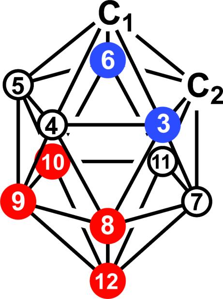
Theoretically, radioiodination of the closo-o-carborane cage can be achieved in three ways. The first method employs a “closo-cluster reconstruction” from nido-o-carborane via treatment with n-butyl lithium (n-BuLi) followed by boron triiodide (BI3) to generate iodinated closo-o-carborane.48,49 The B-iodocarborane synthesized in this way has iodine either at B3 or at B6 (Figure 2). The second method is the direct introduction of iodine to the closo-o-carborane by means of a Friedel Crafts-type (electrophilic) halogenation using I2 or ICl as iodine sources.48,50,51 The electrophilic attack of positively charged species on o-carborane is regiospecific based on the charge distribution in the cluster.48,50,51 Electrophilic halogenation occurs first at the most electron rich boron atoms 9 and 12 (Figure 2), which are furthest away from both carbon atoms followed by the boron atoms 8 and 10.48,50,51 Thus, reaction of closo-o-carboranes with halogenating species can result in mono-, di-, tri- or tetraiodination depending on the applied reaction conditions and stoichiometry of halogenation species.48,49 A disadvantage of both methods discussed above is that 125I2, 125ICl, or B125I3, which would be necessary for the synthesis of 125I-labeled 3 (N5-125I, 13a/13b, Scheme 3), are not readily available from commercial sources.
Figure 2.
Atom numbering and electron density distribution in closo-o-carborane. Boron atoms shown in red are the most electron rich whereas the boron atoms shown in blue are the most electron deficient.
The third method is a halogen/isotope-exchange reaction using a “cold” B-iodocarborane as the starting material that is treated with a nucleophilic radiohalogen source in of the presence of a suitable catalyst.6,8–10 The advantage of this method is that the radiohalogen source required for this strategy (Na125I) is commercially available. This strategy was therefore chosen for the synthesis of 13a/13b.
For the synthesis of 13a/13b by means of isotope exchange, the “cold” precursor, 4a/4b, must be synthesized first. Two methods were explored for its synthesis: 1) direct iodination of carborane cage of 3 using either ICl or I2 in the presence of a Lewis acid in a Friedel-Crafts halogenation strategy already discussed above and 2) initial monoiodination of unsubstituted closo-o-carborane with ICl followed by its functionalization to 4a/4b using conventional synthetic organic methods.28
Direct iodination of preformed 3 [Strategy 1]
A new strategy for the synthesis of 3 was applied (Scheme 1). The reaction of 6-heptynyl-tosylate (1)29 with decaborane (B10H14) in a biphasic mixture of toluene and the ionic liquid, 1-butyl-3-methylimidazolium chloride (bmim+ Cl−),52 yielded the o-carboranyl tosylate 228 in 55% yield. The reaction of compound 2 with dThd in DMF/acetone in the presence of K2CO3 produced compound 3 in 56% yield, as described previously by Lunato et al.28 Overall, the yields for both compounds 2 and 3 described here are comparable with those reported previously for the same compounds.28 However, the synthesis of 3 is much shorter, far more convenient, and also safer than the procedure described previously involving the synthesis and isolation of an intermediate decaborane/acetonitrile complex.28
Several different reaction conditions were explored to optimize the synthesis of 4a/4b via direct iodination of 3, as shown in Scheme 1 and Table 1. The treatment of 3 with one equivalent of ICl in the absence of Lewis acid (AlCl3) at room temperature for 1 h in CH2Cl2 proved to be detrimental to the nucleoside scaffold resulting in complete decomposition. When 3 was treated with 1 equivalents of ICl in the presence of 10 equivalents of AlCl3 in CH2Cl2 at 0 °C for 2 h, the reaction mixture contained 40 % of 4a/4b, ~ 45 % of unreacted 3, as well as minor quantities of diiodinated 3 (N5-I2) and decomposition products (Table 1). The reaction of 3 with 5 equivalents of ICl and 10 equivalents of AlCl3 at 40 °C in CH2Cl2 for 48 h resulted in the formation of ~ 55 % of compound 5 (N5-I4) along with smaller quantities triiodinated 3 (N5-I3) and decomposition products in the reaction mixture (Table 1). Preliminary analytical data indicated that the decomposition products may include nucleobases and pyranosyl nucleosides (data not shown). In general, compounds 4a/4b, 5, N5-I2, and N5-I3 were crudely separated from decomposition products (primarily nucleobases) and inorganic impurities by column chromatography followed by complete purification via semi-preparative HPLC. When AlCl3/I2 instead of AlCl3/ICl was used in these reactions, overall similar product ratios were observed (data not shown).
Table 1.
Reaction conditions to control the degree of iodination by Strategy 1.
| Reactions (Scheme 1) | % in Reaction Mixturesa |
|||||
|---|---|---|---|---|---|---|
| Decomp. Prods. | 3 (recov.) | 4 | N5-I2 | N5-I3 | 5 | |
| c | ~10 | ~45 | ~40 | ~5 | 0 | 0 |
| d | ~35 | 0 | 0 | 0 | ~10 | ~55 |
| e | 100 | 0 | 0 | 0 | 0 | 0 |
Estimations based on TLC- and 1H-NMR analyses of reaction mixtures.
Initial monoiodination of o-carborane followed by functionalization to compounds 4a/4b [Strategy 2]
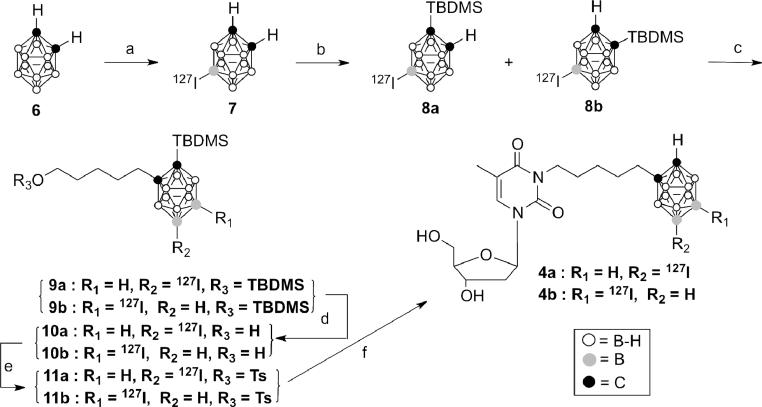
Scheme 2 describes the synthesis of 4a/4b according to strategy 2. The reaction of o-carborane with ICl in the absence of any Lewis acids under reflux condition in CH2Cl2 furnished 9-iodo-o-carborane (7)30,31,53 in 70 % yield without producing detectable amounts of di-, tri-, or tetraiodinated o-carboranes. This monoiodination of o-carborane was operationally very convenient and 7 was the only product obtained even when an excess of ICl was used. It should be noted that in the absence of Lewis acid, I2 did not cause any iodination of o-carborane (data not shown), whereas both ICl and I2 were far more reactive in the presence of AlCl3 causing even tetraiodination of 3, as discussed above.
Scheme 2.
Reagents and conditions: a) ICl, DCM, 40°C, 5 h; b) n-BuLi, TBDMSCl, THF, 66°C, 12 h; c) n-BuLi, 5-(tert-butyldimethylsilyloxy)pentyl 4-methylbenzenesulfonate; THF, 66°C, 12 h; d) TBAF, THF, −78°C to 4°C, 30 min; ii) 10% methanolic HCl, 4°C, 30 min; e) TsCl, Et3N, DMAP, 0°C to rt, 7 h; f) dThd, K2CO3, DMF/acetone (1:1), 40°C, 2.5 h.
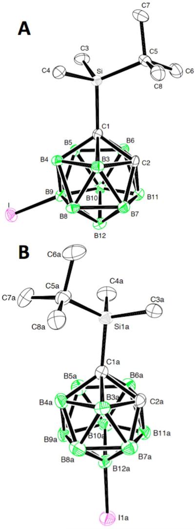
Deprotonation of the carboranyl C-H of 7 by n-BuLi, followed by the reaction with tertbutyldimethylsilyl chloride (TBDMSCl), resulted in the formation of a mixture of structural isomers 8a and 8b in 66 % overall yield with iodine at either B9 or B12 (Scheme 2, Figure 2). This protection of one of the carboranyl carbons of 7 with the TBDMS group was carried out to guarantee the subsequent monofunctionalization of carboranyl carbon.54,55 Unfortunately, the column chromatographic separation of larger quantities of 8a and 8b from their mixture was very tedious even though they have distinct Rf. values (see Experimental Section). The same was true for all other compound mixtures described in Schemes 1, 2, and 3 because of very close Rf. values (see Experimental Section). Therefore, we decided to carry out all follow up reactions, the optimization of the halogen/isotope reactions, and all biological evaluation with isomeric mixtures because this would not affect most of the major objectives of this study. As already discussed, the carborane cage of 3CTAs may be located outside the hTK1 substrate-binding site. Therefore, the regioselectivity of monoiodination (B9-I vs B12-I) should not affect the phosphorylation by hTK1 significantly (Figure 1). It was, however, important to confirm the formation of structural isomers with iodine and TBDMS group in meta- and para-orientation, respectively, and to assure that nucleophilic displacement/exchange of iodine did not occur after the treatment of 9-iodo-o-carborane with n-BuLi (Figure 4). Therefore, a small quantity of 8a and 8b was separated by silica gel chromatography for a thorough analysis by 1H-, 13C-, 11B- NMR, X-ray crystallography (Figure 4), and HRMS.
Figure 4.
The ORTEP plots of 8a and 8b were drawn with 50% probability displacement ellipsoids. The hydrogen atoms are omitted for clarity. Only one of the four molecules in the asymmetric unit, molecule A, is shown for structure 8b.
In the next reaction step, the carboranyl C-Hs of isomeric 8a/8b were deprotonated by n-BuLi followed by the reaction with 5-(tert-butyldimethylsilyloxy)pentyl 4-methylbenzenesulfonate to afford 9a/9b in 66% yield. Complete desilylation of 9a/9b was accomplished by reaction with tetrabutylammonium fluoride (TBAF)/THF for 30 min at 0 °C followed by addition of 10% methanolic-HCl56 and continued stirring for 30 min at room temperature to obtain 10a/10b in 76 % yield. Reaction with TBAF alone resulted in complete deprotection of the C-TBDMS group of 9a/9b but only in partial removal of the O-TBDMS group. The complete cleavage of the O-TBDMS group by TBAF using longer reaction times, higher temperatures, and/or higher equivalents (2.2-2.5 ×) of TBAF was not explored as there was a possibility of degradation of the closo-o-carborane cage to nido-carborane.48,57 On the other hand, reaction of 9a/9b with 10% methanolic-HCl56 alone for 30 min at room temperature led to the complete removal of the O-TBDMS group whereas the C-TBDMS remained unaffected. The free hydroxyl groups of 10a/10b were functionalized to the corresponding tosylates (11a/11b) in 70 % yield, which were then subjected to treatment with dThd in the presence of K2CO3 in DMF:acetone (1:1) to afford 4a/4b in 60 % yield after purification by column chromatography. All compounds described in Schemes 1 and 2 were analyzed by 1H-, 13C-, 11B-NMR and HRMS.
Halogen/ Isotope exchange reactions with 4a/4b
In order to optimize the reaction conditions for the final isotope exchange reaction (Scheme 3, reaction b), compounds 4a/4b were initially subjected to a halogen exchange reaction using 10 equivalents of NaBr in presence of Herrmann's catalyst (25 mol %) in DMF at 110 °C for 1 h (Scheme 3, reaction a). This resulted in 100 % halogen exchange to afford N5-79/81Br (12a/12b) in 52 % yield following column chromatographic purification, as indicated by 1H NMR, 11B NMR, and mass spectrometry. Commercial Na125I is available in the form of an aqueous alkaline solution (pH 8–11). Strong bases such as the methoxide ion, ammonia, alkylamines, and piperidine are known to degrade closo-o-carboranes into the corresponding nido-carboranes.48,57 In order to measure the possible extent of degradation of 4a/4b during the isotope exchange reaction using an alkaline solution of Na125I, 4a/4b were also treated with 0.5 equivalents of NaBr and 5 mol % of NaOH in DMF at 110 °C for 1 h. 1H NMR and 11B NMR spectroscopy did not indicate the formation of measurable quantities of the corresponding nido-species during this pilot reaction. Additional pilot reactions for the optimization of halogen/isotope exchange with different iodo-o-carboranyl compounds are described in Supplemental Information.
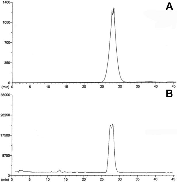
The isotope exchange reaction of 4a/4b was carried out with Na125I in presence of 10 mol % of Herrmann's catalyst in DMF at 110 °C for 1 h followed by semi-preparative HPLC purification. The reaction was also monitored by radioTLC (Supplemental Information). Both, the analytical HPLC UV chromatogram and radioanalytical trace of the purified product (13a/13b) displayed split peaks, which is consistent with the presence of two structural isomers (Figure 3). Specific activity, radiochemical yield, and radiochemical purity of 13a/13b were 2.55 μCi/mg, 8%, and 98 %, respectively. The specific activity obtained may be sufficient for preliminary in vitro uptake studies of 13a/13b.58 The primary intention of this isotope exchange reaction was proof-of-concept and no attempts were made to improve the specific activity and the radiochemical yield. This could be accomplished by varying reaction conditions and stoichiometries of reagents. Furthermore, the specific activity of the final product could be improved significantly by employing a “reversed” halogen exchange (B-79/81Br to B-125I) followed by the chromatographic separation of the halogenated species. Preliminary HPLC studies indicated that it may be possible to separate N5-127I (4a/b) from N5-79/81Br (12a/12b) (See Supporting Information).
Figure 3.
HPLC analysis of N5-125I (13a/13b). A: Radioanalytical trace; B: UV chromatogram. Split peaks are likely due to the presence of two positional isomers.
X-RAY CRYSTALLOGRAPHY
X-ray crystallography confirmed the formation of the structural isomers 8a and 8b with iodine and TBDMS group in meta- and para-orientation, respectively (Figure 4). Compound 8a crystallized with one molecule in the asymmetric unit (Z'=1), whereas compound 8b was obtained as a collection of four slightly different rotamers (Z'= 4 and labeled as A, B, C and D) (see Table 2 for crystallographic details and Figure S2 [Supplementary Information] for structures). The four rotamers of 8b have different torsion angles for C5-Si1-C1-C2, which are −131.8(2)° (A), −140.6(2)° (B), 138.5(2)° (C), and 142.1(2)° (D). Interestingly, the C5-Si1-C1-C2 torsion angle found for 8a was 6.3(2)°. Structure 8a does not show any significant I⋯I halogen bond-type interactions, whereas structure 8b does form two of these. Both structures show significant Ccarb-H⋯··-B interactions (see Table 3). These types of intermolecular interactions are similar to those observed by Puga et al. in the crystal lattices of iodinated o-carboranes.53 Table 3 lists selected bond lengths in 8a and in all four rotamers of 8b along with the Ccarb- H⋯··-B and I⋯··I interactions. The bond lengths found for both crystal structures were in agreement with those reported by Puga et al. for similar compounds.53
BIOLOGICAL STUDIES
Phosphoryl Transfer Assays
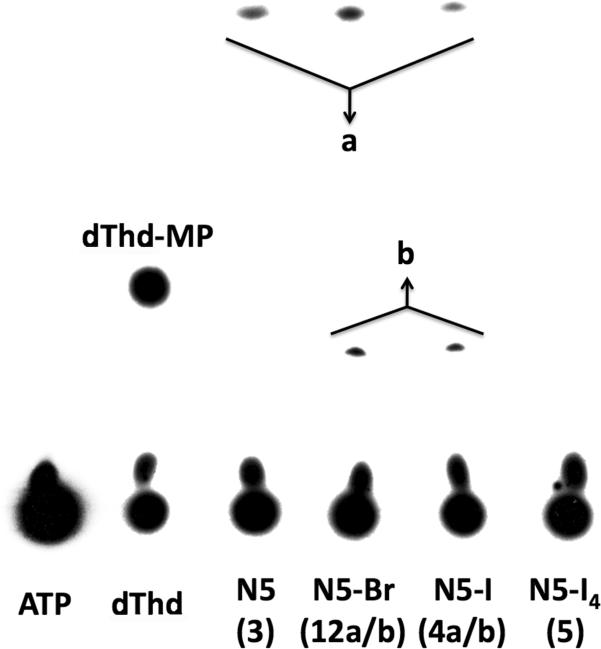
In order to evaluate their hTK1 substrate characteristics, phosphoryl transfer assays (PTAs)28,44 with recombinant hTK1 were carried with N5 (3), N5-127I (4a/4b), N5-79/81Br (12a/12b) and N5-I4(5). The results are shown in Figure 5 and Table 4. The phosphorylation rate of dThd, being a natural substrate of hTK1, is set to 100% and the phosphorylation rates of the test compounds are expressed relative to that of dThd. They indicate that substitution of hydrogen in the carborane cluster of 3 by either iodine (4a/4b) or bromine (12a/12b) only had minimal effects on the phosphorylation rates (38.0 % for 3 vs. 36.9 % for 12a/12b, vs. 29.6 % for 4a/4b). The observed phosphorylation rate for 3 was comparable to that reported previously for the same compound.28 The moderate decrease in phosphorylation of 4a/4b and 12a/12b as compared with 3 substantiates our hypothesis that the carborane cage of 3CTAs is located far away from the site of phosphorylation and that introduction of one iodine or bromine at the carborane cage did not significantly affect hTK1 substrate characteristics. On the other hand, the introduction of four iodine atoms at the carborane cage, as in compound 5, affected binding to hTK1 resulting in markedly decreased phosphorylation, which could not be accurately determined (Figure 5). The chromatogram shown in Figure 5 displays additional spots for two unidentified 32P-containing compounds (b) that were formed during phosphoryl transfer or the following work-up. It is conceivable that these spots were the monophosphates of the nido-carboranyl forms of 4a/4b and 12a/12b, since halogenated carboranes may be somewhat more susceptible to nido-formation than non-halogenated carboranes.48
Figure 5.
Phosphorylation of dThd, N5 (3), N5-Br (12a/b), N5-I (4a/b), and N5-I4 (5) by recombinant TK1. Assay products were separated by PEI–cellulose TLC.
a: Monophosphates of N5 (3) N5-I (4a/b), and N5-Br (12a/b).
b: Unidentified phosphorylation products with relative phosphorylation rates of 14 ± 0.7 % (N5-I lane) and 25 ±1.1 % (N5-Br lane)
Table 4.
Human TK1 phosphorylation rates (PRs) of N5 (3), N5-I (4a/b), and N5-Br (12a/b), relative to that dThd and in vitro uptake data of N5 (3), N5-I (4a/b), N5-Br (12a/b), and BPA.a
| Compounds | Phosphorylation Rate (%) | Uptake (μg B/109 cells) |
|
|---|---|---|---|
| L929 TK1 (+) | L929 TK1 (−) | ||
| dThd | 100 | nd | nd |
| N5(3) | 38.0 ± 2.2 | 77.6 ± 6.2 *** | 38.6±3.3 |
| N5-Br (12a/b) | 36.9 ± 0.4 | 76.0 ± 9.0 * | 54.0 ± 5.3 |
| N5-I (4a/b) | 29.6 ± 0.5 | 118.2 ± 24.3 ** | 65.5 ± 5.3 |
| BPA b | ndb | 48.0 ± 8.9 | 42.1 ± 13.6 |
Data represent the means of four replicates ± SD for Phosphorylation rates (PR), three replicates ± SD for cellular uptake studies with 3, 12 a/b, 5, and BPA, and 6 replicates ± SD for cellular uptake studies with 4 a/b.
P<0.05,
P<0.01,
P<0.001, compared with L929 TK1(−).
BPA: Boronophenylalanine, nd: not determined. BPA is not a nucleoside analogue. Hence PR determination was not applicable.
Cellular uptake studies
Uptake of 3, 12a/b, and 4a/b by L929 TK1 (+) cells was quantified by determining boron concentrations by means of ICP-OES (Table 4). These were 2 ×, 1.4 ×, and 1.8 × greater than those of L929 TK1 (−) cells indicating that TK1 activity may play a major role in the in vitro uptake of these compounds. However, there was no obvious correlation with the hTK1 phosphorylation rates of these compounds. We do not have an explanation for the uptake data of 4a/4b both in L929 TK1 (+) and TK1 (−) cells, which was higher than those of 3 and 12a/12b in the same cell lines. Compound 5 was not evaluated in the cellular uptake studies because of lack of material. However, cellular uptake of boronophenylalanine (BPA), a drug used clinically for BNCT,60 did not differ significantly significantly (P-value = 0.563) between L929 TK1 (+) and L929 TK1 (−), as determined by a two-sample t-test. This was expected since BPA is a boron-containing amino acid that is not a substrate of TK1. Uptake of 3, 12a/b, and 4a/b in L929 TK1 (−) cells was also very high indicating that other factors, such as compound lipophilicity, causing non-specific retention in cellular membranes, or nucleoside/nucleotide influx and/or efflux, may have played important roles in the uptake and retention of these compounds.
SUMMARY and CONCLUSIONS
Both synthetic strategies explored for the synthesis of 4a/4b were successful. However, even under optimized reaction conditions, strategy 1 generated a complex reaction mixture containing decomposition products, diiodinated material, and substantial quantities of unreacted 3. This was a major disadvantage of strategy 1 because it necessitated the purification of 4a/4b not only by conventional column chromatography but also by semi-preparative HPLC. On the other hand, strategy 1 also generated the tetraiodinated compound 5. Phosphoryl transfer assays indicated that this compound might not be a good substrate of hTK1, presumably because of increased bulkiness at the carborane cage. However, tetra-radioiodination potentially may be an important approach for other closo-carboranyl radiopharmaceuticals, especially those that might be used for cancer treatment. From this perspective, it is important to develop a general strategy for the tetraiodination of closo-carboranyl biomolecules, as reported in this study. In general, strategy 2 allowed for a more controlled synthesis of 4a/4b. Potentially, the separation of the isomers 4a and 4b is also possible using this strategy on the level of compound 8a and 8b. Radiohalogenation of 4a/4b to obtain 13a/13b was successful. Further optimization of reaction conditions should result in improved specific activity and radiochemical yield of 13a/13b. To the best of our knowledge, this is the first report of halogen- and isotope exchange reactions at a B-halocarborane, which is conjugated to a complex biomolecule such as dThd. Noteworthy is the observed stability of the B-I bond under a variety of reaction conditions. In general, the synthetic technology developed at the examples of 12a/12b and 13a/13b may pave the way for the synthesis of a wide range of carborane cage radiohalogenated therapeutics and diagnostics.
Only small differences in hTK1 phosphorylation rates were found between 3, 12a/12b, and 4a/4b. This supports our original assumption that monohalogenation of 3 will not drastically change its hTK1 substrate characteristics. In contrast, tetraiodination of 3, as in compound 5, notably decreased the hTK1 phosphorylation rate, indicating that there is a limit for size increase at the carborane cage. These findings also reinforce our initial hypothesis that mono(radio)iodination at the carborane cage of 3 is at least an appropriate tool to study the real time pharmacokinetics of this 3CTA. The phosphoryltransfer rates obtained for 3, 12a/12b, and 4a/4b were supported by a higher uptake in L929 TK1 (+) than in L929 TK1 (−) cells. However, uptake of all three compounds in L929 TK1 (−) cells was also substantial indicating that mechanisms other than KMT, such as passive diffusion, may have contributed to their accumulation.
Supplementary Material
ACKNOWLEDGMENT
The present studies were supported by funds from The Ohio State University College of Pharmacy, the Dipartimento di Chimica Generale e Chimica Organica Studi di Torino, Italy, NIH grant 1 R01 CA127935-01A2, and the National Center for Research Resources award UL1RR025755. The content is solely the responsibility of the authors and does not necessarily represent the official views of the National Center for Research Resources or the National Institutes of Health. The authors thank Drs. John Byrd and Fengting Yang from the Division of Hematology and Oncology, Department of Internal Medicine, at The Ohio State University for generously availing their laboratory facilities and assistance for work related to hTK1 expression, -purification, and -phosphoryl transfer assays. The authors thank George Hinkle, RPh, MS,□BCNP, FASHP, FAPhA, Director of Nuclear Pharmacy Services at The Ohio State University Medical Center, as well as Drs. Staffan Eriksson and Elena Sjuvarsson from the Biomedical Centre in Uppsala, Sweden, for helpful discussions.
Footnotes
SUPPORTING INFORMATION
Additional data, including additional general experimental information, 13C NMR chemical shifts, supporting synthetic, computational, chromatographic, and radioanalytical studies, as well as detailed X-ray crystallographic and spectroscopic information, are available in the Supporting Information. This material is available free of charge via the Internet at http://pubs.acs.org.
REFERENCES
- (1).This work was presented in part at the ACS National Meeting; Salt Lake City, UT, United States. March 22–26, 2009; and the XIX International Round Table on Nucleosides, Nucleotides and Nucleic Acids; Lyon, France, August 29 – September 3, 2010. [Google Scholar]
- (2).Adam MJ, Wilbur DS. Chem. Soc. Rev. 2005;34:153–163. doi: 10.1039/b313872k. [DOI] [PubMed] [Google Scholar]
- (3).Agarwal HK, Hasabelnaby S, Tiwari R, Tjarks W. Boron cluster (radio)halogenation in biomedical research. In: Hosmane NS, editor. Boron Science: New Technologies and Applications. CRC Press; Boca Raton: 2011. pp. 108–144. [Google Scholar]
- (4).Kassiou M, Loc'h C, Dolle F, Musachio JL, Dolci L, Crouzel C, Dannals RF, Mazière B. Appl. Rad. Isot. 2002;57:713–717. doi: 10.1016/s0969-8043(02)00187-2. [DOI] [PubMed] [Google Scholar]
- (5).Neumeyer JL, Wang S, Milius RA, Baldwin RM, Zea-Ponce Y, Hoffer PB, Sybirska E, Al-Tikriti M, Charney DS, Malisons RT, Laruelle M, Innis RB. J. Med. Chem. 1991;34:3144–3146. doi: 10.1021/jm00114a027. [DOI] [PubMed] [Google Scholar]
- (6).Marshall WJ, Young RJ, Jr., Grushin VV. Organometallics. 2001;20:523–533. [Google Scholar]
- (7).Shchukin E, Orlova A, Korsakov M, Sjoeberg S, Tolmachev V. J. Radioanal. Nucl. Chem. 2004;260:295–299. [Google Scholar]
- (8).Stanko VI, Iroshnikova NG. Zh. Obshch. Khim. 1970;40:311–315. [Google Scholar]
- (9).Winberg KJ, Barbera G, Eriksson L, Teixidor F, Tolmachev V, Vinas C, Sjoberg S. J. Organomet. Chem. 2003;680:188–192. [Google Scholar]
- (10).Winberg KJ, Mume E, Tolmachev V, Sjoeberg S. J. Labelled Comp. Radiopharm. 2005;48:195–202. [Google Scholar]
- (11).Winberg KJ, Persson M, Malmstrom P-U, Sjoeberg S, Tolmachev V. Nucl. Med. Biol. 2004;31:425–433. doi: 10.1016/j.nucmedbio.2003.11.007. [DOI] [PubMed] [Google Scholar]
- (12).Byun Y, Yan J, Al-Madhoun AS, Johnsamuel J, Yang W, Barth RF, Eriksson S, Tjarks W. J. Med. Chem. 2005;48:1188–1198. doi: 10.1021/jm0491896. [DOI] [PubMed] [Google Scholar]
- (13).Aspland SE, Ballatore C, Castillo R, Desharnais J, Eustaquio T, Goelet P, Guo Z, Li Q, Nelson D, Sun C, Castellino AJ, Newman MJ. Bioorg. Med. Chem. Lett. 2006;16:5194–5198. doi: 10.1016/j.bmcl.2006.07.003. [DOI] [PubMed] [Google Scholar]
- (14).Barth RF, Yang W, Al-Madhoun AS, Johnsamuel J, Byun Y, Chandra S, Smith DR, Tjarks W, Eriksson S. Cancer Res. 2004;64:6287–6295. doi: 10.1158/0008-5472.CAN-04-0437. [DOI] [PubMed] [Google Scholar]
- (15).Celen S, de Groot T, Balzarini J, Vunckx K, Terwinghe C, Vermaelen P, Van Berckelaer L, Vanbilloen H, Nuyts J, Mortelmans L, Verbruggen A, Bormans G. Nucl. Med. Biol. 2007;34:283–291. doi: 10.1016/j.nucmedbio.2007.01.003. [DOI] [PubMed] [Google Scholar]
- (16).Desbouis D, Struthers H, Spiwok V, Kuster T, Schibli R. J. Med. Chem. 2008;51:6689–6698. doi: 10.1021/jm800530p. [DOI] [PubMed] [Google Scholar]
- (17).Ghosh P, Pal A, Shavrin A, Bornmann W, Gelovani JG, Alauddin MM. Med. Chem. 2008;4:503–512. doi: 10.2174/157340608785700252. [DOI] [PubMed] [Google Scholar]
- (18).Struthers H, Viertl D, Kosinski M, Spingler B, Buchegger F, Schibli R. Bioconjugate Chem. 2010;21:622–634. doi: 10.1021/bc900380n. [DOI] [PubMed] [Google Scholar]
- (19).Tjarks W, Tiwari R, Byun Y, Narayanasamy S, Barth RF. Chem. Commun. 2007:4978–4991. doi: 10.1039/b707257k. [DOI] [PubMed] [Google Scholar]
- (20).Toyohara J, Hayashi A, Gogami A, Fujibayashi Y. Nucl. Med. Biol. 2006;33:765–772. doi: 10.1016/j.nucmedbio.2006.06.001. [DOI] [PubMed] [Google Scholar]
- (21).Toyohara J, Hayashi A, Gogami A, Hamada M, Hamashima Y, Katoh T, Node M, Fujibayashi Y. Nucl. Med. Biol. 2006;33:751–764. doi: 10.1016/j.nucmedbio.2006.06.003. [DOI] [PubMed] [Google Scholar]
- (22).Barth RF, Yang W, Wu G, Swindall M, Byun Y, Narayanasamy S, Tjarks W, Tordoff K, Moeschberger ML, Eriksson S, Binns PJ, Riley KJ. Proc. Natl. Acad. Sci. U. S. A. 2008;105:17493–17497. doi: 10.1073/pnas.0809569105. [DOI] [PMC free article] [PubMed] [Google Scholar]
- (23).Al-Madhoun AS, Johnsamuel J, Barth RF, Tjarks W, Eriksson S. Cancer Res. 2004;64:6280–6286. doi: 10.1158/0008-5472.CAN-04-0197. [DOI] [PubMed] [Google Scholar]
- (24).Bodei L, Kassis AI, Adelstein SJ, Mariani G. Cancer Biother. Radiopharm. 2003;18:861–877. doi: 10.1089/108497803322702833. [DOI] [PubMed] [Google Scholar]
- (25).Kassis AI, Adelstein SJ. J. Nucl. Med. 1996;37:10S–12S. [PubMed] [Google Scholar]
- (26).Kassis AI, Adelstein SJ, Mariani G. Q. J. Nucl. Med. 1996;40:301–319. [PubMed] [Google Scholar]
- (27).Kassis AI, Kirichian AM, Wang K, Semnani ES, Adelstein SJ. Int. J. Radiat. Biol. 2004;80:941–946. doi: 10.1080/09553000400017671. [DOI] [PubMed] [Google Scholar]
- (28).Lunato AJ, Wang J, Woollard JE, Anisuzzaman AK, Ji W, Rong FG, Ikeda S, Soloway AH, Eriksson S, Ives DH, Blue TE, Tjarks W. J. Med. Chem. 1999;42:3378–3389. doi: 10.1021/jm990125i. [DOI] [PubMed] [Google Scholar]
- (29).Knapp FF, Srivastava PC, Callahan AP, Cunningham EB, Kabalka GW, Sastry KAR. J. Med. Chem. 1984;27:57–63. doi: 10.1021/jm00367a011. [DOI] [PubMed] [Google Scholar]
- (30).Andrews JS, Zayas J, Jones M., Jr. Inorg. Chem. 1985;24:3715–3716. [Google Scholar]
- (31).Barbera G, Vaca A, Teixidor F, Sillanpaa R, Kivekas R, Vinas C. Inorg. Chem. 2008;47:7309–7316. doi: 10.1021/ic800362z. [DOI] [PubMed] [Google Scholar]
- (32).Otwinowski Z, Minor W. Methods Enzymol. 1997;276:307–326. doi: 10.1016/S0076-6879(97)76066-X. [DOI] [PubMed] [Google Scholar]
- (33).Blessing RH. Cryst. Rev. 1987;1:3–58. [Google Scholar]
- (34).Blessing RH. J. Appl. Cryst. 1989;22:396–397. [Google Scholar]
- (35).Sheldrick GM. Acta Crystallogr. 2008;A64:112–122. doi: 10.1107/S0108767307043930. [DOI] [PubMed] [Google Scholar]
- (36).Farrugia LJ. J. Appl. Crystallogr. 1999;32:837–838. [Google Scholar]
- (37).International Tables for Crystallography. Vol C. Kluwer Academic Publishers; Dortrecht, The Netherlands: 1992. [Google Scholar]
- (38).Bernardinelli G, Flack HD. Acta Crystallogr. 1985;A41:500–511. [Google Scholar]
- (39).Flack HD. Acta Crystallogr. 1983;A39:876–881. [Google Scholar]
- (40).Jain AN. J. Med. Chem. 2003;46:499–511. doi: 10.1021/jm020406h. [DOI] [PubMed] [Google Scholar]
- (41).Segura-Pena D, Lutz S, Monnerjahn C, Konrad M, Lavie A. J. Mol. Biol. 2007;369:129–141. doi: 10.1016/j.jmb.2007.02.104. [DOI] [PMC free article] [PubMed] [Google Scholar]
- (42).Arnold K, Bordoli L, Kopp J, Schwede T. Bioinformatics. 2006;22:195–201. doi: 10.1093/bioinformatics/bti770. [DOI] [PubMed] [Google Scholar]
- (43).Tiwari R, Mahasenan K, Pavlovicz R, Li C, Tjarks W. J. Chem. Inf. Model. 2009;49:1581–1589. doi: 10.1021/ci900031y. [DOI] [PMC free article] [PubMed] [Google Scholar]
- (44).Wang L, Munch-Petersen B, Herrström Sjöberg A, Hellman U, Bergman T, Jörnvall H, Eriksson S. FEBS lett. 1999;443:170–174. doi: 10.1016/s0014-5793(98)01711-6. [DOI] [PubMed] [Google Scholar]
- (45).Narayanasamy S, Thirumamagal BTS, Johnsamuel J, Byun Y, Al-Madhoun AS, Usova E, Cosquer GY, Yan J, Bandyopadhyaya AK, Tiwari R, Eriksson S, Tjarks W. Bioorg. Med. Chem. 2006;14:6886–6899. doi: 10.1016/j.bmc.2006.06.039. [DOI] [PubMed] [Google Scholar]
- (46).Segura-Pena D, Lichter J, Trani M, Konrad M, Lavie A, Lutz S. Structure. 2007;15:1555–1566. doi: 10.1016/j.str.2007.09.025. [DOI] [PMC free article] [PubMed] [Google Scholar]
- (47).Byun Y, Thirumamagal BTS, Yang W, Eriksson S, Barth RF, Tjarks W. J. Med. Chem. 2006;49:5513–5523. doi: 10.1021/jm060413w. [DOI] [PubMed] [Google Scholar]
- (48).Grimes RN. Carboranes. 2nd ed. Elsevier; Amsterdam: 2011. [Google Scholar]
- (49).Yamazaki H, Ohta K, Endo Y. Tetrahedron Lett. 2005;46:3119–3122. [Google Scholar]
- (50).Potenza JA, Lipscomb WN. Inorg. Chem. 1966;5:1483–1488. [Google Scholar]
- (51).Potenza JA, Lipscomb WN. Inorg. Chem. 1966;5:1478–1482. [Google Scholar]
- (52).Kusari U, Li Y, Bradley MG, Sneddon LG. J. Am. Chem. Soc. 2004;126:8662–8663. doi: 10.1021/ja048018n. [DOI] [PubMed] [Google Scholar]
- (53).Puga AV, Teixidor F, Sillanpää R, Kivekäs R, Viñas C. Chem.-Eur. J. 2009;15:9764–9772. doi: 10.1002/chem.200900926. [DOI] [PubMed] [Google Scholar]
- (54).Gomez FA, Hawthorne MF. J. Org. Chem. 1992;57:1384–1390. [Google Scholar]
- (55).Gomez FA, Johnson SE, Hawthorne MF. J. Am. Chem. Soc. 1991;113:5915–5917. [Google Scholar]
- (56).Wuts PGM, Greene TW. Greene's Protective Groups in Organic Synthesis. 4th ed. John Wiley & Sons; New Jersey: 2007. [Google Scholar]
- (57).Fox MA, Wade K. J. Organomet. Chem. 1999;573:279–291. [Google Scholar]
- (58).Maini A, Hillman G, Haas GP, Wang CY, Montecillo E, Hamzavi F, Pontes JE, Leland P, Pastan I, Debinski W, Puri RK. J. Urol. 1997;158:948–953. doi: 10.1097/00005392-199709000-00077. [DOI] [PubMed] [Google Scholar]
- (59).Macrae CF, Bruno IJ, Chisholm JA, Edgington PR, McCabe P, Pidcock E, Rodriguez-Monge L, Taylor R, van de Streek J, Wood PA. J. Appl. Crystallogr. 2008;41:466–470. [Google Scholar]
- (60).Barth RF, Coderre JA, Vicente MGH, Blue TE. Clin. Cancer Res. 2005;11:3987–4002. doi: 10.1158/1078-0432.CCR-05-0035. [DOI] [PubMed] [Google Scholar]
Associated Data
This section collects any data citations, data availability statements, or supplementary materials included in this article.


