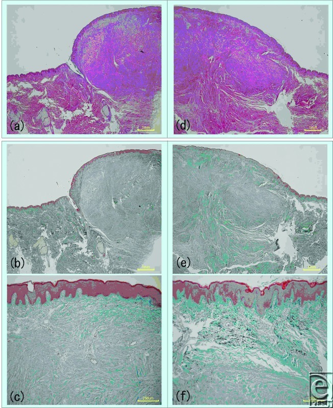
Figure 4.
Changes in keloid tissue after laser irradiation, as observed by light microscopy. Half of the precordial keloid of a 26-year-old woman was irradiated with laser. Three days later, the unirradiated (left) and irradiated keloid halves (right) were excised and examined by hematoxylin and eosin staining (top images) and Elastica Masson-Goldner (middle and bottom images) staining. Structural changes were observed in the collagen of the irradiated tissues. Intense inflammatory cell infiltration was observed by hematoxylin and eosin staining.

