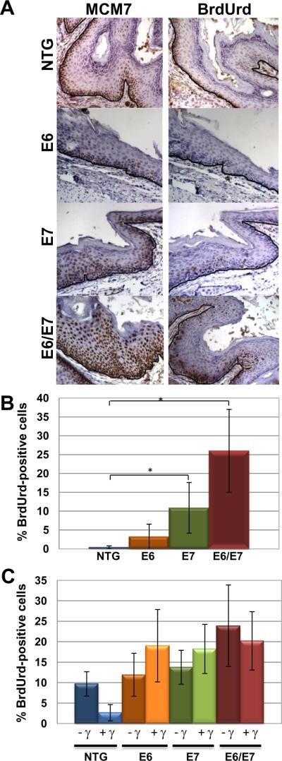
Figure 1. Acute phenotypes of HPV16 transgenic mouse anal epithelium.
Panel A. Shown is MCM7-specific and BrdUrd immunohistochemical staining of sections from the anus of nontransgenic (NTG), E6, E7 and E6/E7 transgenic mice. Strong induction of MCM7 occurs in E7 and E6/E7 anal epithelium compared to NTG and E6 anal tissue. Similar staining is seen with BrdUrd staining with suprabasal DNA synthesis occurring more frequently in E7 and E6/E7 mice. Brown nuclei indicate positive staining which is expected along the basement membrane (outlined in black) as seen in NTG mice. Panel B. Quantification of the percentage of suprabasal cells with BrdUrd-positive nuclei. Panel C. Response of anal epithelium to radiation-induced growth arrest. Shown is the quantification of the frequency of BrdUrd-positive basal cells in anal epithelia of HPV transgenic mice that were (+) or were not (−) treated with 12Gy (γ) external radiation.

