Abstract
Mutations in DNMT3A encoding DNA methyltransferase 3A were recently described in patients with acute myeloid leukemia. To assess their prognostic significance, we determined the mutational status of DNMT3A exon 23 in 288 patients with AML excluding acute promyelocytic leukemia, aged from 18 to 65 years and treated in Toulouse University Hospital. A mutation was detected in 39 patients (13.5%). All DNMT3A exon 23+ patients had intermediate-risk cytogenetics. Mutations significantly correlated with a higher WBC count (p<0.001), NPM1 (p<0.001) and FLT3-ITD mutations (p=0.027). DNMT3A mutations were conserved through xenotransplantation in immunodeficient mice. No difference in outcome between DNMT3A exon 23+ and DNMT3A exon 23- patients was found even if the results were stratified by NPM1 or FLT3-ITD status. However, DNMT3A exon 23+ patients had better median DFS (not reached vs 11.6 months, p=0.009) and OS (not reached vs 14.3 months, p=0.005) as compared to DNMT3A exon 23- patients when treated with idarubicin, whereas patients treated with daunorubicin had similar outcome regardless the DNMT3A status. This study shows that DNMT3A mutations have no impact on outcome but could be a predictive factor for response to idarubicin and thus, could have a direct influence in the way AML patients should be managed.
Keywords: DNMT3A, acute leukemia, idarubicin, NOD/SCID, xenograft
INTRODUCTION
Acute myeloid leukemia (AML), most widespread acute leukemia in adults, is characterized by clonal proliferation of oncogenic event-transformed hematopoietic stem cells or progenitors. Despite a high rate of complete remission after treatment with intensive chemotherapy combining cytarabine and anthracyclines in schemas that have little changed during the past 30 years, relapse rates are very high, resulting in a poor outcome in most cases with 5-year overall survival of 40% in younger adults and only 10-15% in elderly patients [1]. Nevertheless, therapeutic results drastically vary regarding the main prognostic factors including age, performance status, cytogenetic and molecular abnormalities. Indeed, the knowledge of molecular basis of AML has considerably increased in the past few years mostly through the identification of recurrent mutational events occurring in a substantial number of patients [2].
The leukemic clone emerges from normal hematopoietic stem cells or more mature myeloid progenitors after acquisition of gene mutations affecting cell differentiation and self-renewal (such as the fusion genes PML-RARA, AML1-ETO, CBFb-MYH11 or point mutations targeting the functions of CEBPA, RUNX1, MLL or NPM1), cell signaling (FLT3, RAS or KIT), epigenetic machinery (TET2) and cell metabolism (IDH1 or IDH2) [3-4]. These mutations particularly occur in AML without chromosomal abnormalities detected (normal karyotypes) and represent a major issue in the clinical management of patients since they could provide targets for both therapy (i.e., tyrosine kinase inhibitors in FLT3-ITD positive AML) and molecular monitoring of residual disease. However, the major impact of these mutations resides in the stratification of consolidation therapy once complete response is achieved following induction chemotherapy. It is now generally accepted that patients with normal karyotype and favorable genotype (i.e., NPM1 or CEBPA mutations without FLT3-ITD mutations) are no longer referred to allogeneic hematopoietic stem-cell transplantation (HSCT) in first complete response since their outcome with chemotherapy alone is similar to those receiving HSCT [5]. In addition, patients with an unfavorable genotype are more readily directed to allograft from unrelated donors or cord blood units since their prognosis with chemotherapy alone is dismal. Thus, FLT3-ITD, CEBPA, NPM1 mutations are now part of the initial work up of all AML patients with intermediate cytogenetic risk who undergo intensive treatments [6]. However, newly discovered mutations such as isocitrate dehydrogenase 1 and 2 (IDH1/2) mutations have been already proposed to refine this molecular stratification although their prognostic impact remains to be definitely established [7-8].
Even more recently, by sequencing the genome of leukemic cells from a patient with AML, Ley et al have detected somatic mutations in the gene of a DNA methyltransferase (MTase), DNMT3A [9]. DNMT3A is a member of the DNA MTases family including DNMT1, DNMT2, DNMT3A and DNMT3B that are involved in the methylation of CpG islands [10]. The hypermethylation of CpG islands that contributes to the downregulation of gene expression, and notably of tumor suppressor genes, is also a hallmark of AML [11]. In a series of 281 de novo AML, DNMT3A mutations were found in 22% of cases, clustering in intermediate-risk cytogenetic group and associated with a poor outcome. Most of the DNMT3A mutations (60%) found in AML samples were missense mutations localized on the R882 amino acid of the MTase domain. It is noteworthy that R882 mutations were significantly associated with a high white blood cell count as compared with other DNMT3A mutations. The aim of our study was to confirm the prognostic impact of R882 DNMT3A mutations in a series of 288 AML patients treated in our institution and to evaluate its screening usefulness in the medical diagnosis process, in a similar way to KIT exon 17 screening.
PATIENTS AND METHODS
Patients and treatments
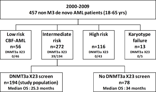
From 2000 to 2009, 457 consecutive patients (18-65 years) with untreated de novo AML (excluding acute promyelocytic leukemia and secondary AML) were admitted for intensive chemotherapy induction at the Hematology department of Toulouse University hospital. In this cohort of patients, 288 samples were available for genetic screening. Since DNMT3A mutations were only observed in the intermediate-cytogenetic risk group, the therapeutic outcome was assessed in this population (n=194). Figure 1 shows the flow chart of all AML patients in this period of time. From this cohort of 194 intermediate-risk patients, those ≤ 60 years (n=156) received either daunorubicin (dnr) (60 mg/m2, d1-3) (n=112) or idarubicin (ida) (8 mg/m2, d1-5) (n=44) with cytarabine (AraC) (200 mg/m2, d1-7). A second course (ida, 8mg/m2 or dnr, 35mg/m2 d17-18 and AraC, 1g/m2/12h d17-20) was delivered if more than 5% marrow blasts persisted on d15 [12]. Patients were treated according to institutional guidelines using daunorubicin as main anthracycline while some of them received idarubicin as part of a randomized clinical trial [12]. Patients in complete response (CR) with HLA-identical sibling were planned to receive allo-HSCT: (i) after a myeloablative conditioning regimen consisting of oral busulfan (16 mg/kg over 4 days) and cyclophosphamide (120 mg/kg over 2 days) for patients less than 51 years; (ii) after one course of intensive consolidation described below and reduced intensity conditioning, (oral busulfan, 8 mg/kg over 2 days, fludarabine, 120 mg/m2 over 4 days, rabbit anti-thymocyte globulins 2,5 mg/kg on d-4 and d-3) for patients between 51 and 60 years. Patients with no HLA-identical sibling received a consolidation regimen (ida, 12 mg/m2 or dnr, 60 mg/m2 d1-2 and AraC 3 g/m2/12h d1-4) then autologous HSCT (auto-HSCT) prepared with busulfan (4 mg/kg, d-6 to d-3) and melphalan (140 mg/m2, d-2) or two courses of high-dose AraC (HiDAC). Since 2008, patients with intermediate-risk cytogenetic and favorable genotype (mutation of NPM1 or CEBPA without FLT3-ITD mutation) were no longer allocated to allo-HSCT in first CR and mainly received three cycles of HiDAC as consolidation. The induction chemotherapy for patients older than 60 years (n=38) combined ida (8 mg/m2, d1-5), AraC (100 mg/m2, d1-7) with (n=17) or without (n=21) lomustine (200 mg/m2, d1). Patients achieving CR received a consolidation with ida (8 mg/m2, d1-3) and AraC (50 mg/m2/12h, d1-5) then maintenance therapy with ida at d1 only and the same scheme of AraC. Bone marrow AML samples were obtained after informed consent in accordance with the Declaration of Helsinki. All samples were stored in the HIMIP tumor bank of the U1037 Inserm department (n°DC-2008-307-CPTP1 HIMIP). The study was approved by the institutional review board.
Figure 1. Flow chart of AML patients treated by intensive chemotherapy between 2000 and 2009.
From 2000 to 2009, 457 consecutive patients with de novo AML were treated by intensive chemotherapy. Patients with acute promyelocytic leukemia or with secondary AML were excluded from this study.
Mutation screening
The mutational status of DNMT3A exon 23 along with 3 additional mutations (FLT3 TKD exon 20, IDH1 and IDH2 exons 4) were analyzed by high-resolution melting (HRM) PCR using the LightCycler 480 with High Melting Resolution Master Mix 1X (Roche Applied Science), with 10 ng genomic DNA, 0.1 μmol/l of each primer (see below), and 25 mmol/l MgCl2. HRM PCR cycling conditions were initial denaturation at 95°C for 10 minutes, followed by 50 cycles at 95°C for 10 seconds, 50 cycles at 63°C for 15 seconds, and 50 cycles at 72°C for 25 seconds. Melting curve was measured from 72°C to 95°C, with 25 acquisitions per degree centigrade. Primer sequences were as follows: DNMT3A_X23_F1, 5′-CTG GCC AGC ACT CAC CCT-3′; DNMT3A_X23_R1, 5′-TGT TTA ACT TTG TGT CGC TAC CTC A-3′; FLT3_X20_F2, 5′-TCA CAG AGA CCT GGC CGC-3′; FLT3_X20_R1, 5′-TGC CCC TGA CAA CAT AGT TGG-3′; IDH1_X4_F1, 5′-GGC TTG TGA GTG GAT GGG TAA-3′; IDH1_X4_R2, 5′-GCA TTT CTC AAT TTC ATA CCT TGC TTA-3′; IDH2_X4_R140_F1, 5′-GAA AGA TGG CGG CTG CAG T-3′; IDH2_X4_R140_R3, 5′-TGT TTT TGC AGA TGA TGG GC-3′; IDH2_X4_R172_F4, 5′-GAT GTG GAA AAG TCC CAA TGG A-3′; IDH2_X4_R172_R2, 5′-CAC CCT GGC CTA CCT GGT C-3′. Positive cases detected by the HRM analysis were sequenced to confirm the mutation. FLT3 exon 13 ITD and NPM1 mutation screening were performed using a multiplex PCR using Gold Taq DNA polymerase (Applied Biosystems) and the following primers: HsNPM1_X12_F1, 5′-GAA GTG TTG TGG TTC CTT AAC-3′; HsNPM1_X12_R1FAM, 5′(FAM)-TGG ACA ACA CAT TCT TGG CA-3′; FLT3_NEM_E, 5′-TGG TGT TTG TCT CCT CTT CAT TGT-3′; and FLT3_NEM_Qned, 5′(NED)-GTT GCG TTC ATC ACT TTT CCA A-3′. PCR products were analyzed on a sequencer using sizing fragment analysis. CEBPA screening was performed according to Pabst et al [13].
NOD/SCID mice xenograft
Adult NOD/SCID mice (6-8 weeks old) were sublethally irradiated with 250 cGy of total body irradiation 24 h before injection of leukemic cells. Leukemia samples were thawed at room temperature, washed twice and suspended in PBS at a final concentration of 1-2 million cells per 200 μL of PBS per mouse for IV injection. Daily monitoring of mice for symptoms of disease (ruffled coat, hunched back, weakness, or reduced motility) determined the time of killing for injected animals with signs of distress. If no sign of distress was seen, mice were analyzed 12 weeks after injection except as otherwise noted. For assessment of leukemic engraftment, NOD/SCID mice were humanely killed in accordance with IACUC protocols. Bone marrow (mixed from tibias and femurs) and spleen were dissected in a sterile environment, flushed in PBS and made into single cell suspensions for analysis by flow cytometry (FACS Calibur, BD Biosciences, San Jose, CA USA).
Statistical analysis
Comparisons of patient characteristics (covariates) were performed using the Mann-Whitney test for continuous variables and the Fisher's exact test for categorical variables. Complete remission (CR) was defined following Cheson criteria [14]. In univariate analysis, covariates associated with response to induction therapy or outcome were identified using Fisher's exact test then included in a multivariable logistic model. Overall survival (OS) and disease-free survival (DFS) rates were measured from the date of diagnosis until death and from the date of CR until death or relapse, respectively. Patients alive were censored at the time of last contact. OS and DFS were estimated by the Kaplan-Meier method and compared using the log-rank test. All calculations were performed using GraphPad Prism software, version 5.0 (GraphPad Software Inc., La Jolla, CA). Survival-time data (DFS and OS) and covariates were analyzed using the backward method of Cox proportional hazards regression.
RESULTS
DNMT3A exon 23 mutation is a frequent event in adult AML
DNMT3A exon 23 screening was performed on available samples coming from 288 AML patients aged from 18 to 65-year old and treated in Toulouse between 2000 and 2009. DNMT3A exon 23 mutations were detected in 39 patients (13.5%) occurring almost exclusively at the R882 codon (23 R882H; 13 R882C; 2 R882P) and in one patient at the W893 codon (W893S). No association between DNMT3A exon 23 mutations and age or sex was detected.
DNMT3A exon 23 mutations occur exclusively in intermediate cytogenetic risk group
Patients were cytogenetically subdivided according to the MRC 2010 classification resulting in 46 patients with low risk, 194 with intermediate risk and 43 with high risk whereas five patients were not classified in absence of karyotypes [15]. Patients with a DNMT3A exon 23 mutation were exclusively identified in the intermediate-risk group (39/194, 20%) as compared to CBF and high-risk AML (p<0.001), 27 with a normal karyotype and 12 with various associated abnormalities, the most frequent being an additional copy of the chromosome 8 (3 patients) or 13 (2 patients). No DNMT3A exon 23 mutation was detected in high or low risk groups. Consequently, we focused the analysis exclusively on the 194 patients with intermediate-risk.
DNMT3A exon 23 mutations are associated with FAB M4/M5 subtypes and a higher WBC count
The characteristics of intermediate-risk patients according to DNMT3A mutational status are listed in table 1. A bias toward monocytic differentiation (FAB groups M4 and M5) was identified in DNMT3A exon 23+ samples (p<0.0001). A strong association was found between DNMT3A exon 23+ and a higher white blood cell count (p=0.001). Platelet count, hemoglobin concentration and marrow blast percentage at diagnosis were similar in DNMT3A exon 23+ and – groups.
Table 1. Characteristics of AML patients with intermediate-risk according to DNMT3A exon 23 mutation.
| No DNMT3A exon 23 mutation | DNMT3A exon 23 mutation | p | |
|---|---|---|---|
| Sex - no. | |||
| Male | 78 | 17 | 0.48 |
| Female | 77 | 22 | |
| Age - y | |||
| Median | 53 | 47 | 0.051 |
| Range | 18-65 | 20-63 | |
| WBC - G/L | |||
| Median | 14.6 | 52 | <0.0001 |
| Range | 0.8-356 | 1.0-250 | |
| Hb - g/dL | |||
| Median | 9.6 | 10.4 | 0.22 |
| Range | 4.3-16 | 4.8-13.2 | |
| Platelets - G/L | |||
| Median | 70 | 62 | 0.54 |
| Range | 5-964 | 8-814 | |
| FAB AML subtypes - no. | |||
| M0/M1/M2 | 4/43/45 | 0/3/6 | <0.0001 |
| M4 /M5 | 26/27 | 17/11 | |
| M6/M7 | 0/0 | 0/0 | |
| Unclassified | 10 | 1 | |
| Normal Karyotype no. (%) | 96 (62) | 27 (69) | 0.46 |
| Mutations - no. /total no. (%) | |||
| FLT3-ITD | 37/155 (24) | 17/39 (44) | 0.027 |
| FLT3-TKD | 3/62 (5) | 1/14 (7) | 0.57 |
| NPM1 | 49/155 (32) | 29/39 (74) | <0.0001 |
| CEBPA | 20/155 (13) | 2/39 (5) | 0.26 |
| IDH1 | 16/155 (10) | 8/39 (21) | 0.10 |
| IDH2 | 16/155 (10) | 5/39 (13) | 0.77 |
| KIT exon 17 | 2/155 (1) | 0/39 (0) | - |
| Complete response - no. (%) | 126/155 (81) | 34/39 (87) | 0.48 |
| Allogeneic SCT - no./total no. (%) | 40/155 (26) | 16/39 (41) | 0.075 |
| Autologous SCT - no./total no. (%) | 28/155 (18) | 7/39 (18) | 1.00 |
| Relapse - no./total no. (%) | 63/126 (50) | 14/34 (41) | 0.36 |
| 5-year survival (%) | 36.5 | 47.3 | - |
WBC, white blood cell count; FAB, French American British; SCT, stem-cell transplantation.
DNMT3A exon 23 mutations are associated with NPM1 and FLT3-ITD mutations
DNMT3A exon 23 mutations were significantly associated with FLT3-ITD (17 FLT3-ITD/39 DNMT3A exon 23+, 44% vs. 37/155 DNMT3A exon 23-, 24%, p=0.027), NPM1 mutations (29 NPM1c/39 DNMT3A exon 23+, 74% vs. 49/155 DNMT3A exon 23-, 32%, p< 0.001). No relationship was identified between DNMT3A exon 23 mutations and CEBPA mutations, IDH1 R132, IDH2 R140 and R172 mutations, FLT3 tyrosine kinase domain or KIT exon 17 mutations. The significant relationship between DNMT3A exon 23 mutations and NPM1 mutations was independent of the FLT3-ITD mutation status (p<0.001 in FLT3-ITD+ or FLT3-ITD- in both groups). In contrast, the relationship between DNMT3A exon 23 mutations and FLT3-ITD mutations was lost in the NPM1 mutation subgroups (p=0.700 in NPM1c - and p=0.043 in NPM1c + patients), suggesting that the relationship between DNMT3A and NPM1 mutations was stronger than the relationship between DNMT3A and FLT3-ITD mutations.
DNMT3A exon 23 mutations are conserved in xenograft mice models
We next analyzed the mutational status of nine primary AML samples which showed engraftment capacities in xenograft NOD/SCID mice model. The presence of FLT3-ITD (seven out of nine, data not shown) correlated with the ability to engraft in these mice as it has been shown earlier [16-18]. Furthermore, we have also observed that four of these nine specimens carried the DNMT3A exon 23+ mutations that were associated with FLT3-ITD and NPM1 mutations (Table 2). For these four samples, we also analyzed their post-transplantation mutational status and found that DNMT3A exon 23+ mutations were conserved in NOD/SCID mice. Overall, these data show a stability of the DNMT3A mutations in AML engrafted mice and suggest a preferential engraftment of primary AML specimens with triple FLT3-ITD/NPM1/DNMT3A mutations in immunodeficient mice.
Table 2. Distribution of FLT3-ITD, NPM1 and DNMT3A exon 23 mutations in AML samples sorted from NOD/SCID mice.
| Patient# | Tx status | Engraftment | Gender | FAB | Karyotype | Status | DNMT3A | FLT3 | NPM1 | IDH1 | IDH2 | IDH2 | CEBPA | KIT |
|---|---|---|---|---|---|---|---|---|---|---|---|---|---|---|
| mean% | Dx/Rel | R132 | R140 | R172 | ||||||||||
| LAM018 | Pre | M | 5 | normal | Dx | + | ITD | + | WT | WT | WT | WT | WT | |
| Post | 83 | + | ITD | + | WT | WT | WT | WT | WT | |||||
| LAM016 | Pre | M | 5 | normal | Rel | + | ITD | WT | WT | WT | WT | WT | WT | |
| Post | 81 | + | ITD | WT | WT | WT | WT | WT | WT | |||||
| LAM002 | Pre | M | 1 | normal | Dx | + | ITD | + | WT | WT | WT | WT | WT | |
| Post | 38 | + | ITD | + | WT | WT | WT | WT | WT | |||||
| LAM007 | Pre | F | 4 | normal | Dx | + | ITD | + | WT | WT | WT | WT | WT | |
| Post | 7 | + | ITD | + | WT | WT | WT | WT | WT |
Tx, transplantation; FAB, French American British; Dx, diagnosis; Rel, relapse; WT, wild type; ND, not done.
DNMT3A exon 23 mutations have no prognostic impact in adult AML with intermediate-risk cytogenetics
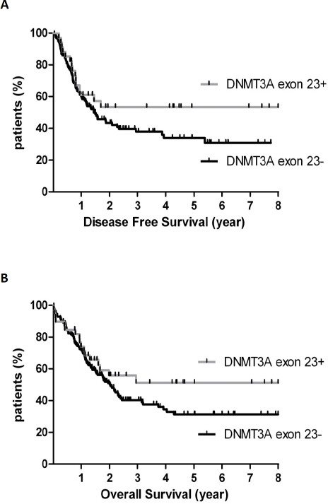
Analysis of the therapeutic outcome was performed for the 194 intermediate-risk patients. Of these patients, 160/194 (83%) achieved CR after induction chemotherapy. The CR rate did not differ according to DNMT3A exon 23 mutational status, with 34/39 (87%) and 126/155 (81%) CR in the DNMT3A exon 23+ group and DNMT3A exon 23- group, respectively (p=0.48). CR rate was significantly influenced by leukocytosis with a cut-off at WBC>30 G/L (95% CI, 0.14-0.68, OR, 0.3, p=0.01) but not by age, CEBPA, NPM1 or FLT3-ITD mutations. In the DNMT3A exon 23+ group, 16 (41%) and 7 (18%) patients in first CR received allo-SCT or auto-SCT as consolidation therapy, not significantly different from the DNMT3A exon 23- group in which 40 (26%) and 28 (18%) received allo-SCT or auto-SCT, respectively. In these complete responders, 15 events were observed in the DNMT3A exon 23+ group and 72 events in the DNMT3A exon 23- group (44% and 57% of CR patients, respectively). Disease-free survival was not significantly different between DNMT3A exon 23+ (median not reached) and DNMT3A exon 23- patients (median DFS, 17.6 months) (95% CI, 0.87-2.33, HR 1.42, p=0.16) (figure 2A). There were 17 deaths (44%) in the DNMT3A exon 23+ group and 88 (57%) in the DNMT3A exon 23- group. Overall survival was not significantly different between DNMT3A exon 23+ (median not reached) and DNMT3A exon 23- patients (median OS, 24.7 months) (95% CI, 0.87-2.19, HR 1.38, p=0.17) (figure 2B). Among age, WBC count, DNMT3A exon 23 mutations and FLT3/NPM1 genotypes, the only factor that significantly influenced both DFS and OS was the FLT3wt/NPM1c genotype (not shown).
Figure 2. Prognostic impact of DNMT3A exon 23 mutations in 194 AML patients with intermediate-cytogenetic risk.
(A) Disease-free (DFS) and (B) overall survival (OS) in patients with or without DNMT3A exon 23 mutations. DFS and OS were not different between DNMT3A exon 23+ and DNMT3A exon 23- patients (p=0.16 and p=0.17, respectively).
AML patients with DNMT3A exon 23 mutations may benefit from idarubicin as compared to daunorubicin
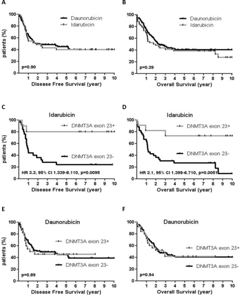
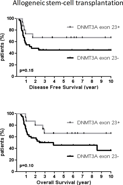
As we did not find any prognostic impact of DNMT3A exon 23+ mutations in contrast to previous studies, we tried to find out whether treatments received by DNMT3A exons 23+ patients could explain these differences.[9;19-20] Because patients who were older than 60 received idarubicin and lomustin but also because they were much less frequently allografted, we focalized our analysis on younger patients (60y or less). The characteristics of patients according to the type of anthracycline used at induction are reported in table 3. There was no difference between patients treated by daunorubicin and those treated by idarubicin in terms of DFS (median DFS, 22.7 months for DNR vs 22.8 months for Ida, p=0.90) and OS (median OS, 28.5 months for DNR vs 24.4 months for Ida, p=0.39). Analysis of covariates associated with both DFS and OS are shown in table S1-S2. However, there was a significant impact of DNMT3A exon 23 mutations in patients treated with idarubicin. DNMT3A exons 23+ patients had better DFS (not reached vs 11.6 months, HR 3.3, 95% CI, 1.34-8.11, p=0.009) and OS (not reached vs 14.3 months, HR 3.1, 95% CI, 1.39-6.71, p=0.005) as compared to DNMT3A exons 23- patients when treated with idarubicin whereas patients treated with daunorubicin had similar outcome regardless the DNMT3A mutational status (figure 3). Conversely, the outcome of patients with NPM1+/FLT3wt genotype was not impacted by the type of anthracycline used in induction (Figure S1). In patients who received allo-SCT (n=53), there was a trend for improved outcome in DNMT3A exon 23+ patients (DFS and OS medians not reached) as compared to DNMT3A exon 23- patients (median DFS, 19.1 months, p=0.15; median OS, 29.2 months, p=0.1) but the difference did not reach statistical significance (figure 4). To better address the effects of idarubicin vs. daunorubicin along with other variables in DNMT3A mutated patients, we performed univariate and multivariate analysis. As shown in table 4, idarubicin had an independent favorable prognostic effect on OS (HR 0.27, 95% CI, 0.08-0.97, p=0.046) when considering age, karyotype, NPM1/FLT3wt genotype, WBC and allo-SCT in DNMT3A mutated patients.
Table 3. Characteristics of patients with intermediate-cytogenetic risk according to the type of anthracycline used at induction treatment.
| No DNMT3A exon 23 mutation | DNMT3A exon 23 mutation | p | |||
|---|---|---|---|---|---|
| DNR | IDA | DNR | IDA | ||
| Sex - no. | |||||
| Male | 39 | 20 | 8 | 6 | 0.16 |
| Female | 48 | 13 | 17 | 5 | |
| Age - y | |||||
| Median | 49 | 50 | 47 | 46 | 0.81 |
| Range | 21-60 | 18-60 | 20-60 | 35-60 | |
| WBC - G/L | |||||
| Median | 14.5 | 17.9 | 52 | 41.3 | 0.0037 |
| Range | 0.8-356 | 1.8-220 | 1.7-249 | 1.0-250 | |
| Mutations - no. /total no. (%) | |||||
| FLT3-ITD | 20/87 (23) | 11/33 (33) | 11/25 (44) | 4/11 (36) | 0.19 |
| NPM1 | 30/87 (34) | 5/33 (15) | 19/25 (76) | 8/11 (73) | <0.0001 |
| CEBPA | 12/87 (13) | 6/33 (18) | 2/25 (8) | 0/11 (0) | 0.38 |
| IDH1 | 11/87 (13) | 2/33 (6) | 6/25 (24) | 2/11 (18) | 0.24 |
| IDH2 | 10/87 (11) | 2/33 (6) | 3/25 (12) | 1/11 (9) | 0.83 |
| Complete response - no. (%) | 73/87 (84) | 25/33 (76) | 22/25 (88) | 10/11 (91) | 0.53 |
| Allogeneic SCT - no./total no. (%) | 27/73 (36) | 11/25 (44) | 10/22 (45) | 5/10 (50) | 0.71 |
| Relapse - no./total no. (%) | 32/73 (44) | 17/25 (68) | 12/22 (55) | 1/10 (10) | 0.014 |
| 5-year survival (%) | 40.1 | 26.9 | 41.4 | 72.7 | - |
DNR, daunorubicin; IDA, idarubicin; WBC, white blood cell count; SCT, stem cell transplantation.
Figure 3. Impact of idarubicin in 156 AML patients ≤ 60y according to DNMT3A exon 23 mutations.
DFS (A) and OS (B) according to daunorubicin or idarubicin treatment. DFS (C) and OS (D) in patients treated by idarubicin according to DNMT3A exon 23 mutations. DFS (E) and OS (F) in patients treated by daunorubicin according to DNMT3A exon 23 mutations.
Figure 4. Outcome of allografted patients according to DNMT3A exon 23 mutations.
DFS (A) and OS (B) in DNMT3A exon 23+ patients (n = 15) and DNMT3A exon 23- patients (n=38) who were allografted in first complete response.
Table 4. Univariate and Multivariate Analysis for DFS and OS in DNMT3A exon 23 mutated patients.
Analysis of covariates associated with DFS and OS. P of the univariate analysis is the p value of the Log rank test. HR is the value of the hazard ratio. 95% CI is the 95% confident interval of the hazard ratio. Data of AML patients with DNMT3A exon 23 mutations were complete and were included in the Cox proportional-hazards regression. NPM1: Nucleophosmin; FLT3-ITD: internal tandem duplication of the FLT3 gene; Allo-SCT: Allogeneic stem-cell transplantation; Ida: idarubicin; WBC: white blood cell count.
| Univariate analysis | Multivariate analysis | |||||
|---|---|---|---|---|---|---|
| DFS | p | HR | 95% CI | p | HR | 95% CI |
| Age > 50y | 0.94 | 1.04 | 0.35-3.09 | >0.1 | ||
| NPM1+/FLT3-ITD- | 0.49 | 0.70 | 0.25-1.95 | >0.1 | ||
| Normal karyotype | 0.95 | 1.04 | 0.33-3.24 | >0.1 | ||
| Allo-SCT | 0.13 | 0.45 | 0.16-1.26 | >0.1 | ||
| Ida treatment | 0.036 | 0.32 | 0.11-0.93 | 0.052 | 0.21 | 0.05-1.01 |
| WBC count > 30G/L | 0.55 | 1.43 | 0.45-4.56 | >0.1 | ||
| OS | ||||||
| Age > 50y | 0.76 | 1.17 | 0.43-3.14 | >0.1 | ||
| NPM1+/FLT3-ITD- | 0.52 | 0.74 | 0.29-1.89 | >0.1 | ||
| Normal karyotype | 0.88 | 1.08 | 0.39-2.99 | >0.1 | ||
| Allo-SCT | 0.06 | 0.41 | 0.16-1.04 | 0.053 | 0.36 | 0.12-1.01 |
| Ida treatment | 0.046 | 0.37 | 0.14-0.98 | 0.046 | 0.27 | 0.08-0.97 |
| WBC count > 30G/L | 0.2 | 2.06 | 0.69-6.13 | >0.1 | ||
DISCUSSION
In accordance with the first study assessing DNMT3A mutations in AML, we confirm here the high prevalence of DNMT3A exon 23 mutations in a series of 288 de novo adults AML [9]. We have also observed that these mutations were exclusively found within the intermediate-risk cytogenetic group, associated with M4/M5 FAB subtypes and hyperleukocytosis. At the molecular level, DNMT3A exon 23 mutations were strongly associated with NPM1 mutations and to a lesser extent with FLT3-ITD mutations whereas no correlation was found with CEBPA, KIT or IDH1/2 mutations.
However, in contrast with previous studies, we failed to show any prognostic impact of DNMT3A exon 23 mutations in AML with intermediate-risk cytogenetic [9;19-20]. We acknowledge that our patients had been treated over a long period of time (nine years) and this could have potentially introduced some bias. Indeed, two major changes in the management of AML patients had been undertaken in our center during this period of time: the introduction of primary prophylaxis of invasive fungal infections using azoles (voriconazole from 2003 to 2008, posaconazole thereafter) [21] and the stratification of allo-HSCT indication according to the new molecular classification of intermediate-risk AML using CEBPA, FLT3 and NPM1 mutations. Indeed, patients with wild type FLT3 and CEBPA or NPM1 mutations were no longer allocated to allo-HSCT in first CR since the results published by Schlenk et al.[5]. However, since these two measures were applied in 2003 and 2008 respectively, their impact on outcome, if any, could have been negligible in the whole cohort of patients. Furthermore, our induction and consolidation regimen have little changed for fifteen years, particularly the dosing of anthracyclines, with daunorubicin always given at 60 mg/m2/day for three days at time of the induction chemotherapy. By contrast, the doses of anthracyclines are not fully described in the study of Ley et al, in which several induction regimen were used, some of them using the infra optimal dose of 45 mg/m2/d [22-24]. Moreover, some patients received hypomethylating agents or lenalidomide which generally induce fewer responses than intensive chemotherapy in AML patients [25-26]. Also, complete response and early death rates were not mentioned and this could be of interest as patients with R882 DNMT3A mutations usually have a high white blood cell count, a recognized risk factor for early death. In our study, DNMT3A exon 23 mutations did not impact on both complete response and early death rates. Moreover, we found that patients with DNMT3A exon 23 mutations could specifically benefit from idarubicin although the small number of patients in our study requires confirmation in larger cohorts. The strong impact of DNMT3A mutations on the response to idarubicin as compared to daunorubicin is quite unexpected. However, it has been recently shown that DNMT3A could play a role in anthracyclines-induced apoptosis of colorectal cancer cells. Indeed, the expression of DNMT3A is upregulated at apoptosis-inducing concentrations of doxorubicin and involved in p21 repression thereby blocking senescence [27]. Whether this program is induced by idarubicin but not daunorubicin in DNMT3A mutated/haploinsufficient cells remains to be determined. Alternatively, DNMT3A mutations could specifically impact on the expression of genes involved in idarubicin metabolism as compared to daunorubicin. The broader spectrum of activity of idarubicin has been attributed to increased lipophilicity, cellular uptake and improved stabilization of a ternary drug-topoisomerase II-DNA complex [28]. Thus, the discrepancy in therapeutic outcome between our series and those previously described could be due to differences in treatment intensity or altered metabolism of anthracyclines. Several clinical trials have already assessed the impact of daunorubicin dose intensification or compared idarubicin vs. daunorubicin in large cohorts of AML patients. Reassessing results of these controlled studies in light of DNMT3A mutational status could easily confirm or not our preliminary observations [12;22-23;29-30]. It remains also to be determined if DNMT3A mutations affect in a similar way the metabolism of other compounds that intercalates DNA and inhibits topoisomerase II such as the new quinolone derivative vosaroxin which is currently assessed in AML [31].
In addition, we were also able to demonstrate for the first time that DNMT3A mutations are also found in leukemic samples engrafted in immunodeficient mice. Although we could not demonstrate that the DNMT3A exon 23 mutation is a surrogate marker of engraftment in NOD/SCID mice, it should be noted that all mutated samples tested recapitulated the initial disease in mice. By comparison, it has been shown that only 50% of intermediate-risk cytogenetic samples display engraftment properties in NOD/SCID mice [32]. This suggests that engraftment properties of AML samples should be assessed in light of specific molecular lesions. It has been shown that engraftment in xenograft NOD/SCID-IL2Rγc−/− mice model did not correlate with French-American-British subtype or cytogenetic abnormalities but with the presence of FLT3-ITD [17]. More recently, Sarry et al. have observed that a high engraftment level occurred within human primary AML samples carrying at least two mutations (seven out of nine samples had FLT3-ITD and NPM1 mutations) and that these mutations were strongly conserved in the different leukemic stem cell populations sorted as well as still present after the first transplantation into NOD/SCID-IL2Rγc−/− mice [18]. In the present study, we show that the DNMT3A mutation is also conserved through the NOD/SCID xenograft model and is associated with higher leukemic engraftment level in cooperation with FLT3-ITD and NPM1 mutations, suggesting that this mutation occurs in the early leukemogenic events and belongs to the main leukemic clone over the course of the disease progression.
In summary, our data confirm that DNMT3A exon 23 mutations can be easily evaluated in medical practice and represent a frequent molecular event in intermediate-risk AML associated with NPM1 and FLT3-ITD mutations but have no clear impact on disease-free and overall survival. However, although this finding needs to be confirmed in largest cohort of patients, DNMT3A mutations could be a predictive factor for response to idarubicin and, thus could have a direct influence in the way AML patients should be managed.
Supplementary Figure and Tables
Acknowledgments
This work was supported by grants from the GREMS association (Groupe de Recherche et d'Enseignement des Maladies du Sang), and the McLaughlin Foundation, Quebec, Canada. The Centre de Ressource Biologique des Hémopathies Malignes de l'INSERM Midi-Pyrénées (CRB HIMIP) provided all AML samples.
Footnotes
O.L designed, performed the research, analyzed data; and wrote the paper; S.B collected all clinical data; F.V performed statistical analysis; JE.S, L.C, A.P and A.K performed in vivo xenograft transplantation assays; V.D.M and C.D centralized the cytological review, performed molecular analysis and provided AML samples from the HIMIP collection; C.C and AC.S performed molecular analysis; A.S collected clinical data; F.H and A.H treated patients; E.D and C.R. designed, controlled, analyzed data, and wrote the paper. All authors checked the final version of the manuscript.
CR declared consultancy and/or advisory board from Celgene, is an advisory board member of Genzyme and received grant support from Celgene and Pierre Fabre. Other authors declare no competing financial interests.
REFERENCES
- 1.Tallman MS. New strategies for the treatment of acute myeloid leukemia including antibodies and other novel agents. Hematology Am Soc Hematol Educ Program. 2005:143–50. doi: 10.1182/asheducation-2005.1.143. [DOI] [PubMed] [Google Scholar]
- 2.Foran JM. New prognostic markers in acute myeloid leukemia: perspective from the clinic. Hematology Am Soc Hematol Educ Program. 2010;2010:47–55. doi: 10.1182/asheducation-2010.1.47. [DOI] [PubMed] [Google Scholar]
- 3.Marcucci G, Haferlach T, Dohner H. Molecular genetics of adult acute myeloid leukemia: prognostic and therapeutic implications. J Clin Oncol. 2011;29:475–86. doi: 10.1200/JCO.2010.30.2554. [DOI] [PubMed] [Google Scholar]
- 4.Renneville A, Roumier C, Biggio V, Nibourel O, Boissel N, Fenaux P, Preudhomme C. Cooperating gene mutations in acute myeloid leukemia: a review of the literature. Leukemia. 2008;22:915–31. doi: 10.1038/leu.2008.19. [DOI] [PubMed] [Google Scholar]
- 5.Schlenk RF, Dohner K, Krauter J, Frohling S, Corbacioglu A, Bullinger L, Habdank M, Spath D, Morgan M, Benner A, Schlegelberger B, Heil G, Ganser A, Dohner H. Mutations and treatment outcome in cytogenetically normal acute myeloid leukemia. N Engl J Med. 2008;358:1909–18. doi: 10.1056/NEJMoa074306. [DOI] [PubMed] [Google Scholar]
- 6.Dohner H, Estey EH, Amadori S, Appelbaum FR, Buchner T, Burnett AK, Dombret H, Fenaux P, Grimwade D, Larson RA, Lo-Coco F, Naoe T, Niederwieser D, Ossenkoppele GJ, Sanz MA, Sierra J, et al. Diagnosis and management of acute myeloid leukemia in adults: recommendations from an international expert panel, on behalf of the European LeukemiaNet. Blood. 2010;115:453–74. doi: 10.1182/blood-2009-07-235358. [DOI] [PubMed] [Google Scholar]
- 7.Mardis ER, Ding L, Dooling DJ, Larson DE, McLellan MD, Chen K, Koboldt DC, Fulton RS, Delehaunty KD, McGrath SD, Fulton LA, Locke DP, Magrini VJ, Abbott RM, Vickery TL, Reed JS, et al. Recurring mutations found by sequencing an acute myeloid leukemia genome. N Engl J Med. 2009;361:1058–66. doi: 10.1056/NEJMoa0903840. [DOI] [PMC free article] [PubMed] [Google Scholar]
- 8.Dang L, Jin S, Su SM. IDH mutations in glioma and acute myeloid leukemia. Trends Mol Med. 2010;16:387–97. doi: 10.1016/j.molmed.2010.07.002. [DOI] [PubMed] [Google Scholar]
- 9.Ley TJ, Ding L, Walter MJ, McLellan MD, Lamprecht T, Larson DE, Kandoth C, Payton JE, Baty J, Welch J, Harris CC, Lichti CF, Townsend RR, Fulton RS, Dooling DJ, Koboldt DC, et al. DNMT3A mutations in acute myeloid leukemia. N Engl J Med. 2010;363:2424–33. doi: 10.1056/NEJMoa1005143. [DOI] [PMC free article] [PubMed] [Google Scholar]
- 10.Xu F, Mao C, Ding Y, Rui C, Wu L, Shi A, Zhang H, Zhang L, Xu Z. Molecular and enzymatic profiles of mammalian DNA methyltransferases: structures and targets for drugs. Curr Med Chem. 2010;17:4052–71. doi: 10.2174/092986710793205372. [DOI] [PMC free article] [PubMed] [Google Scholar]
- 11.Figueroa ME, Lugthart S, Li Y, Erpelinck-Verschueren C, Deng X, Christos PJ, Schifano E, Booth J, van Putten W, Skrabanek L, Campagne F, Mazumdar M, Greally JM, Valk PJ, Lowenberg B, Delwel R, et al. DNA methylation signatures identify biologically distinct subtypes in acute myeloid leukemia. Cancer Cell. 2010;17:13–27. doi: 10.1016/j.ccr.2009.11.020. [DOI] [PMC free article] [PubMed] [Google Scholar]
- 12.Chevallier P, Fornecker L, Lioure B, Bene MC, Pigneux A, Recher C, Witz B, Fegueux N, Bulabois CE, Daliphard S, Bouscary D, Vey N, Delain M, Bay JO, Turlure P, Bernard M, et al. Tandem versus single autologous peripheral blood stem cell transplantation as post-remission therapy in adult acute myeloid leukemia patients under 60 in first complete remission: results of the multicenter prospective phase III GOELAMS LAM-2001 trial. Leukemia. 2010;24:1380–5. doi: 10.1038/leu.2010.111. [DOI] [PubMed] [Google Scholar]
- 13.Pabst T, Mueller BU, Zhang P, Radomska HS, Narravula S, Schnittger S, Behre G, Hiddemann W, Tenen DG. Dominant-negative mutations of CEBPA, encoding CCAAT/enhancer binding protein-alpha (C/EBPalpha), in acute myeloid leukemia. Nat Genet. 2001;27:263–70. doi: 10.1038/85820. [DOI] [PubMed] [Google Scholar]
- 14.Cheson BD, Bennett JM, Kopecky KJ, Buchner T, Willman CL, Estey EH, Schiffer CA, Doehner H, Tallman MS, Lister TA, Lo-Coco F, Willemze R, Biondi A, Hiddemann W, Larson RA, Lowenberg B, et al. Revised recommendations of the International Working Group for Diagnosis, Standardization of Response Criteria, Treatment Outcomes, and Reporting Standards for Therapeutic Trials in Acute Myeloid Leukemia. J Clin Oncol. 2003;21:4642–9. doi: 10.1200/JCO.2003.04.036. [DOI] [PubMed] [Google Scholar]
- 15.Grimwade D, Hills RK, Moorman AV, Walker H, Chatters S, Goldstone AH, Wheatley K, Harrison CJ, Burnett AK. Refinement of cytogenetic classification in acute myeloid leukemia: determination of prognostic significance of rare recurring chromosomal abnormalities among 5876 younger adult patients treated in the United Kingdom Medical Research Council trials. Blood. 2010;116:354–65. doi: 10.1182/blood-2009-11-254441. [DOI] [PubMed] [Google Scholar]
- 16.Rombouts WJ, Martens AC, Ploemacher RE. Identification of variables determining the engraftment potential of human acute myeloid leukemia in the immunodeficient NOD/SCID human chimera model. Leukemia. 2000;14:889–97. doi: 10.1038/sj.leu.2401777. [DOI] [PubMed] [Google Scholar]
- 17.Sanchez PV, Perry RL, Sarry JE, Perl AE, Murphy K, Swider CR, Bagg A, Choi JK, Biegel JA, Danet-Desnoyers G, Carroll M. A robust xenotransplantation model for acute myeloid leukemia. Leukemia. 2009;23:2109–17. doi: 10.1038/leu.2009.143. [DOI] [PMC free article] [PubMed] [Google Scholar]
- 18.Sarry JE, Murphy K, Perry R, Sanchez PV, Secreto A, Keefer C, Swider CR, Strzelecki AC, Cavelier C, Recher C, Mansat-De Mas V, Delabesse E, Danet-Desnoyers G, Carroll M. Human acute myelogenous leukemia stem cells are rare and heterogeneous when assayed in NOD/SCID/IL2Rgammac-deficient mice. J Clin Invest. 2011;121:384–95. doi: 10.1172/JCI41495. [DOI] [PMC free article] [PubMed] [Google Scholar]
- 19.Yan XJ, Xu J, Gu ZH, Pan CM, Lu G, Shen Y, Shi JY, Zhu YM, Tang L, Zhang XW, Liang WX, Mi JQ, Song HD, Li KQ, Chen Z, Chen SJ. Exome sequencing identifies somatic mutations of DNA methyltransferase gene DNMT3A in acute monocytic leukemia. Nat Genet. 2011;43:309–15. doi: 10.1038/ng.788. [DOI] [PubMed] [Google Scholar]
- 20.Thol F, Damm F, Ludeking A, Winschel C, Wagner K, Morgan M, Yun H, Gohring G, Schlegelberger B, Hoelzer D, Lubbert M, Kanz L, Fiedler W, Kirchner H, Heil G, Krauter J, et al. Incidence and prognostic influence of DNMT3A mutations in acute myeloid leukemia. J Clin Oncol. 2011;29:2889–96. doi: 10.1200/JCO.2011.35.4894. [DOI] [PubMed] [Google Scholar]
- 21.Chabrol A, Cuzin L, Huguet F, Alvarez M, Verdeil X, Linas MD, Cassaing S, Giron J, Tetu L, Attal M, Recher C. Prophylaxis of invasive aspergillosis with voriconazole or caspofungin during building work in patients with acute leukemia. Haematologica. 2010;95:996–1003. doi: 10.3324/haematol.2009.012633. [DOI] [PMC free article] [PubMed] [Google Scholar]
- 22.Lowenberg B, Ossenkoppele GJ, van Putten W, Schouten HC, Graux C, Ferrant A, Sonneveld P, Maertens J, Jongen-Lavrencic M, von Lilienfeld-Toal M, Biemond BJ, Vellenga E, van Marwijk Kooy M, Verdonck LF, Beck J, Dohner H, et al. High-dose daunorubicin in older patients with acute myeloid leukemia. N Engl J Med. 2009;361:1235–48. doi: 10.1056/NEJMoa0901409. [DOI] [PubMed] [Google Scholar]
- 23.Fernandez HF, Sun Z, Yao X, Litzow MR, Luger SM, Paietta EM, Racevskis J, Dewald GW, Ketterling RP, Bennett JM, Rowe JM, Lazarus HM, Tallman MS. Anthracycline dose intensification in acute myeloid leukemia. N Engl J Med. 2009;361:1249–59. doi: 10.1056/NEJMoa0904544. [DOI] [PMC free article] [PubMed] [Google Scholar]
- 24.Moore JO, George SL, Dodge RK, Amrein PC, Powell BL, Kolitz JE, Baer MR, Davey FR, Bloomfield CD, Larson RA, Schiffer CA. Sequential multiagent chemotherapy is not superior to high-dose cytarabine alone as postremission intensification therapy for acute myeloid leukemia in adults under 60 years of age: Cancer and Leukemia Group B Study 9222. Blood. 2005;105:3420–7. doi: 10.1182/blood-2004-08-2977. [DOI] [PMC free article] [PubMed] [Google Scholar]
- 25.Fenaux P, Mufti GJ, Hellstrom-Lindberg E, Santini V, Gattermann N, Germing U, Sanz G, List AF, Gore S, Seymour JF, Dombret H, Backstrom J, Zimmerman L, McKenzie D, Beach CL, Silverman LR. Azacitidine prolongs overall survival compared with conventional care regimens in elderly patients with low bone marrow blast count acute myeloid leukemia. J Clin Oncol. 2010;28:562–9. doi: 10.1200/JCO.2009.23.8329. [DOI] [PubMed] [Google Scholar]
- 26.Fehniger TA, Uy GL, Trinkaus K, Nelson AD, Demland J, Abboud CN, Cashen AF, Stockerl-Goldstein KE, Westervelt P, DiPersio JF, Vij R. A phase 2 study of high-dose lenalidomide as initial therapy for older patients with acute myeloid leukemia. Blood. 2011;117:1828–33. doi: 10.1182/blood-2010-07-297143. [DOI] [PMC free article] [PubMed] [Google Scholar]
- 27.Zhang Y, Gao Y, Zhang G, Huang S, Dong Z, Kong C, Su D, Du J, Zhu S, Liang Q, Zhang J, Lu J, Huang B. DNMT3a plays a role in switches between doxorubicin-induced senescence and apoptosis of colorectal cancer cells. Int J Cancer. 2011;128:551–61. doi: 10.1002/ijc.25365. [DOI] [PubMed] [Google Scholar]
- 28.Minotti G, Menna P, Salvatorelli E, Cairo G, Gianni L. Anthracyclines: molecular advances and pharmacologic developments in antitumor activity and cardiotoxicity. Pharmacol Rev. 2004;56:185–229. doi: 10.1124/pr.56.2.6. [DOI] [PubMed] [Google Scholar]
- 29.Pautas C, Merabet F, Thomas X, Raffoux E, Gardin C, Corm S, Bourhis JH, Reman O, Turlure P, Contentin N, de Revel T, Rousselot P, Preudhomme C, Bordessoule D, Fenaux P, Terre C, et al. Randomized study of intensified anthracycline doses for induction and recombinant interleukin-2 for maintenance in patients with acute myeloid leukemia age 50 to 70 years: results of the ALFA-9801 study. J Clin Oncol. 2010;28:808–14. doi: 10.1200/JCO.2009.23.2652. [DOI] [PubMed] [Google Scholar]
- 30.Ohtake S, Miyawaki S, Fujita H, Kiyoi H, Shinagawa K, Usui N, Okumura H, Miyamura K, Nakaseko C, Miyazaki Y, Fujieda A, Nagai T, Yamane T, Taniwaki M, Takahashi M, Yagasaki F, et al. Randomized study of induction therapy comparing standard-dose idarubicin with high-dose daunorubicin in adult patients with previously untreated acute myeloid leukemia: the JALSG AML201 Study. Blood. 2011;117:2358–65. doi: 10.1182/blood-2010-03-273243. [DOI] [PubMed] [Google Scholar]
- 31.Hawtin RE, Stockett DE, Wong OK, Lundin C, Helleday T, Fox JA. Homologous recombination repair is essential for repair of vosaroxin-induced DNA double-strand breaks. Oncotarget. 2010;1:606–19. doi: 10.18632/oncotarget.195. [DOI] [PMC free article] [PubMed] [Google Scholar]
- 32.Pearce DJ, Taussig D, Zibara K, Smith LL, Ridler CM, Preudhomme C, Young BD, Rohatiner AZ, Lister TA, Bonnet D. AML engraftment in the NOD/SCID assay reflects the outcome of AML: implications for our understanding of the heterogeneity of AML. Blood. 2006;107:1166–73. doi: 10.1182/blood-2005-06-2325. [DOI] [PMC free article] [PubMed] [Google Scholar]
Associated Data
This section collects any data citations, data availability statements, or supplementary materials included in this article.


