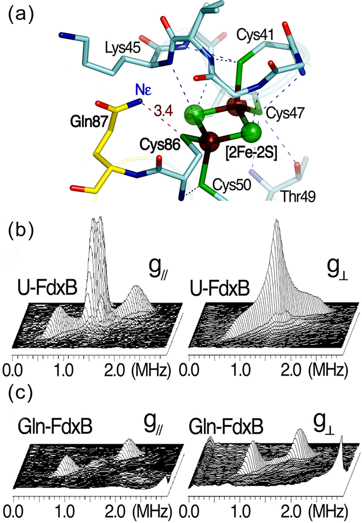
Figure 6.
The X-ray crystal structure and 2D pulsed EPR studies on the [2Fe-2S](Cys)4 cluster binding site of P. putida FdxB. (a) The hydrogen bond network around the [2Fe-2S](Cys)4 cluster binding site in the 1.90-Å structure of P. putida FdxB (PDB code: 3AH7) [19], (b) the 2D ESEEM characterization of weakly coupled 15N nuclei in uniformly 15N-labeled FdxB (U-FdxB) and (c) selectively Gln15Nε-labeled FdxB (Gln-FdxB) in the reduced state. The 2D ESEEM spectra in 3D stacked presentation (b,c) were measured at the low-field edge of the EPR line near g// (342.5 mT, left) and the intermediate position at g⊥ (357 mT, right). The cross-peaks for 15Nε of Gln87, which is involved in the N-H…S hydrogen bond with Cys86 Sγ (a), were clearly resolved with the hfi splitting of ~0.9 MHz ((2.01, 0.92) MHz and 15A=1.09 MHz near g// (left); (1.95, 1.13) MHz and 15A=0.82 MHz at g⊥ (right)) in the Gln-FdxB spectra (c).

