Abstract
Osteomas of the facial bones are a rare entity and very few cases have been reported in the literature. Osteomas are benign neoplasms, often asymptomatic and consist of well-differentiated matured bone. There are three varieties of osteomas- the central type arising from the endosteum, the peripheral type arising from the periosteum, and the extra-skeletal soft tissue osteomas which usually develops within the muscle. In the facial bones, both central and peripheral osteomas have been described. Peripheral osteomas have been described to occur in the frontal, ethmoid, and maxillary sinuses, but are not common in jawbones. We describe a rare case of symptomatic peripheral osteoma of mandible in a middle-aged female patient.
Keywords: Benign neoplasm, mandible, osteoma, peripheral type, symptomatic
INTRODUCTION

Osteoma is a benign osteogenic lesion characterized by proliferation of compact or cancellous bone. It can be central, peripheral, or of an extraskeletal type. The central osteoma arises from the endosteum, the peripheral osteoma from the periosteum, and the extraskeletal soft tissue osteoma usually develops within a muscle.[1]
Peripheral osteoma is an uncommon lesion, mostly occurring in young adults, which affects equally men and women. It mainly affects the frontal bone, mandible, and paranasal sinuses. Mandibular cases occur in the angle or condyle, followed by the molar area of the mandibular body and ascending ramus.[2]
Intraoral cases occur frequently in the lingual molar-premolar area .and rarely in anterior lingual alveolar cortical plate of the mandible.[3]
Clinically, most of the lesions are pedunculated unilateral masses. Occlusal dysfunction and facial asymmetry are the most common findings in condylar osteomas. Multiple osteomas of the jaws are commonly observed in Gardner syndrome.[2]
Although trauma, inflammatory or infectious processes are the causes commonly cited in literature, no etiological factor can be associated with this case.[4]
CASE REPORT
A 34-year-old female patient visited the Department of Oral Medicine and Radiology, with the chief complaint of swelling on the left side of the face that had persisted for 6 months [Figure 1]. Two years prior to this, the patient had pain in the left body of the mandible, which was dull, continuous and radiating to the temple region. Patient gave no history of trauma to the region and no evidence of pathology was noted during the clinical examination and radiological investigations. She was advised regular follow-up, however, patient did not adhere to this instruction.
Figure 1.
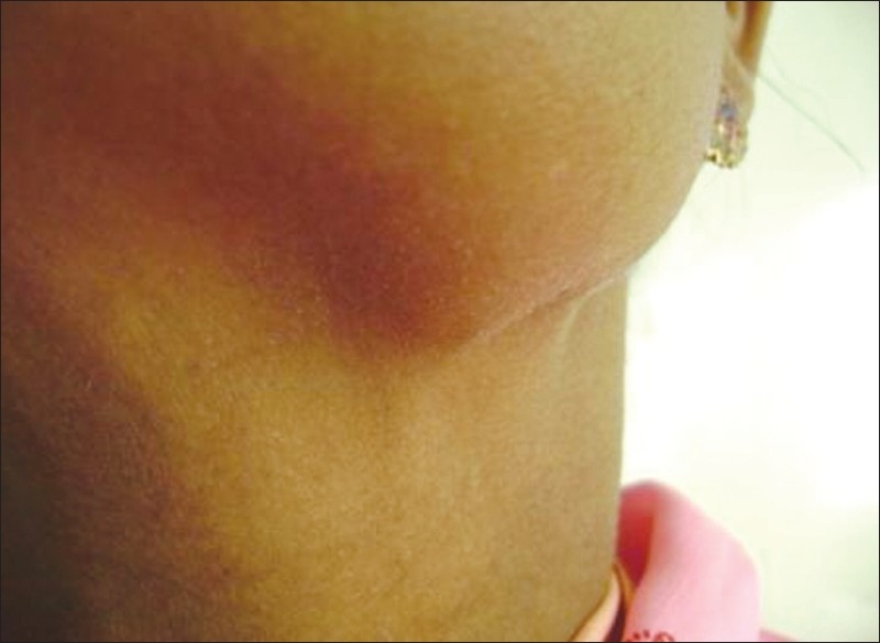
A 34-year-old female patient with a swelling on the left side of the face.
The current lesion presented as a well-defined swelling over the left lower border of the body of the mandible, 1 cm in front of the angle, 1×1 cm in size, oval in shape, bony hard, tender, and attached to the lower border of mandible, with no local rise in temperature. The overlying skin was normal in color and was not attached to underlying structure. Intraoral examination revealed lingually tilted tooth number 35 and periodontal pocket in relation to teeth 37 and 38 with no significant finding with respect to the swelling [Figure 2].
Figure 2.
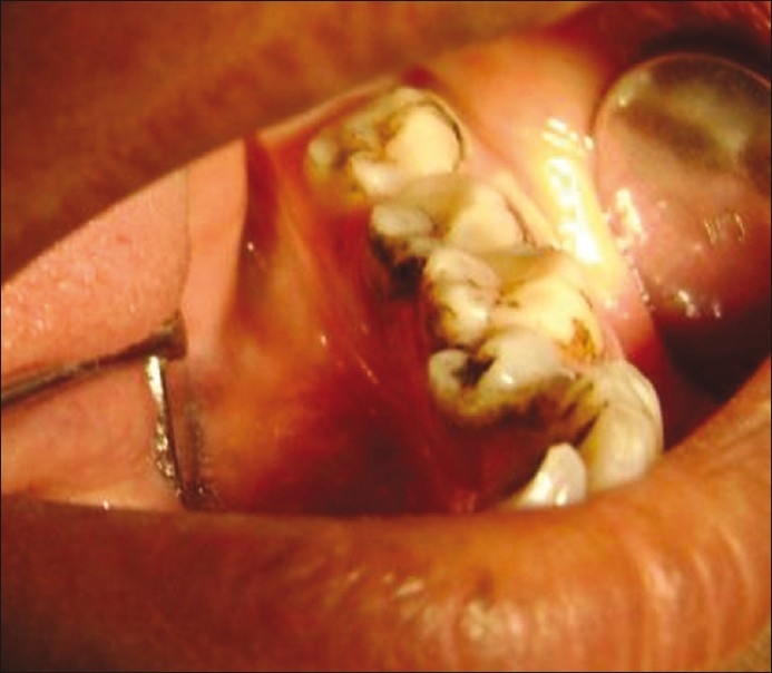
Intraoral view of 34-year-old female shows lingually tilted tooth 35 and periodontal pocket in relation to teeth 37 and 38.
On the basis of history and clinical examination, provisional diagnosis of osteoma of left body of mandible was given. Differential diagnosis of exostosis and osteoblastoma was considered. The patient was subjected to routine hematological and radiographical examination. The orthopantamograph [Figure 3] view revealed a well-defined radiopaque mass, round in shape, measuring 1×1 cm over the left inferior border of the mandible.
Figure 3.
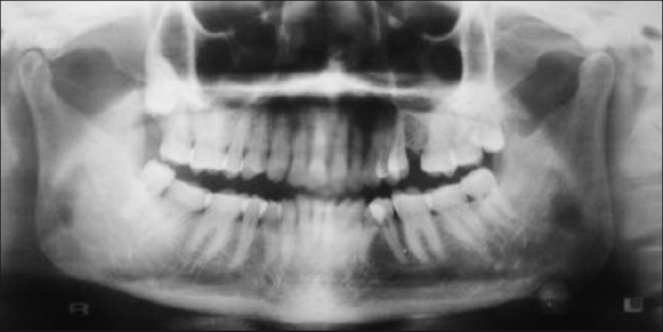
The orthopantamograph of 34-year-old female view revealed a well-defined radiopaque mass in relation to left lower border of mandible.
CT view revealed a well-defined radio-dense area attached to the left medial aspect of lower border of mandible [Figures 4 and 5]. On the basis of radiographical examination, diagnosis of osteoma of left body of mandible was given. An excisional biopsy of the lesion and subsequent histopathological analysis revealed well-differentiated mature bone with proliferation of cancellous bone, thus confirming the lesion as osteoma.
Figure 4.
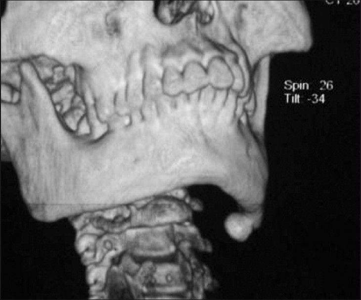
CT with 3D reconstruction view of 34-year-old female revealed a well-defined radio-dense area attached to the left medial aspect of lower border of mandible.
Figure 5.
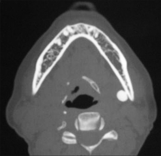
CT view revealed a well-defined radio-dense area attached to the left medial aspect of lower border of mandible of 34-year-old female.
DISCUSSION
Osteoma is a benign neoplasm consisting of well-differentiated compact or cancellous bone that increases in size by continuous osseous growth.[5–10] Peripheral osteoma occurs most frequently in the frontal, ethmoid, and maxillary sinuses but are not common in jawbones.[5,8,9]
A review of the literature of the last 30 years revealed only 16 well-documented cases: 15 cases of peripheral osteoma in the mandible and 1 case in the body of the maxilla. Kaplan et al reported that the age at which the lesions are first identified ranges between 15 and 75 years, the majority being noticed after the age of 25.[8] The duration of the lesions varies between 1 and 22 years. It is reported that it is more predominant in females by a ratio of 3:1.[5]
The pathogenesis of peripheral osteoma is still unknown.[5,7,9] Some investigators classified it as a reactive condition triggered by trauma, because peripheral osteomas are generally located on the lower border or buccal aspect of the mandible which are traumatized areas. Trauma may be minor, that is unlikely to be remembered by the patient years later, and causes subperiosteal bleeding or edema that causes an osteogenic reaction.[5,7–9] Others believe it is a true benign neoplasm.[8]
Regarding sign and symptoms, peripheral osteoma causes some degree of asymmetry while paranasal osteoma causes headache, neuralgia, exopthalmus, and diplopia.[3] Bony hyperplasia associated with muscle traction is a documented phenomena. Authors have suggested that a combination of trauma and muscle traction may play a role in development of osteomas. Either one or both might initiate an osteogenic reaction that could be perpetuated by the continuous muscle traction in the masseter muscle.[5,8]
The discovery of an osteoma of the facial skeleton should raise the possibility of Gardener′s syndrome.[5] Patients with Gardner's syndrome may present with symptoms of rectal bleeding, diarrhea, and abdominal pain. The triad of colorectal polyposis, skeletal abnormalities, and multiple impacted or supernumerary teeth is consistent with this syndrome. Onset occurs in the second decade, with malignant transformation of the colorectal polyps approaching 100% by age 40. The skeletal involvement includes both peripheral and endosteal osteomas, which can occur in any bone but are found more frequently in the skull, ethmoid sinuses, mandible, and maxilla. Controversy also exists as to the adequate terminology for soft tissue lesions. Choristoma is a more accepted term and it is used first time from Krolls et al.[10]
In the presented case, the age, sex, and site of the lesion are in agreement with the earlier reports of osteomas. In our patient, there was no history of trauma, but there could be a chance that patient experienced minor trauma which she is not aware of. It is believed that masseter traction, in particular, plays a role in the occurrence of such lesions. The lesion in our case is an isolated one and no corroborating syndromal features were found.
Footnotes
Source of Support: Nil
Conflict of Interest: None declared.
Available FREE in open access from: http://www.clinicalimagingscience.org/text.asp?2011/1/1/56/90483
REFERENCES
- 1.Woldenberg Y, Nash M, Bodner L. Peripheral osteoma of the maxillofacial region.Diagnosis and management: A study of 14 cases. Med Oral Patol Oral Cir Bucal. 2005;10 Suppl 2:E139–42. [PubMed] [Google Scholar]
- 2.Mancini JC, Woltmann M, Felix VB, Freitas RR. Peripheral osteoma of the mandibular condyle Int, J Oral Maxillofac Surg 2005;34:92-3. Int J Oral Maxillofac Surg. 2005;34:92–3. doi: 10.1016/j.ijom.2004.01.028. [DOI] [PubMed] [Google Scholar]
- 3.Rattan V, Gautam R. Peripheral osteoma of mandible arising from anterior lingual alveolar plate-A case report. J Indian Soc Pedod Prev Dent. 1999;17:132–4. [PubMed] [Google Scholar]
- 4.Bessho K, Murakami K, Iizuka T, Ono T. Osteoma in mandibular condyle. Int J Oral Maxillofac Surg. 1987;16:372–5. doi: 10.1016/s0901-5027(87)80162-5. [DOI] [PubMed] [Google Scholar]
- 5.Kashima K, Rahman OI, Sakoda S, Shiba R. Unusual peripheral osteoma of the mandible: Report of 2 cases. J Oral Maxillofac Surg. 2000;58:911–3. doi: 10.1053/joms.2000.8223. [DOI] [PubMed] [Google Scholar]
- 6.Scheneider LC, Dolinsky HB, Grodjesk JE. Solitary peripheral osteoma of the jaws: Report of case and review of literature. J Oral Surg. 1980;38:452–5. [PubMed] [Google Scholar]
- 7.Richards HE, Strider JW, Jr, Short SG, Theisen FC, Larson WJ. Large peripheral osteoma arising from the genial tubercle area. Oral Surg Oral Med Oral Pathol. 1986;61:268–71. doi: 10.1016/0030-4220(86)90373-7. [DOI] [PubMed] [Google Scholar]
- 8.Kaplan I, Calderon S, Buchner A. Peripheral osteoma of the mandible: A study of 10 new cases and analysis of the literature. J Oral Maxillofac Surg. 1994;52:467–70. doi: 10.1016/0278-2391(94)90342-5. [DOI] [PubMed] [Google Scholar]
- 9.Bodner L, Gatot A, Sion-Vardy N, Fliss DM. Peripheral osteoma of the mandibular ascending ramus. J Oral Maxillofac Surg. 1998;56:1446–9. doi: 10.1016/s0278-2391(98)90414-1. [DOI] [PubMed] [Google Scholar]
- 10.Krolls SO, Jacoway JR, Alexander WN. Osseous choristomas (osteomas) of intraoral soft tissues. Oral Surg Oral Med Oral Pathol. 1971;32:588–95. doi: 10.1016/0030-4220(71)90324-0. [DOI] [PubMed] [Google Scholar]


