Abstract
A case series of four patients who presented with large surface vascular tumors and low platelet count and their management is reported. Medical management was done with steroids, propranolol and vincristine in different combinations. The final response was excellent without surgery.
KEY WORDS: Congenital hemangioma, Kaposiform hemangioendothelioma, Kasabach–Merritt syndrome, propranolol, steroids, vincristine
INTRODUCTION
Kasabach–Merritt syndrome (KMS) is a potentially life-threatening coagulopathy characterized by enlarging hemangioma with severe thrombocytopenia.[1] KMS is associated with kaposiform hemangioendothelioma (KHE), tufted angiomas and rarely with congenital hemangiomas (CHs).[2] Almost 200 cases have been reported in the literature since Kasabach and Merritt described the first case in 1940. More than 80% of cases occur within the first year of life.[3] We report four cases of KMS, three associated with KHE and one associated with CH, which have been successfully treated with steroids, propranolol, and vincristine in varying combinations.
CASE REPORTS
Case 1
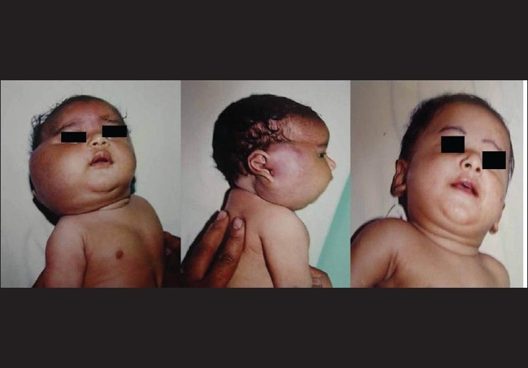
In 1998, a newborn girl was admitted with a swelling in the right parotid region [Figure 1]. It was a tense infiltrating vascular lesion with thickening of the skin. A clinical diagnosis of hemangioma involving the right parotid extending on to the right temporal area was made. An ultrasound confirmed the diagnosis. The platelet count was low and a diagnosis of KHE with KMS was made.
Figure 1.

Kaposiform Hemangioendothelioma with Kasabach–Merritt Syndrome involving the parotid region, treated with steroids – complete regression
The platelet count was 10,000 and she was started on steroids at 1 mg/kg. The platelet count improved and the steroids were continued for 3 months. The lesion started regressing and the whole lesion had regressed completely by 1 year of age [Figure 1].
Case 2
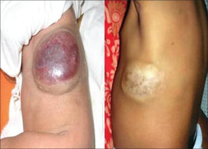
A full-term male baby was referred at 4 days of age with a large CH on the left side of the chest wall. On examination, the baby was a healthy newborn with a violaceous swelling of 7 × 6 cm on the lateral chest wall [Figure 2]. An ultrasound had shown a heterogeneous mass with increased vascularity and a differential diagnosis of CH/fibrosarcoma was made. Magnetic resonance imaging (MRI) was suggestive of CH. The child was icteric with a platelet count of 45,000. D-dimer was positive and suggestive of consumptive coagulopathy. A diagnosis of KMS with CH was made. Treatment was started with compression bandage and steroids. The platelet count did not improve and hence the child was started on propranolol. The swelling started regressing in size and the platelet count normalized in 5 days, and by 10 days propranolol was tapered as there was hypoglycemia. The steroids were continued and then tapered after 6 months. The lesion had completely regressed with minimal skin wrinkling by 10 months of age [Figure 2].
Figure 2.

Chest wall congenital hemangioma with Kasabach–Merritt Syndrome – treated with steroids and propranolol
Case 3
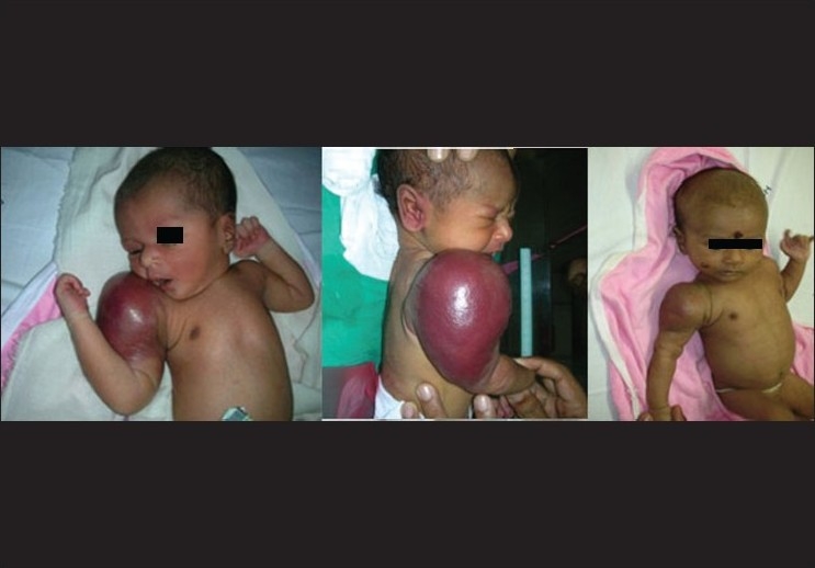
A full-term male baby was referred at 7 hours of life with a large swelling involving the right arm. The antenatal scans were normal. On examination, there was an 8 × 10 cm, red, tense, shiny, warm swelling over the right shoulder, extending to the upper two-thirds of the right arm [Figure 3]. There was no distal neurovascular deficit. Systemic examination was normal. A diagnosis of KHE/fibrosarcoma was made.
Figure 3.

Kaposiform Hemangioendothelioma with Kasabach–Merritt Syndrome – right arm treated with steroids and propranolol
There was thrombocytopenia (60,000) and the coagulation profile was deranged [prothrombin time 23/15 seconds, activated partial thromboplastin time 46/30 seconds and international normalized ratio (INR) 2.1]. A radiograph of the right arm showed no bony abnormality. An ultrasonogram of the neck and shoulder revealed a large soft tissue mass involving subcutaneous and muscular plane with increased vascularity within and arterialization of the veins. The ultrasonogram of the abdomen and echocardiogram were normal. MRI of the right shoulder revealed a diffuse, large circumferential, soft tissue mass lesion in the right arm around the humeral shaft, extending over the shoulder up to the right suprascapular region and elbow joint caudally. The D-dimer was positive (>0.9 mcg FEU/ml) and suggestive of consumptive coagulopathy. A diagnosis of KMS with KHE was made.
The baby was initially treated with steroids but the platelets dropped to 13,000 cells/mm, and hence was started on platelet transfusions. After transfusion of two units of platelet concentrates, the swelling appeared to shrink, but the platelet count dropped to 10,000 and the baby developed small purpuric spots over trunk and a subcutaneous hematoma (3 × 3 cm) over the left calf. A possibility of coagulopathy aggravated by repeated platelet transfusion was considered. Hence, platelet transfusions were discontinued and propranolol was started. From the 10th day, the swelling started regressing and the platelet count started to rise. The baby was subsequently discharged on oral steroids and propranolol. After a week, there was a steady rise in platelet count (platelets 70,000 cells/mm). Propranolol was tapered after a month and steroids were continued and then tapered. The hemangioma slowly diminished in size. When last seen at 1 year of age, there was faint skin staining at the site of the original hemangioma [Figure 3].
Case 4
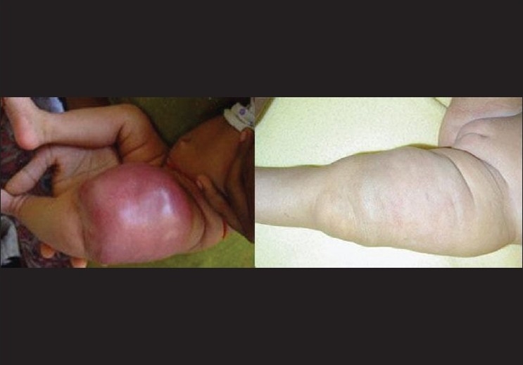
A newborn girl child was born with a large shiny swelling involving the left thigh. As there was severe thrombocytopenia, a diagnosis of KHE with KMS was made. As per the protocol, steroids were started but there was no regression. There was no improvement in the platelet count and hence propranolol was added. There was no visible improvement in the clinical scenario, and due to increasing size, biopsy was advised. Parents refused biopsy and injection Vincristine was started at a dose of 0.1 mg/kg. There was dramatic improvement and hence weekly injections were continued for 27 weeks. At the age of 8 months, there was 90% regression and the baby is on compression bandage [Figure 4].
Figure 4.

Kaposiform Hemangioendothelioma with Kasabach–Merritt Syndrome – right thigh after a course of vincristine
DISCUSSION
KMS with KHE is a well-known entity. KMS occurs during the first few weeks of life with a mortality of 20–30%.[4] KMS shows a wide variation in its response to different treatment modalities. Currently, there are no known treatment guidelines.[5]
The aims of treatment of KMS are twofold – involution of the tumor and correction of the life-threatening coagulopathy. Different interventions are recommended including use of steroids, compression, embolization, use of interferon, laser therapy, sclerotherapy, chemotherapy, radiation or surgery.[5] In each case, the treating physician must decide the suitable treatment to achieve maximum involution of the lesion and preservation of organ function with the least toxicity. Corticosteroids have been the traditional first-line therapy for KMS when surgical excision is not possible.[5,6] Several researchers agree that most patients with KMS respond to steroids within a few days of treatment.
The first baby was a classical case of KMS with KHE, wherein the hemangioma responded to prednisolone which is the first line of therapy. However, one-third may not respond to conventional dose of prednisolone (2 mg/kg/day) and a mega dose of 5 mg/kg/day may be effective.
There have been few reports of CH with KMS, but the platelet count does not drop below 40,000. Due to the fear of intracranial bleed, propranolol was added in the second patient. Propranolol could have played a role in the improvement of hemangioma, but was stopped due to hypoglycemia. The third patient who had a diagnosis of KHE with KMS had severe symptomatic thrombocytopenia. There was a dramatic response in the platelet count with regression of the tumor after commencing propranolol.
The most recent option available for treatment of infantile hemangiomas is propranolol. In a short span of less than 2 years, propranolol therapy has gone from a serendipitous discovery to first-line therapy in some hemangiomas requiring medical treatment.[2] Propranolol is a non-selective beta blocker with a long history of use in infants for cardiac problems.[2] In infantile hemangiomas, the possible mechanism of action of propranolol is vasoconstriction, decreased expression of vascular endothelial growth factor (VEGF) and basic fibroblast growth factor (bFGF) genes through the down-regulation of the RAF-mitogen-activated protein kinase pathway (which explains the progressive improvement of the hemangioma) and the triggering of apoptosis of capillary endothelial cells.[7] Propranolol is a revolutionary treatment for life-threatening, endangering and disfiguring hemangiomas which acts by stopping the growth and hastening involution.[2] It is associated with some severe systemic complications and side effects and infants need to be closely monitored.[5] The most common reported adverse effects of propranolol include hypotension, bradycardia, hypoglycemia and bronchospasm.[2,7,8] Infants with very large hemangiomas or military hemangiomatosis are at risk for high-output cardiac compromise. Propranolol may mask the clinical signs of early cardiac failure and diminish cardiac performance. Propranolol may also blunt the clinical features of hypoglycemia. Sustained hypoglycemia in infancy has been associated with long-term neurologic sequelae.[8]
Propranolol's familiarity, long track record, tolerability and dramatic efficacy relative to current standard of care are causes for both excitement and caution. Most experiences with propranolol are in very different clinical contexts – sick infants, closely monitored and in hospital drug initiation. As we broaden propranolol indication to include a typically outpatient condition, restraint and prudence are paramount.[2] The use of propranolol in KHE is still in its infancy. Hermans et al. state that the use of propranolol may be a new indication in KHE with KMS.[9] A treatment protocol has been developed to optimize safety: baseline echocardiography and 48-hour hospitalization or home nursing visits to monitor vital signs and blood glucose levels. Medication is given every 8 hours, with an initial dose of 0.16 mg/kg of body weight. If the vital signs and glucose levels remain normal, the dose is incrementally doubled to a maximum of 0.67 mg/kg (to a maximum daily dose of 2.0 mg/kg). Propranolol should be gradually tapered over a period of 2 weeks.
The fourth baby has been successfully treated with injection Vincristine. Vincristine is an anti-microtubule chemotherapeutic drug of which there is an increasing evidence showing its efficacy in the treatment of KMS.[6,10] Using vincristine and corticosteroids as first-line treatment of KMS in KHE has been reported.[11] Potential adverse effects associated with vincristine use are reversible alopecia, nausea, vomiting, autonomic neuropathy, peripheral paresthesia, jaw pain, ataxia and hyponatremia owing to syndrome of inappropriate antidiuretic hormone hypersecretion (SIADH).[6,12]
When a baby presents with KHE and KMS, the first line of management in our hospital has been steroids at a dose of 2 mg/kg/day. If there is no response, propranolol is added at a maximum dose of 2 mg/kg/day. Injection Vincristine has been used as a last resort at a dose of 0.1 mg/kg/day.
CONCLUSION
Vascular tumors in infancy may present with KMS and require aggressive treatment. All our patients responded to medical management. The use of propranolol is new and encouraging, but the infants have to be monitored for the complications.
Footnotes
Source of Support: Nil
Conflict of Interest: None declared.
REFERENCES
- 1.Kasabach HH. Merritt. Capillary hemangioma with extensive purpura: Report of a case. Am J Dis Child. 1940;59:1063–70. [Google Scholar]
- 2.Sidbury R. Update on vascular tumors of infancy. Curr Opin Pediatr. 2010;22:432–7. doi: 10.1097/MOP.0b013e32833bb764. [DOI] [PubMed] [Google Scholar]
- 3.Hesselmann S, Micke O, Marquardt T, Baas S, Bramswig JH, Harms E, et al. Case report: Kasabach-Merritt syndrome: A review of the therapeutic options and a case report of successful treatment with radiotherapy and interferon alpha. Br J Radiol. 2002;75:180–4. doi: 10.1259/bjr.75.890.750180. [DOI] [PubMed] [Google Scholar]
- 4.López V, Martí N, Pereda C, Martín JM, Ramón D, Mayordomo E, et al. Successful management of Kaposiform hemangioendothelioma with Kasabach-Merritt phenomenon using vincristine and ticlopidine. Pediatr Dermatol. 2009;26:365–6. doi: 10.1111/j.1525-1470.2009.00923.x. [DOI] [PubMed] [Google Scholar]
- 5.Abass K, Saad H, Kherala M, Abd-Elsayed AA. Successful treatment of kasabach-Merritt syndrome with vincristine and surgery: A case report and review of literature. Cases J. 2008;23:1–9. doi: 10.1186/1757-1626-1-9. [DOI] [PMC free article] [PubMed] [Google Scholar]
- 6.Drucker AM, Pope E, Mahant S, Weinstein M. Vincristine and corticosteroids as first-line treatment of Kasabach-Merritt syndrome in kaposiform hemangioendothelioma. J Cutan Med Surg. 2009;13:155–9. doi: 10.2310/7750.2008.08006. [DOI] [PubMed] [Google Scholar]
- 7.Guldbakke KK, Rørdam OM, Huldt-Nystrøm T, Hanssen HK, Høivik F. Propanolol used in treatment of infantile hemangioma. Tidsskr Nor Laegeforen. 2010;130:1822–4. doi: 10.4045/tidsskr.09.1435. [DOI] [PubMed] [Google Scholar]
- 8.Siegfried EC, Keenan WJ, Al-Jureidini S. More on propranolol for hemangiomas of infancy. N Engl J Med. 2008;359:2846. doi: 10.1056/NEJMc086443. [DOI] [PubMed] [Google Scholar]
- 9.Hermans DJ, van Beynum IM, van der Vijver RJ, Kool LJ, de Blaauw I, van der Vleuten CJ. Kaposiform hemangioendothelioma with Kasabach-Merritt syndrome: A new indication for propranolol treatment. J Pediatr Hematol Oncol. 2011;33:e171–3. doi: 10.1097/MPH.0b013e3182152e4e. [DOI] [PubMed] [Google Scholar]
- 10.Fahrtash F, McCahon E, Arbuckle S. Successful treatment of kaposiform hemangioendothelioma and tufted angioma with vincristine. J Pediatr Hematol Oncol. 2010;32:506–10. doi: 10.1097/MPH.0b013e3181e001a9. [DOI] [PubMed] [Google Scholar]
- 11.Drucker AM, Pope E, Mahant S, Weinstein M. Vincristine and corticosteroids as first-line treatment of Kasabach-Merritt syndrome in kaposiform hemangioendothelioma. J Cutan Med Surg. 2009;13:155–9. doi: 10.2310/7750.2008.08006. [DOI] [PubMed] [Google Scholar]
- 12.Vin-Christian K, McCalmont TH, Frieden IJ. Kaposiform hemangioendothelioma.An aggressive, locally invasive vascular tumor that can mimic hemangioma of infancy. Arch Dermatol. 1997;133:1573–8. doi: 10.1001/archderm.133.12.1573. [DOI] [PubMed] [Google Scholar]


