Abstract
Hemangioendothelioma is an uncommon vascular lesion that usually occurs in the liver, bone, lung, skin, and other organs with unknown etiology. A rare form of this lesion has been reported in the eyelid. We report the case of a 27-year-old female with right lower eyelid mass simulating chalazion of 3 weeks duration. The histopathologic examination of the excised nodule confirmed the diagnosis. To our knowledge, this is the fourth case of eyelid epithelioid hemangioendothelioma reported in the English literature.
Keywords: Chalazion, eyelid, hemangioendothelioma
Introduction
Vascular neoplasms or malformations are frequently reported in adnexal tissues that usually possess benign behavior (hemangiomas) or rarely frank malignant characteristics (angiosarcomas). A rare vascular tumor entity with intermediate malignant potential is the hemangioendothelioma which is derived from vascular endothelium.[1] It was initially described by Weiss and Enzinger in 1982 and later discussed as differential diagnosis of masquerade eyelid tumors by de Keizer and Scheffer in 1989.[2,3] It can affect many organs; however, eyelid involvement is extremely rare.[4]
Case Report
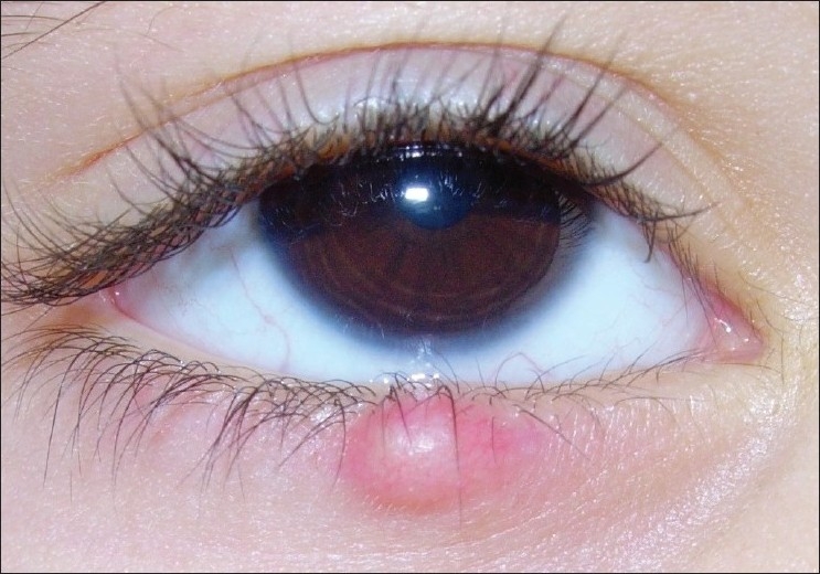
A 27-year-old lady presented with a 3-week history of painless right lower eyelid swelling with no history of trauma or evidence of systemic disease. The nodule was small, firm, fixed to the tarsus and pinkish in color [Figure 1]. Complete ocular examination showed 20/20 visual acuity in both eyes, normal anterior and posterior segment findings, no proptosis, and full extraocular motility. A provisional diagnosis of chalazion was made with a surgical plan to incise and curette the lesion. Intraoperatively, identification of abnormal content encouraged us to perform complete excision under local anesthesia. Unlike chalazion, the content was fleshy, reddish, and bloody with gritty sensation.
Figure 1.

Clinical feature of epithelioid hemangioendothelioma of the right lower eyelid shows small, firm, pinkish nodule that mimics chalazion
The excised specimen was gray tan soft tissue and measured 0.5 × 0.3 cm. Histopathological examination of hematoxylin and eosin-stained slides revealed densely packed spindle to ovoid, unremarkable endothelial cells with newly formed slits that contain red blood cells [Figures 2a and b] with free margins. Immunohistochemistry study showed a positive staining of the neoplastic cells by the endothelial cell marker CD31 supporting the diagnosis of epithelioid hemangioendothelioma of the eyelid [Figure 2c].
Figure 2.

Histological examination of hematoxylin and eosin (H and E) stained slides revealed densely packed spindle to ovoid, unremarkable endothelial cells (arrows) with newly formed slits that contain red blood cells. (a) H and E stain, ×40. (b) H and E stain, ×200. (c) Immunohistochemistry study showed a positive staining of the neoplastic cells by the endothelial cell marker CD31 (arrows)
At this stage, complete blood count, urinalysis, renal and liver function tests, chest radiography, and abdominal ultrasound were ordered and all were within normal limits. After more than 2 years of follow-up, there has been no evidence of local recurrence or distant metastasis.
Discussion
According to the updated classification by the International Society for the Study of Vascular Anomalies, hemangioendothelioma has been subclassified into Kaposiform, spindle cell, epithelioid, composite, retiform, and polymorphous hemangioendotheliomas, lymphangioendotheliomatosis, and Dabska tumor.[5] The histopathologic features of our reported lesion were consistent with epithelioid hemangioendothelioma without atypia.
Epithelioid hemangioendothelioma is a rare vascular tumor believed to be in the middle of the spectrum of epithelioid vascular tumors between benign epithelioid hemangioma and highly aggressive epithelioid angiosarcoma.[6] It is seen mainly in middle-aged adults and usually occurs as a solitary lesion with variable clinical behavior.[1] It can affect many organs such as the liver, bone, skin, lung, and soft tissues; however, eyelid involvement is exceedingly rare. To our knowledge, this is the fourth report of an epithelioid hemangioendothelioma arising on the eyelid reported in the English literature.[7–9]
Because the eyelid epithelioid hemangioendothelioma is a low-grade malignant lesion that shows a high rate of local recurrence but rarely metastasizes,[1,6] complete excision (not biopsy) should be performed to avoid recurrence. As our patient underwent complete excision of the eyelid hemangioendothelioma, she required no therapy other than local excision. Moreover, adjuvant radiotherapy or chemotherapy has not proven to be beneficial.[6,10] In fact, because epithelioid hemangioendothelioma is rare and almost always discovered after surgical excision for a supposed benign cause, few patients have undergone complete preoperative investigation to detect the presence of possible metastasis. In addition, it has been reported that possible metastasis might not become evident for many years because of the slow growth of this tumor.[6] Therefore, follow-up with computed tomography scan is recommended to study regional lymph nodes and lungs, which are the most involved sites. So, although our patient has shown no evidence of recurrence so far, careful follow-up will be necessary to detect possible late recurrence of her epithelioid hemangioendothelioma.
In summary, hemangioendothelioma can masquerade as a chalazion and any abnormal content observed during chalazion evacuation should encourage complete excision.
Footnotes
Source of Support: Nil
Conflict of Interest: None declared.
References
- 1.Kumar V, Abbas AK, Fausto N, Aster JC. Robbins and Cotran Pathologic Basis of Disease. 8th ed. Vol. 11. Philadelphia: Saunders, Elsevier Inc; 2010. p. 524. [Google Scholar]
- 2.Weiss SW, Enzinger FM. Epithelioid hemangioendothelioma a vascular tumor often mistaken for a carcinoma. Cancer. 1982;50:970–8. doi: 10.1002/1097-0142(19820901)50:5<970::aid-cncr2820500527>3.0.co;2-z. [DOI] [PubMed] [Google Scholar]
- 3.De Keizer RJ, Scheffer E. Masquerade of eyelid tumours. Doc Ophthalmol. 1989;72:309–21. doi: 10.1007/BF00153498. [DOI] [PubMed] [Google Scholar]
- 4.Bollinger BK, Laskin WB, Knight CB. Epithelioid hemangioendothelioma with multiple site involvement. Literature review and observations. Cancer. 1994;73:610–5. doi: 10.1002/1097-0142(19940201)73:3<610::aid-cncr2820730318>3.0.co;2-3. [DOI] [PubMed] [Google Scholar]
- 5.Enjolras O, Wassef M, Chapot R. In: Color atlas of vascular tumors and vascular malformations. New York: Cambridge University Press; 2007. Introduction: ISSVA classification; pp. 1–11. [Google Scholar]
- 6.Enzinger FM, Weiss SW. Hemangioendothelioma: Vascular tumors of intermediate malignancy. In: Enzinger FM, editor. Soft Tissue Tumors. St. Louis: Mosby; 1995. pp. 891–914. [Google Scholar]
- 7.Wolter JR, Lewis RG. Endovascular hemangioendothelioma of the eyelid. Am J Ophthalmol. 1974;78:727–9. doi: 10.1016/s0002-9394(14)76315-9. [DOI] [PubMed] [Google Scholar]
- 8.Cho SH, Na KS. Haemangioendothelioma on the conjunctiva of the upper eyelid. Clin Experiment Ophthalmol. 2006;34:794–6. doi: 10.1111/j.1442-9071.2006.01320.x. [DOI] [PubMed] [Google Scholar]
- 9.Tsuji H, Kanda H, Kashiwagi H. Primary epithelioid haemangioendothelioma of the eyelid. Br J Ophthalmol. 2010;94:261–2. doi: 10.1136/bjo.2008.156497. [DOI] [PubMed] [Google Scholar]
- 10.Mentzel T, Beham A, Calonje E, Katenkamp D, Fletcher CD. Epithelioid hemangioendothelioma of skin and soft tissues: Clinicopathologic and immunohistochemical study of 30 cases. Am J Surg Pathol. 1997;21:363–74. doi: 10.1097/00000478-199704000-00001. [DOI] [PubMed] [Google Scholar]


