Dear Editor,
We congratulate Mahesh et al., for their article on “video indirect ophthalmoscopy using handheld video camera.”[1] Documentation of clinical findings is a crucial part of an ophthalmologist's work and is important for communication with col-leagues and patients. The quality of an image depends as much as on the photographer's knowledge of the anatomy and physiology of the eye, as it does on photographic techniques and technology. In this regard, we would like to describe certain techniques in anterior segment photography which would greatly enhance the quality of the images obtained using an inexpensive personal digital camera.
The settings we recommend for external photography (for lids and adnexal structures) are:
Most cameras have “auto focus” mode and the system automatically adjusts to select the best focus. For clinical photography it is better to switch off the auto focus mode and to select “central focus” mode.
Flash is optional, depending on ambient lighting. It is better to capture he image with and without flash and choose the better one later.
ISO settings should be set as “auto” unless the photographer is experienced with different ISO speeds of camera.
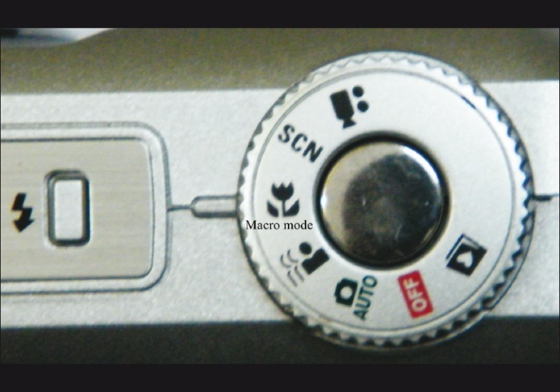
An important setting is the “macro mode” function denoted by “flower symbol” [Fig. 1]. It allows the camera to focus on objects as close as 2-4 inches.[2] With this, finer details of lid and adnexal lesions like surface irregularity, vascularity, and pigmentation can be documented, preserving clarity even on zooming. With the same technique gross details of the anterior segment can also be photographed.
Proper focus is obtained by pressing the shutter button halfway, then adjusting the camera to obtain clear focus as seen on the LCD viewfinder. On obtaining clear focus, the yellow-colored rectangle will change to green in the LCD viewfinder.
Figure 1.

Photograph showing “macro mode” in the camera
For slit-lamp photography of the anterior segment, the digital camera should be placed in one of the slit-lamp oculars after adjusting focus.
Cameras with 3 megapixel (MP) resolution cater to most of our needs in obtaining clinical photographs. Higher resolution cameras (>3 MP) are not required for this purpose. Choose cameras with the objective piece protruding out so that it can snugly fit into slit-lamp oculars.
Settings recommended are centre focus, macro mode, flash off, auto ISO.
Camera focus can be adjusted by depressing shutter halfway down until the green box appears. Yellow box indicates that the camera has to be repositioned to obtain better focus.
Most anterior segment photographs require an external light source which is usually provided by ambient room lighting. Without an external light source, the camera makes poor choice regarding exposure, resulting in the image being too dark or too bright. However, for ocular findings like keratic precipitates, iris transillumination defects, or lens opacities, external light source is not necessary.
Anterior segment photographs are obtained better with wide illumination with diffuser on.
Good-quality photographs make scientific publications more convincing. They can also be used as evidence in medico-legal cases. Techniques of obtaining clinical photographs should be part of resident training. This method may help ophthalmologists working in resource-constraint situations to document ophthalmic findings without expensive photography equipments.
References
- 1.Shanmugham PM. Video indirect ophthalmoscopy using a hand held video camera. Indian J Ophthalmol. 2011;59:53–5. doi: 10.4103/0301-4738.73718. [DOI] [PMC free article] [PubMed] [Google Scholar]
- 2.Fogla R, Rao SK. Ophthalmic photography using a digital camera. Indian J Ophthalmol. 2003;51:269–72. [PubMed] [Google Scholar]


