Dear Editor,
Dirofialria repens (D. repens) is a nematode which occasionally causes ocular infestation. Commonly known as the dog heartworm, it is a nematode of veterinary importance. It infects man as an accidental host through vectors such as the anopheles and culex mosquitoes.[1
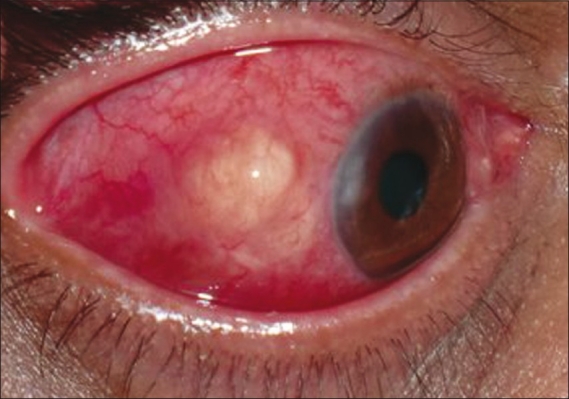
A 48-year-old male from New Mumbai, Western India presented to the Laxmi Eye Institute, Panvel, a tertiary referral centre in Western India, with complaints of a painless swelling and redness over the white portion of the right eye noticed since 18 days. There was no history of similar episodes in the past, ocular trauma or foreign body. There was no history of any systemic illness and recent travel (in the past one year) out of Mumbai. The right eye (OD) revealed localized conjunctival congestion with a yellowish cream, firm, non-mobile, non-tender, scleral nodule measuring 4 mm × 4 mm situated 3 mm away from the temporal limbus. There were no abnormal feeder vessels. Temporal cornea showed dellen [Fig. 1]. Rest of the ocular examination was normal. Ocular examination of the left eye was essentially normal. A provisional diagnosis of OD atypical infective scleritis was made and a scleral deroofing with excision biopsy of the nodule was planned. At operation, after reflecting the overlying conjunctiva, two live thread-like worms were seen; they were coiled amidst a thick granulation tissue. Most of the conjunctiva was also adherent to the granulation tissue below. The worms were retrieved entirely and the conjunctival space was explored all around to rule out presence of any more worms. The whole granulation tissue was excised and sent in 10% formalin for histopathological analysis to the department of microbiology, King Edward Memorial Hospital, Parel, Mumbai. The resultant conjunctival defect was covered with a conjunctival autograft using fibrin glue.
Figure 1.

Scleral nodule with corneal dellen
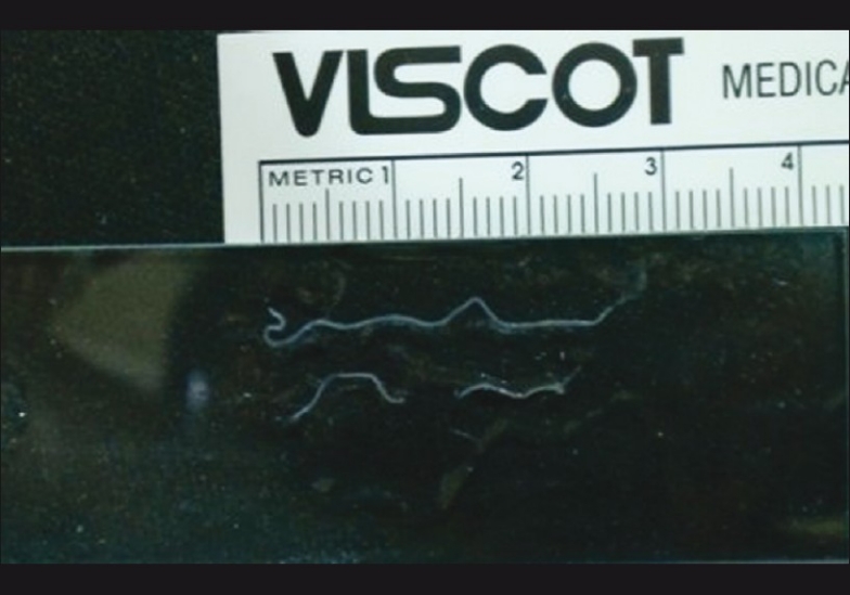
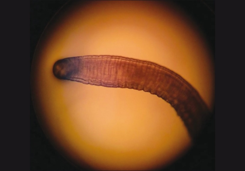
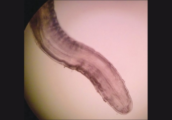
Blood examination revealed normal hemogram. Peripheral smears did not reveal any microfilariae and stool examination was normal. Analysis of the worms revealed presence of two white worms approximately 3.5 cm long with a maximum thickness of 0.7 mm [Fig. 2]. A thick cuticle with fine transverse striations was present throughout the length of the worms [Fig. 3]. The species were identified as female D. repens based on the presence of longitudinal striae and a thick muscle coat[2] [Fig. 4] and the presence of a prominent uterine pore. The histopathological analysis of the tissue showed a large number of eosinophils.
Figure 2.

Naked eye appearance of worms of D. repens
Figure 3.

Microscopic appearance of D. repens showing transverse striations and longitudinal ridge
Figure 4.

Microscopic appearance of D. repens showing thick muscle coat
The patient was started on prednisolone acetate 1% eye drops in tapering dose for one month and ofloxacin 0.3% eye drops for one week. The patient had a well-integrated conjunctival autograft with a quiet eye when he followed up after one month.
Our case is the first reported case of ocular dirofilariasis from Western India and was managed with adequate surgical excision with conjunctival autograft. This first indigenous, case of ocular dirofilaria from Western India highlights the importance of being aware of this infection for ophthalmologists from this part of the country; this can help in increased recognition and reporting of such infections.
Acknowledgments
The authors wish to thank Dr Madhav Sathe, Dept of Microbiology, Seth G S Medical College, K E M Hospital and Dr Leena Gajbar, Aditya Labs, Panvel for their assistance in microbiological identification of the worm and Dr Maninder Setia, Dept of Clinical research and Epidemiology, Laxmi Eye Institute, Panvel, for editorial comments.
References
- 1.Rani PA, Irwin PJ, Gatne M, Coleman GT, McInnes LM, Traub RJ. A survey of canine filarial diseases of veterinary and public health significance in India. Parasit Vectors. 2010;3:30. doi: 10.1186/1756-3305-3-30. [DOI] [PMC free article] [PubMed] [Google Scholar]
- 2.Nath R, Gogoi R, Bordoloi N, Gogoi T. Ocular Dirofilariasis. Indian J Pathol Microbiol. 2010;53:157–9. doi: 10.4103/0377-4929.59213. [DOI] [PubMed] [Google Scholar]


